Search results (1066 results)
-
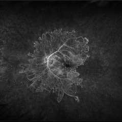
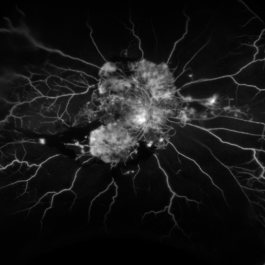
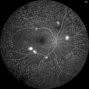
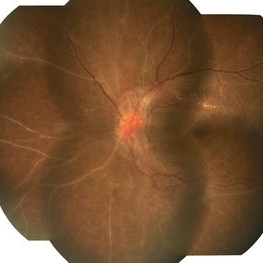
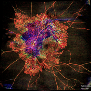 Flame of the Forest
Apr 9 2020 by Daraius N Shroff, MS FMRF FRCS
A 54-year-old man with DM for 15 years. The left eye had a visual acuity of 20/40. Wide field swept source OCTA revealed branching out central neovascular trunk vessels from the disc with terminal loops, along with exuberant proliferation of irregular small-calibre fine new vessels. The patient underwent OCTA guided pan retinal photocoagulation.
Photographer: Anuj Choudhary, Shroff Eye Centre, New Delhi
Imaging device: Zeiss Plex Elite 9000
Condition/keywords: proliferative diabetic retinopathy (PDR)
-
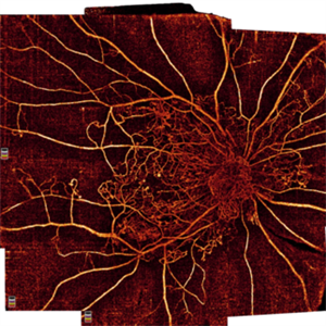
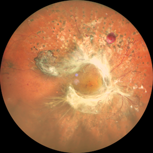
 Proliferative Diabetic Retinopathy with Choroidal Effusion Status Post PRP
Dec 15 2020 by Manish Nagpal, MD, FRCS (UK), FASRS
A 17-year-old juvenile diabetic patient came to us with extensive neovascular proliferations and PRP done a week back and had developed 360 degree choroidal effusion as seen in this wide field montage image
Photographer: Sham Talati, Retina Fellow , Retina Foundation, Ahmedabad, India
Imaging device: Mirante CSLO
Condition/keywords: choroidal effusion, diabetic retinopathy, proliferative diabetic retinopathy (PDR)
-
 Tractional Retinal Detachment
Dec 4 2019 by Janet Brazil
Fundus photograph of a 32-year-old female with severe end-stage diabetic tractional retinal detachment.
Photographer: Janet Atkinson, Eye Associates of New Mexico, Albuquerque, NM
Imaging device: Topcon TRC- 50EX
Condition/keywords: diabetes, proliferative diabetic retinopathy (PDR), tractional retinal detachment
-
 Aurora Borealis in Retina
Apr 25 2025 by Poornachandra B, MS, FVRS
Fundus picture of 54 year old male with proliferative diabetic retinopathy with fluorescent blood clot in vitreous cavity.
Photographer: Mr Dhikshith
Imaging device: Optos daytona
Condition/keywords: blood, proliferative diabetic retinopathy (PDR)
-
 Venous Beading
Nov 4 2021 by Stefanie Palmer
Venous Beading in a patient with both PDR and CRVO.
Photographer: Stefanie Palmer, CRA
Imaging device: Topcon
Condition/keywords: central retinal vein occlusion (CRVO), diabetic retinopathy, proliferative diabetic retinopathy (PDR), venous beading
-
 Proliferative Diabetic Retinopathy
Sep 17 2012 by Michael P. Kelly, FOPS
Retinal fundus photograph of a patient with PDR and NVD.
Photographer: Michael P. Kelly, FOPS Director, Duke Eye Labs, Duke University Hospital, Duke Eye Center
Imaging device: Topcon
Condition/keywords: neovascularization of the disc (NVD)
-
 Ocular Ischemic Syndrome/ Severe NPDR
Oct 6 2021 by Becca Harris
53 year old female with Severe NPDR and Ocular Ischemic Syndrome.
Photographer: Becca Harris
Imaging device: Optos California
Condition/keywords: Diabetic Retinopathy, left eye, nonproliferative diabetic retinopathy, ocular ischemic syndrome, optos, retinal ischemia
-
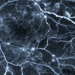
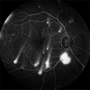 Shooting Stars
Jul 9 2025 by Majda Hadziahmetovic, MD
Fluorescein angiography image demonstrating multiple areas of neovascularization in a middle-aged male patient with long-standing diabetes.
Condition/keywords: proliferative diabetic retinopathy (PDR)
-
 Bilateral CRVO and PDR
Nov 4 2021 by Stefanie Palmer
Patient with both PDR and CRVO, 34 year old female, post-COVID.
Photographer: Stefanie Palmer, CRA
Imaging device: Topcon
Condition/keywords: central retinal vein occlusion (CRVO), COVID-19, diabetic retinopathy, proliferative diabetic retinopathy (PDR), venous beading
-
 PRP laser
Mar 29 2013 by Henry J. Kaplan, MD
Right after PRP laser in PDR.
Condition/keywords: laser photocoagulation, pan-retinal photocoagulation (PRP)
-
 Bilateral CRVO and PDR
Nov 4 2021 by Stefanie Palmer
Patient with both PDR and CRVO, 34 year old female, post-COVID.
Photographer: Stefanie Palmer, CRA
Imaging device: Topcon
Condition/keywords: central retinal vein occlusion (CRVO), COVID-19, diabetic retinopathy, proliferative diabetic retinopathy (PDR), venous beading
-
 Idiopathic Uveal Effusion Syndrome with Diabetic TRD
Jul 2 2025 by Virginia Gebhart
53 year old male with prominent choroidal effusions and significant diabetic tractional pathology/detachment. No visible breaks in presence pf Descemet's folds, unclear whether choroidals may be secondary to rhegmatogenous/tractional RD and low IOP, or separate process of exudative detachment/uveitis with some pre-existing diabetic traction and PDR. All labs WNL, pt was started on oral prednisone.
Photographer: Virginia Gebhart, Retina Consultants of Carolina
Imaging device: Optos California
Condition/keywords: Diabetic Tractional Detachment, idiopathic uveal effusion syndrome, TRD, uveal effusion syndrome
-
 Proliferative Diabetic Retinopathy
Aug 16 2022 by Donnie Willis
51 yo female. Uncontrolled Diabetes. Active PDR.
Photographer: Donnie Willis, Tennessee Retina
Imaging device: Optos
Condition/keywords: capillary dropouts, Diabetes, FA, fluorescein angiogram (FA), Optos, proliferative diabetic retinopathy (PDR), vitreomacular traction (VMT)
-
 Proliferative Diabetic Retinopathy
Oct 16 2021 by Timur Shaimov
32 y.o. female with Type 1 Diabetes with no glucose compensation for several years. A manual montage of several 8x8 mm OCT angiograms were obtained for this Widefield OCTA image.
Photographer: Timur Shaimov
Imaging device: RTVue xR Avanti
Condition/keywords: OCT Angiography, proliferative diabetic retinopathy (PDR)
-
 Proliferative Diabetic Retinopathy with Pre-retinal Hemorrhage
Jan 16 2018 by Olivia Rainey
Ultra-wide field pseudo-color image of an 57-year-old male with a large pre-retinal hemorrhage secondary to proliferative diabetic retinopathy affecting his left eye.
Photographer: Olivia Rainey
Imaging device: Optos California
Condition/keywords: color fundus photograph, diabetic mellitus, hemorrhage, left eye, neovascularization (NV), Optos, proliferative diabetic retinopathy (PDR), pseudocolor, ultra-wide field imaging
-
 Venous Beading
Apr 30 2021 by Shivani Reddy, MD
This is a fluorescein angiogram image capturing a beautiful example of different stages of venous beading in diabetic retinopathy all in one frame. This patient also has various microangiopathic findings including microaneurysms, venous loops and capillary dropout. This patient is a 41 y/o male with a history of type 1 diabetes, presenting for his first eye exam in years.
Imaging device: Optos FA
Condition/keywords: capillary dropouts, nonproliferative diabetic retinopathy, proliferative diabetic retinopathy (PDR), retinal ischemia, venous beading
-
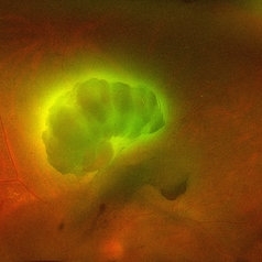
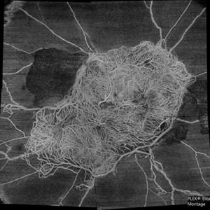 Who Stole My Blood Supply?
Jan 25 2025 by Muna Bhende, MD
Superficial capillary plexus slab montage image of a young female diabetic with florid proliferation . There is no flow in the capillaries anterior to the tangle of new vessels indicating severe retinal ischemia.
Photographer: Mohanapriya L , Medical Research Foundation, Sankara Nethralaya, Chennai, India
Imaging device: PLEX elite 9000
Condition/keywords: florid type PDR, OCTA
-
 Active diabetic retinopathy despite PRP
Oct 30 2022 by Diego Andrés Rodriguez, MD
A 52-year-old patient with active proliferative diabetic retinopathy despite good glycemic control and PRP performed 1 year ago in the right eye
Photographer: Sociedad de Cirugía Ocular
Imaging device: Clarus 700
Condition/keywords: diabetic retinopathy, pan-retinal photocoagulation (PRP), proliferative diabetic retinopathy (PDR), wide angle imaging
-
 Advanced Active PDR
Mar 29 2013 by Henry J. Kaplan, MD
Extensive NVD-FPD and NVE-FPE in a diabetic patient.
Condition/keywords: foveal photoreceptor defect, FPE, neovascularization (NV), neovascularization of the disc (NVD)
-
 Advanced PDR
Mar 29 2013 by Henry J. Kaplan, MD
Large FPD, FPE, NVE and multiple boat shaped subhyaloid hemorrhages inferiorly, ischemic retina and multiple occluded sclerotic vessels.
Condition/keywords: foveal photoreceptor defect, subhyaloid hemorrhage
-
 Advanced PDR RE FFA
Aug 31 2014 by Neha Goel, MS DNB FRCS (Glasg)
Fluorescein angiogram of the right eye.
Photographer: Neha Goel
Imaging device: Zeiss Visucam
Condition/keywords: fibrovascular proliferation, ischaemic diabetic maculopathy, proliferative diabetic retinopathy (PDR)
-
 Advanced PDR-RE
Aug 31 2014 by Neha Goel, MS DNB FRCS (Glasg)
Fundus photograph of the right eye of a 50-year-old diabetic male.
Photographer: Neha Goel
Imaging device: Zeiss Visucam
Condition/keywords: fibrovascular proliferation, ischaemic diabetic maculopathy, proliferative diabetic retinopathy (PDR)
-
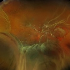
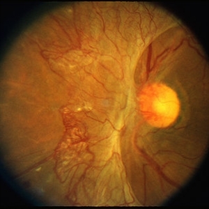
 Advanced Proliferative Diabetic Retinopathy
Nov 4 2017 by Hamid Ahmadieh, MD
Merged color fundus photograph of the left eye of a 30-year-old woman with type1 diabetes since childhood. Note laser scars, severe fibrous proliferation, traction RD and macular dragging.
Photographer: Shabnam Poureh, Negah Eye Center, Tehran, Iran
Condition/keywords: color fundus photograph, diabetes, fibrous proliferation, proliferative diabetic retinopathy (PDR), severe traction
-
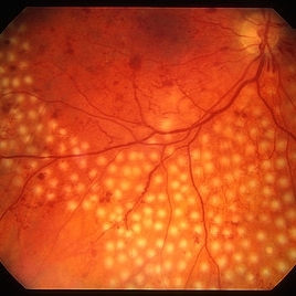
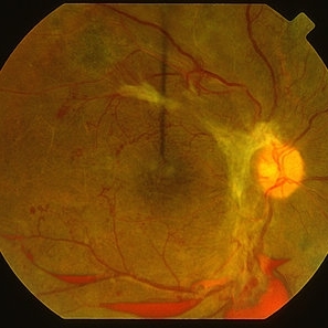
 Advanced Proliferative Diabetic Retinopathy With Fibrovascular Proliferation
Jan 4 2019 by Isha Agarwalla
A 29-year-old female with a long-standing history of diabetes mellitus presented with a fibrovascular membrane (FVM) at the viteroretinal interface due to underlying inflammation and angiogenesis induced by ischemia. FVM involved the disc and extended towards the superior and inferior arcades along with extensive capillary drop out areas due to micro aneurysms.
Condition/keywords: fibrovascular proliferation, proliferative diabetic retinopathy (PDR)
-
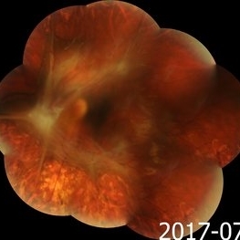
 Annular Tractional Retinal Detachment
Jul 4 2024 by Hector Gabriel Moreno Solano, MD, MHA
52-year-old Hispanic female patient with a diagnosis of type II diabetes mellitus of 15 years of evolution, comes to the retina service for progressive visual loss in the right eye (single functional eye) with visual acuity of 20/100, Fundus examination reveals laser-modified proliferative diabetic retinopathy with activity + annular tractional retinal detachment with macular involvement.
Photographer: Hector Gabriel Moreno Solano, MD, MHA, HGZ #20 IMSS Puebla.
Imaging device: Mirante
Condition/keywords: macular detachment, proliferative diabetic retinopathy (PDR), tractional retinal detachment
 Loading…