Search results (1066 results)
-
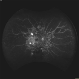
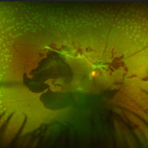
 Proliferative Diabetic Retinopathy
Nov 20 2025 by Oftalmontt Clínica Láser
Multimodal examination of a 39-year-old patient presenting with decreased visual acuity in both eyes due to clear proliferative diabetic retinopathy. Presence of neovascularization at the papillary level, microaneurysms in all four quadrants, a vascular loop in the inferior temporal arcade, and an altered ZAF due to low blood perfusion is observed.
Photographer: Ophthalmic Medical Technologist
Imaging device: Canon cx-1 and Avanti XR Optovue
Condition/keywords: proliferative diabetic retinopathy (PDR)
-
 Napkin on my Retina: The Diabetic TRD
Nov 17 2025 by SHRADDHA RAJ SHRIVASTAVA
Left eye pseudocolor widefield fundus image of a 57 year old patient with poorly controlled diabetes mellitus, presenting with BE tractional retinal detachment (TRD) on a background of high risk proliferative diabetic retinopathy (PDR). The disc, macula and posterior pole anatomy is obscured and distorted by the extensive tractional fibrovascular component, giving the appearance of a crumpled napkin on this patient's retina.
Photographer: Dr. Shraddha Raj Shrivastava
Imaging device: Nidek Mirante SLO/OCT (Confocal scanning/Spectral domain OCT)
Condition/keywords: Diabetic Tractional Detachment, Diabetic Tractional Retinal Detachment involving the Macula, PDR, proliferative diabetic retinopathy (PDR), tractional retinal detachment
-
 Napkin on my Retina: The Diabetic TRD
Nov 17 2025 by SHRADDHA RAJ SHRIVASTAVA
Left eye pseudocolor fundus image of a 57 year old patient with poorly controlled diabetes mellitus, presenting with BE tractional retinal detachment (TRD) on a background of high risk proliferative diabetic retinopathy (PDR). The disc, macula and posterior pole anatomy is obscured and distorted by the extensive tractional fibrovascular component, giving the appearance of a crumpled napkin on this patient's retina.
Photographer: Dr. Shraddha Raj Shrivastava
Imaging device: Nidek Mirante SLO/OCT (Confocal scanning/Spectral domain OCT)
Condition/keywords: Diabetic Tractional Detachment, Diabetic Tractional Retinal Detachment involving the Macula, proliferative diabetic retinopathy (PDR), tractional retinal detachment
-
 Traction Detachment of Retina
Nov 14 2025 by Virginia Gebhart
50 year old female with proliferative diabetic retinopathy, large ridge of traction temporally, and significant band of fibrosis. Subretinal fluid throughout the macula. Due to traction, surgical repair not recommended at this time as it could worsen condition. Will observe closely. BCVA CF @ 1 ft.
Photographer: Virginia Gebhart, Retina Consultants of Carolina
Imaging device: Optos California
Condition/keywords: fibrosis, proliferative diabetic retinopathy (PDR), traction detachment, Traction retinal detachment
-
 Filigree Networks
Nov 14 2025 by Malvika Singh
Fundus photograph of a 31 year old male with type 1 diabetes mellitus showing neovascularisation along the superotemporal arcade.
Photographer: Dr Malvika Singh, Retina Foundation, Ahmedabad, India
Imaging device: Mirante SLO/OCT
Condition/keywords: neovascularization (NV), proliferative diabetic retinopathy (PDR)
-
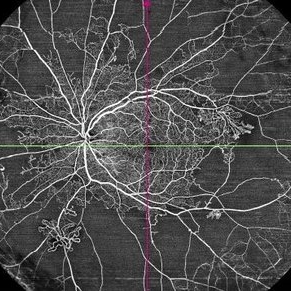
 Proliferative Diabetic Retinopathy
Nov 13 2025 by DR APOORVA JADHAV, MBBS , DNB
Wild Field OCTA of 61 year old male showing us NVD, NVE, CNP areas , macular ischaemia.
Photographer: Dr Apoorva Jadhav
Imaging device: Intalight Dream OCT
Condition/keywords: neovascularization of the disc (NVD), NVE, PDR
-
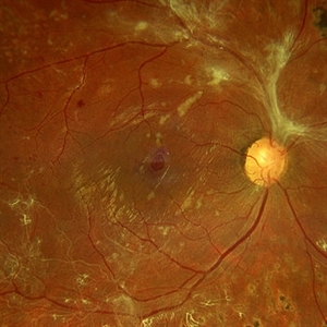
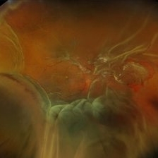
 Neovascular Medusa: A Bad Hair Day at the Optic Disc
Nov 4 2025 by SHRADDHA RAJ SHRIVASTAVA
Left eye pseudocolor fundus photo of 67 year old male, diagnosed with both eyes proliferative diabetic retinopathy, showing hair-like fronds of active neovascularisation at the disc (NVD) extending into the vitreous, giving the medusa-head appearance. There is a band of fibrovascular proliferation nasal to the disc, with presence of hard exudates and dot hemorrhages at the macula.
Photographer: Dr. Shraddha Raj Shrivastava
Imaging device: Nidek Mirante SLO/OCT (Confocal scanning/Spectral domain OCT
Condition/keywords: Diabetic Retinopathy, fibrovascular proliferation, Neovascularisation at the Disc (NVD), proliferative diabetic retinopathy (PDR)
-
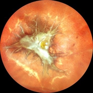
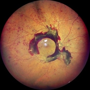
 Fibrovascular Fortress: Disc in Captivity
Nov 4 2025 by SHRADDHA RAJ SHRIVASTAVA
Right eye pseudocolor fundus photo of 67 year old male, diagnosed with both eyes proliferative diabetic retinopathy, showing extensive neovascularisation at the disc (NVD), with table-top configuration of the diabetic tractional retinal detachment (TRD) obscuring the view of the disc and threatening the macula. We can also see a mound of pre-retinal bleed and active neovascular frond nasal to the disc.
Photographer: Dr. Shraddha Raj Shrivastava
Imaging device: Nidek Mirante SLO/OCT (Confocal scanning/Spectral domain OCT
Condition/keywords: Active PDR Tractional retinal Detachment, Diabetic Tractional Detachment, Neovascularisation at the Disc (NVD), proliferative diabetic retinopathy (PDR), TABLE TOP TRD, tractional retinal detachment
-
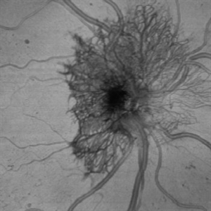
 Proliferative Ring of Fire
Oct 29 2025 by SHRADDHA RAJ SHRIVASTAVA
Right eye color fundus photo of 57 year old male, diagnosed with both eyes high risk proliferative diabetic retinopathy (PDR). Posterior pole reveals Neovascularization of disc (NVD) with extensive fibrovascular proliferations (FVPs) overlying the disc and along the arcades. We can also see a florid network of neovascularization (NVEs), with veins showing looping and beading changes. Hard exudates and dot-blot hemorrhages were seen at the macula.
Photographer: Dr. Shraddha Raj Shrivastava
Imaging device: Nidek Mirante SLO/OCT (Confocal scanning/Spectral domain OCT)
Condition/keywords: fibrovascular proliferation, Neovascularisation elsewhere (NVE), NVE, proliferative diabetic retinopathy (PDR), venous beading
-
 Proliferative Diabetic Retinopathy
Oct 22 2025 by Jeffrey Barker
56 year old Female with Diabetes Mellitus, lost to follow up for a year.
Photographer: Jeffrey P. Barker, B.S.
Condition/keywords: DME, fluorescein angiogram (FA), PDR
-
 Choroidal Detachment Secondary to Pan Retinal Photocoagulation Right Eye
Oct 14 2025 by NIDHI PANWAR, MD FRCS Glasgow FNB FICO
Serous choroidal detachment noted temporally after two sittings of pan retinal photocoagulation in proliferative diabetic retinopathy right eye, which resolved in 1 week of observation and topical steroid drops.
Photographer: NIDHI PANWAR, NMC ROYAL HOSPITAL, SHARJAH
Imaging device: optos
Condition/keywords: choroidal detachment, laser photocoagulation, pan retinal photocoagulation, proliferative diabetic retinopathy (PDR)
-
 Choroidal Detachment Secondary to Pan Retinal Photocoagulation Left Eye
Oct 14 2025 by NIDHI PANWAR, MD FRCS Glasgow FNB FICO
Serous choroidal detachment noted temporally after two sittings of pan retinal photocoagulation in proliferative diabetic retinopathy left eye, which resolved in 1 week of observation and topical steroid drops.
Photographer: NIDHI PANWAR, NMC ROYAL HOSPITAL, SHARJAH
Imaging device: OPTOS
Condition/keywords: choroidal detachment, laser photocoagulation, pan retinal photocoagulation, proliferative diabetic retinopathy (PDR)
-
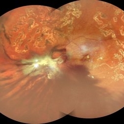 Table Top Tractional Retinal Detachment With Vitreous Hemorrhage in a Case of Proliferative Diabetic Retinopathy
Sep 12 2025 by Akansha Sharma
Color fundus photograph of a 56 year old male with table top tractional retinal detachment with vitreous hemorrhage in a case of proliferative diabetic retinopathy.
Photographer: DR. AKANSHA SHARMA
Condition/keywords: pan-retinal photocoagulation (PRP), PDR, proliferative diabetic retinopathy (PDR), PRP, TABLE TOP TRD, tractional retinal detachment, TRD, VH, vitreous hemorrhage
-
 Macular Hole Due to Proliferative Diabetic Retinopathy
Aug 13 2025 by Ricardo Leitão Guerra
A macular hole formation after anti-VEGF injection prior to vitrectomy for tractional retinal detachment in a patient presenting proliferative diabetic retinopathy.
Photographer: Ricardo Leitão Guerra
Imaging device: ZEISS CLARUS 700
Condition/keywords: macular hole, proliferative diabetic retinopathy (PDR)
-
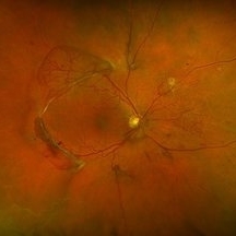
 Proliferative Diabetic Retinopathy
Aug 11 2025 by Marin Shehata
Fundus photograph of a 63 year-old male with diabetic retinopathy has been treated with PRP.
Photographer: Marin Shehata, Retina Consultants of Carolina
Imaging device: Optos California
Condition/keywords: proliferative diabetic retinopathy (PDR), PRP
-
 Unstable PDR s/p Laser
Aug 4 2025 by Anjana Mirajkar, MS Ophthalmology
Fundus photograph of a 60 year old male with an unstable PDR showing traction at the posterior pole with sub hyaloid hemorrhage. Peripheral PRP marks can be seen.
Photographer: Dr. Anjana Mirajkar- HV Desai eye hospital ,Pune
Imaging device: Optos
Condition/keywords: pan-retinal photocoagulation (PRP), proliferative diabetic retinopathy (PDR), subhyaloid hemorrhage, tractional retinal detachment
-
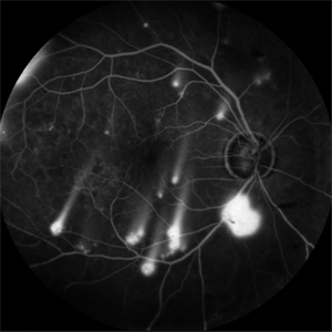 Shooting Stars
Jul 9 2025 by Majda Hadziahmetovic, MD
Fluorescein angiography image demonstrating multiple areas of neovascularization in a middle-aged male patient with long-standing diabetes.
Condition/keywords: proliferative diabetic retinopathy (PDR)
-
 Proliferative Diabetic Retinopathy
Jul 9 2025 by Jeffrey Barker
57 year old male presents with Proliferative Diabetic Retinopathy and Tractional Retina detachment.
Photographer: Jeffrey P. Barker, B.S. Retina Vitreous Surgeons of CNY
Condition/keywords: Diabetes, proliferative diabetic retinopathy (PDR), Traction retinal detachment
-
 Idiopathic Uveal Effusion Syndrome with Diabetic TRD
Jul 2 2025 by Virginia Gebhart
53 year old male with prominent choroidal effusions and significant diabetic tractional pathology/detachment. No visible breaks in presence pf Descemet's folds, unclear whether choroidals may be secondary to rhegmatogenous/tractional RD and low IOP, or separate process of exudative detachment/uveitis with some pre-existing diabetic traction and PDR. All labs WNL, pt was started on oral prednisone.
Photographer: Virginia Gebhart, Retina Consultants of Carolina
Imaging device: Optos California
Condition/keywords: Diabetic Tractional Detachment, idiopathic uveal effusion syndrome, TRD, uveal effusion syndrome
-
 Central Retinal Vein Occlusion With Waldenstroms macroglobulinemia
Jun 18 2025 by Korey Starkey
64-year-old patient presents with CRVO with secondary macular edema in both eyes. Venous beading present in 2/4 quadrants OU. Patient diagnosed with Waldenstroms macroglobulinemia, found on SPEP and bone marrow biopsy. Treatment recommended of anti-vegF intravitreal injections OU.
Photographer: Korey Starkey
Imaging device: Optos
Condition/keywords: attenuated vessels, central retinal vein occlusion (CRVO), CRVO, FA early phase, FLUORESCEIN ANGIOGRAPHY, macular edema, Optomap, OPTOS CALIFORNIA, severe NPDR, venous beading, Waldenstroms macroglobulinemia
-
 Traction in Proliferative Diabetic Retinopathy
Jun 9 2025 by Malvika Singh
Fundus photograph of a 44 year old with uncontrolled diabetes showing fibrovascular proliferation and traction with details of disc and macula obscured with sclerosed vessels in the periphery.
Photographer: Dr Malvika Singh, Retina Foundation, Ahmedabad, India
Imaging device: Mirante SLO/OCT
Condition/keywords: proliferative diabetic retinopathy (PDR), TRACTION
-
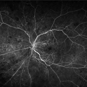
 Neovascularization of the Disc
Jun 3 2025 by Scott D Walter, MD, MSc, FASRS
Near-infrared (NIR) en face OCT image showing neovascularization of the disc (NVD) in a patient with type II diabetes mellitus, complicated by proliferative diabetic retinopathy (PDR).
Imaging device: Heidelberg Spectralis
Condition/keywords: Diabetes, Heidelburg Spectralis, microaneurysms, Neovascularisation at the Disc (NVD), NEOVASCULARISATION OF DISC, OCT EN FACE, proliferative diabetic retinopathy (PDR)
-
 The Dread of the Crimson Red
Jun 2 2025 by Thirumalesh Mochi Basavaraj, MD
Fundus photograph of a 64 year man post laser depicting a regressed NVD in the superior aspect and a Persistent Neo vascularization in the inferior aspect
Photographer: Vivek
Condition/keywords: Neovascularisation at the Disc (NVD), proliferative diabetic retinopathy (PDR)
-
 Proliferative Diabetic Retinopathy
May 29 2025 by KANWALJEET HARJOT MADAN, M.S. (Ophthalmology); FAICO (Vitreous - Retina)
This is widefield optic coherence tomography angiography (WF-OCTA) picture of LE of a diabetic patient. This patient had Proliferative Diabetic Retinopathy and depicts large areas of capillary non perfusion with neovascularization elsewhere.
Photographer: Dr. Kanwaljeet Harjot Madan, Thind Eye Hospital, Jalandhar City (Punjab) INDIA.
Imaging device: Widefield Optic Coherence Tomography Angiography (WF-OCTA).
Condition/keywords: OCTA, proliferative diabetic retinopathy (PDR), ultra-wide field imaging
-
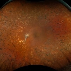
 High Risk Proliferative Diabetic Retinopathy with Sub-hyaloid Hemorrhage
May 13 2025 by Anupama Kiran Kumar
This image shows a case of high risk proliferative diabetic retinopathy. The retina is unlasered with a taut posterior hyaloid and a sub-hyaloid hemorrhage at the macula and along the arcades ,sparing the fovea.
Photographer: Mr Pratap
Imaging device: Mirante SLO/OCT (Nidek Co., Gamagori, Japan)
Condition/keywords: Diabetes, Diabetic Retinopathy, proliferative diabetic retinopathy (PDR), subhyaloid hemorrhage
 Loading…