Search results (436 results)
-
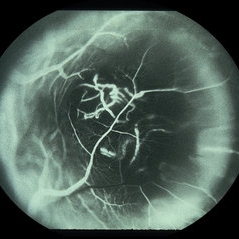
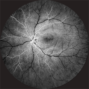
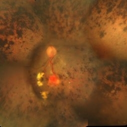 Retinitis Pigmentosa With Hemangioma CF
Dec 15 2016 by Manish Nagpal, MD, FRCS (UK), FASRS
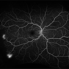
Fluorescein angiography OS of a patient having retinitis pigmentosa with a hemangioma inferiorly.
Condition/keywords: hemangioma, retinitis pigmentosa
-
 Venous Loop
Feb 20 2024 by Soobien Lee
A 77-year-old male with a history of bilateral optic neuropathy from bilateral optic nerve sheath meningiomas S/P radiation/proton-beam therapies. Presented with radiation retinopathy OS and a known venous loop OS.
Photographer: Gavin Bragdon, Elman Retina Group
Imaging device: Optos Ultra-Widefield Fluorescein Angiography
Condition/keywords: fluorescein angiogram (FA), Optos, radiation retinopathy, retinal vascular disease, venous loop
-
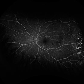
 Acute Posterior Multifocal Placoid Pigment Epitheliopathy
Feb 20 2024 by Soobien Lee
Fluorescein angiogram of a 20-year-old caucasian female with viral prodrome and vision loss OS>OD secondary to Acute Posterior Multifocal Placoid Pigment Epitheliopathy (APPME). Early blockage with late hyperfluorescent leakage can be seen on fluorescein angiography of the left eye.
Photographer: Ashley Metzger, Elman Retina Group
Imaging device: Optos Ultra-Widefield Fluorescein Angiography
Condition/keywords: acute posterior multifocal placoid pigment epitheliopathy (APMPPE), bacilliary layer detachment, FA, FA early phase, fluorescein angiogram (FA), Optos, uveitis, white dot syndrome
-
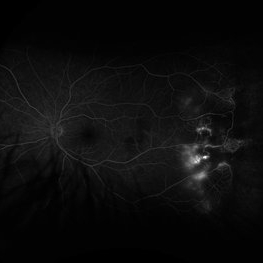
 Acute Posterior Multifocal Placoid Pigment Epitheliopathy
Feb 20 2024 by Soobien Lee
Fluorescein angiogram of a 20-year-old caucasian female with viral prodrome and vision loss OS>OD secondary to Acute Posterior Multifocal Placoid Pigment Epitheliopathy (APPME). Early blockage with late hyperfluorescent leakage can be seen on fluorescein angiography of the left eye.
Photographer: Ashley Metzger, Elman Retina Group
Imaging device: Optos Ultra-Widefield Fluorescein Angiography
Condition/keywords: acute posterior multifocal placoid pigment epitheliopathy (APMPPE), bacilliary layer detachment, FA, FA late phase, FA late phase leakage, fluorescein angiogram (FA), Optos, uveitis, white dot syndrome
-
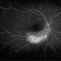
 Coats' Disease
Feb 2 2021 by Niloofar Piri, MD
#2 Fluorescein angiography of the same patient in lamellar arteriovenous phase, demonstrating temporal peripheral telangiectatic vessels, as well as hyperfluorescent aneurysma lesions. Note the anterior capillary non perfusion. Posterior hypofluorescence is secondary to blocking effect from hard exudates.
Condition/keywords: Coats' disease, Leber's miliary aneurysm
-
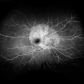
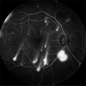 Shooting Stars
Jul 9 2025 by Majda Hadziahmetovic, MD
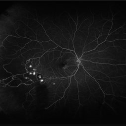
Fluorescein angiography image demonstrating multiple areas of neovascularization in a middle-aged male patient with long-standing diabetes.
Condition/keywords: proliferative diabetic retinopathy (PDR)
-
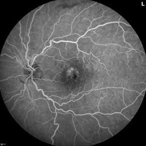
 Lady in a dress
Feb 9 2023 by Shelby Helton
Fluorescein Angiography on a 67-year-old male with significant RPE changes secondary to a severe subretinal hemorrhage that required a vitrectomy with subretinal TPA in 2013.
Photographer: Shelby Helton
Imaging device: Heidelberg Spectralis
Condition/keywords: wet age-related macular degeneration (wet AMD)
-
 Radiation Retinopathy; BRVO with Macular Edema
Apr 26 2023 by Denica Rodriguez
Ultra-wide field fluorescein angiography of a 61 year old male with radiation retinopathy following brachytherapy for choroidal melanoma of his left eye. Following treatment, patient developed a branch retinal vein occlusion both ischemic and non-ischemic. Anti-VEGF injections were recommended. The fine needle biopsy showed a class 2 uveal melanoma. Patient also has diabetic retinopathy affecting both eyes. Patient's vision at the time the image was taken was Dcc 20/80-1.
Photographer: Denica Rodriguez COA, ST
Imaging device: Optos California
Condition/keywords: branch retinal vein occlusion (BRVO), Choroidal melanoma, diabetic retinopathy, FA, fluorescein angiogram (FA), I-125 brachytherapy, macular edema, melanoma, Optos, radiation retinopathy, Retina, ultra-wide field imaging
-
 Choroidal Granuloma
Apr 7 2017 by Manish Nagpal, MD, FRCS (UK), FASRS
Fluorescein angiography of a case of peripapillary choroidal granuloma presenting with exudation and hemorrhages.
Photographer: Pooja Barot
Condition/keywords: choroid, granuloma, inflammation
-
 Choroidal Melanoma
May 28 2014 by Henry J. Kaplan, MD
Fluorescein angiography of a patient with choroidal melanoma clearly shows the double circulation of the retina and whithin the melanoma #2
Imaging device: Fluorescein angiography
Condition/keywords: melanoma
-
 Choroideremia
Jul 12 2021 by Stefanie Palmer
Fluorescein angiography of a 58-year-old female.
Photographer: Stefanie Palmer, CRA
Condition/keywords: choroideremia, retina
-
 Coats' Disease
Feb 2 2021 by Niloofar Piri, MD
#3 Mid AV phase fluorescein angiography of the same patient demonstrating increasing hyper fluorescence of aneurysmal lesions.
Condition/keywords: Coats' disease, Leber's miliary aneurysm
-
 Coats' Disease
Feb 2 2021 by Niloofar Piri, MD
#4 Recirculation phase fluorescein angiography of the same patient demonstrating increased hyperfluorescence and leakage from abnormal vascular lesions in temporal periphery. Note the capillary non perfusion area anteriorly.
Condition/keywords: Coats' disease, Leber's miliary aneurysm
-
 Lupus Retinopathy
Mar 14 2021 by Marco Antonio Sauza
Fluorescein angiography photo of and 13-year-old female with ischemic retinopathy with LES.
Photographer: Marco Sauza
Imaging device: Zeiss fundus camera
Condition/keywords: systemic lupus erythematosus (SLE) retinopathy
-
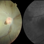 Ophthalmic Artery Occlusion in a 39-Year-Old with Rheumatoid Arthritis
Oct 6 2020 by Michael Izzo, MD
Left image: fundus photograph of a 39-year-old male with rheumatoid arthritis found to have ophthalmic artery occlusion depicting boxcar segmentation of blood in retinal vasculature and macular ischemia demonstrated by retinal whitening without cherry red fovea. Right image: early phase fluorescein angiography demonstrating patchy choroidal filling, arterial non-perfusion and optic nerve head leakage.
Photographer: Karen Rivera, COA; Washington National Eye Center
Condition/keywords: fluorescein angiogram (FA), ophthalmic artery occlusion, rheumatoid arthritis
-
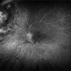
 Proliferative Diabetic Retinopathy with Severe Ischemia
Nov 30 2023 by Gabriel Costa Andrade, PhD
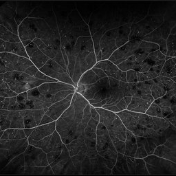
Ultra-widefield fluorescein angiography of the right eye of a 47 year old woman with diabetes mellitus showing macular and nasal retinal capillary dropout and neovascularization of the disc and temporal vascular arcades.
Photographer: Gabriel Andrade
Imaging device: Optos California
Condition/keywords: Diabetic Retinopathy
-
 Proliferative Sickle Cell Retinopathy
Feb 1 2023 by Olivia Rainey
Ultra-widefield fluorescein angiography of a 25-year old male with Proliferative Sickle Cell Retinopathy affecting his right eye. Patient stated that he was born with Sickle disease (SC), and has yearly eye exams. He noted no vision concerns over the last year but has typically experienced sickle attacks about 1-2 per year. The physician noted that the fluorescein obtained showed peripheral nonperfusion affecting the patient's nasal and temporal retina as well as neovascularization affecting his left eye more than his right. He recommended pan retinal photocoagulation in his left eye for his temporal and nasal retina, as as well as his right eye following.
Photographer: Olivia Rainey, OCT-C, COA
Imaging device: Optos California
Condition/keywords: early phase, fluorescein angiogram (FA), fluorescein leakage, neovascularization (NV), non-perfusion, proliferative retinopathy, right eye, sickle cell retinopathy, ultra-wide field imaging, ultra-widefield image
-
 Retinal Hemorrhages
Mar 10 2021 by Kachelle Brown
Ultra widefield Fluorescein Angiography of a 48-year-old female with retinal hemorrhages affecting her right eye. Physician suspect sickle cell due to family history, and has ordered labs to rule out.
Photographer: Kachelle Brown
Imaging device: Optos California
Condition/keywords: fluorescein angiogram (FA), fluorescein leakage, Optos, retinal hemorrhage, sickle cell retinopathy, ultra-wide field imaging
-
 Retinal Lightning
Apr 24 2022 by Mariam Cernichiaro-Espinosa, MD
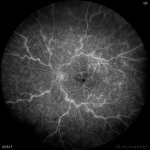
Late phase venous leakage on IV fluorescein angiography from a 32-year-old male with central retinal vein occlusion (CRVO).
Photographer: Mariam Cernichiaro-Espinosa, Asociación para Evitar la Ceguera, I.A.P. Mexico City, Mexico.
Imaging device: Zeiss Clarus
Condition/keywords: central retinal vein occlusion (CRVO)
-
 Roth Spots
Oct 26 2022 by Denica Rodriguez
Roth spots during optos FA on a 68 year old female with retinal hemorrhage effecting her left eye. Patient was referred for non-proliferative diabetic retinopathy without macular edema.
Photographer: Denica Rodriguez & Zachary Seim
Imaging device: Optos California
Condition/keywords: Diabetes, FLUORESCEIN ANGIOGRAPHY, left eye, Optos, Retina, Roth Spots, ultra-wide field imaging
-
 Takayasu Retinopathy
Apr 30 2025 by Vishal Agrawal, MD, FRCS,FACS,FASRS
Fundus fluorescein angiography image of a young girl with diagnosed Takayasu arteritis who presented with complains of diminished vision in both eyes. FFA shows complete absence of venous filling with segmented blood column secondary to CRAO with peripheral avascular area.
Photographer: Dr Ayushi Gupta
Imaging device: Clarus 700
Condition/keywords: calcified drusen, CRAO, takayasu arteritis
-
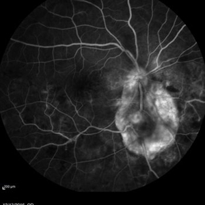
 Acute Syphilitic Posterior Placoid Chorioretinitis
May 4 2021 by RAFAEL REIS PEREIRA, MD
A 31-year-old patient with a complaint of photophobia and low visual acuity OD in the previous three weeks. BCVA was 20/60 and 20/20 The fundus examination revealed a placoid white lesion in the posterior pole and vitreous cells in the right eye. The left eye was unremarkable. Fluorescein angiography reveals hyperfluorescent plaque with distinctive “leopard spots” hypofluorescence.
Imaging device: Opto California
Condition/keywords: acute syphilitic posterior placoid chorioretinitis
-
 Acute Zonal Occult Outer Retinopathy (AZOOR) FA, Fluorescein Angiography, Peripheral Vasculitis
Jan 19 2022 by James B. Soque, CRA, OCT-C, COA, FOPS
Acute Zonal Occult Outer Retinopathy (AZOOR). Peripheral Vasculitis OD. Fluorescein angiography showing vasculitis in the far right periphery 8-10 o'clock. 46-year-old white male, VA CC 20/16, 20/12.5, has had recurrent vasculitis for 11 years. No treatment.
Photographer: James Soque, CRA, OCT-C, COA, FOPS, Island Retina, Shirley, NY
Imaging device: Optos California
Condition/keywords: acute zonal occult outer retinopathy (AZOOR), FA early phase, fluorescein angiogram (FA), Peripheral Vasculitis, ultra-wide field imaging
-
 Branch Retinal Vein Occlusion
Oct 17 2012 by Sharon Fekrat, MD FACS FASRS
Fluorescein angiography of an inferior perfused branch retinal vein occlusion in the left eye of a middle aged male with hypertension. The foveal avascular zone is irregular. Subretinal hemorrhage is present.
Photographer: John Reaves, Ophthalmic Photographer, Durham VA Medical Center Eye Clinic Imaging Suite, Durham, NC
Imaging device: Fluorescein Angiography
Condition/keywords: branch retinal vein occlusion (BRVO), subretinal hemorrhage
-
 Branch Retinal Vein Occlusion with Multifactorial Macular Edema and Epiretinal Membrane
Oct 3 2024 by Logan ryzenga
Fluorescein angiogram of a 62 year old woman with cystoid macular edema from concurrent Epiretinal Membrane and Branch Retinal Vein occlusion. She has an extensive history of anti-VEGF injections with stable but unresolved macular edema. Following angiography, it was determined that an epiretinal membrane peel would be indicated in an attempt to achieve resolution of macular edema.
Photographer: Logan Ryzenga
Imaging device: Heidelberg Spectralis
Condition/keywords: 55-degrees, branch retinal vein occlusion (BRVO), cystoid macular edema (CME), epiretinal membrane (ERM), Fluorescein angiography, heidelberg spectralis, hyperfluorescence, leakage, left eye, OS, wide angle imaging
 Loading…