Search results (11 results)
-
 Vitreoschisis
Sep 3 2020 by J. Sebag, MD, FACS, FRCOphth, FARVO
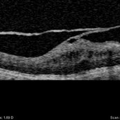
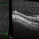
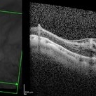
OCT of the left eye in a patient with macular pucker (see SLO image below to right) demonstrates splitting of the posterior vitreous cortex in two separate places. Tangential traction caused thickening of the underlying macula. [For histopathology see: Gupta P, Yee KMP, Garcia P, Rosen RB, Parikh J, Hageman GS, Sadun AA, Sebag J: Vitreoschisis in macular diseases. Brit J Ophthalmol 2011;95(3):376-80]
Condition/keywords: vitreoschisis
-
 Lamellar Structure of the Primate Posterior Vitreous Complex
Sep 3 2020 by J. Sebag, MD, FACS, FRCOphth, FARVO
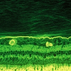
Immunohistochemistry of a monkey eye imaged with fluorescein conjugated ABA lectin staining demonstrates the lamellar structure pf the posterior vitreous cortex. During anomalous PVD, there can be splitting between these lamellae, a phenomenon known as vitreoschisis. (original magnification = 400x)
Condition/keywords: vitreous
-
 Pathophysiology of Vitreoschisis
Sep 1 2020 by J. Sebag, MD, FACS, FRCOphth, FARVO
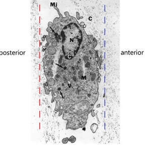
Transmission electron microscopy of human hyalocyte in situ demonstrates embedding within the dense collagen matrix of the posterior vitreous cortex. The retina was to the left (“posterior”) and the anterior segment was to the right (“anterior”). The red dashed line indicates the level of vitreoschisis split that might occur posterior to the level of the hyalocyte monolayer, leaving a thin, hypocellular membrane attached to the macula. The dashed blue line indicates the level of vitreoschisis split that might occur anterior to the level of the hyalocyte monolayer, leaving a thick, hypercellular membrane attached to the macula. The former is more likely to present as macular hole, while the latter as macular pucker (see Figure 12). Mi = microvilli; black C = collagen of posterior vitreous cortex; N = lobulated nucleus typical of mononuclear phagocytes; white C = dense marginal chromatin in nucleus; M = mitochondria; V = vacuoles; arrows = dense granule (original magnification = 11,670) [Modified from Sebag J: Anomalous PVD – a unifying concept in vitreo-retinal diseases. Graefe’s Arch Clin Exp Ophthalmol 2004;242:690-8 and Sebag J, Niemeyer M, Koss M: Anomalous PVD and vitreoschisis. In: Vitreous – in Health & Disease (J. Sebag, ed.) Springer, New York, 2014, pg. 252]
Condition/keywords: pathology, vitreoschisis
-
 Vitreo-Macular Traction Syndrome
Sep 1 2020 by J. Sebag, MD, FACS, FRCOphth, FARVO
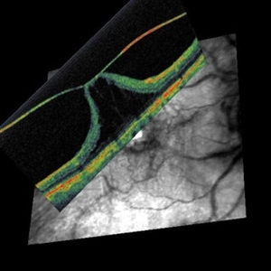
Combined OCT and Scanning laser ophthalmoscopy demonstrate separation of full-thickness posterior vitreous cortex (no vitreoschisis), but persistent adhesion centrally with significant detachment of the fovea. [from Sebag J: Vitreous – in Health & Disease (J. Sebag, ed.) Springer, New York, 2014; image © Springer Nature, reprinted with permission]
Condition/keywords: vitreomacular traction (VMT)
-
 Pathophysiology of Anomalous PVD
Sep 1 2020 by J. Sebag, MD, FACS, FRCOphth, FARVO
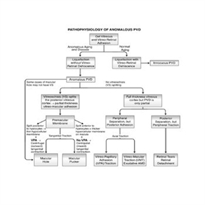
This unifying concept of vitreo-retinopathies hypothesizes that the pathogenesis of several vitreoretinal diseases that were previously considered very disparate, are actually all manifestations of the same underlying pathophysiology – anomalous PVD. Note that vitreo-papillary adhesion (VPA) and traction can cause primary optic neuropathy, but might also play a role in facilitating/promoting cell migration and proliferation during pathologic neovascularization of the optic disc. Further, VPA seems to alter the vector of tangential forces exerted by a membrane, in some cases full-thickness posterior vitreous cortex and in some cases the outer layer of the posterior vitreous cortex left attached to the macula after vitreoschisis. While not all cases of macular holes have vitreoschisis, they feature vitreomacular adhesion and traction almost always with VPA. [From Sebag J: Anomalous PVD – a unifying concept in vitreo-retinal diseases. Graefe’s Arch Clin Exp Ophthalmol 2004;242:690-8 and Sebag J, Niemeyer M, Koss M: Anomalous PVD and vitreoschisis. In: Vitreous – in Health & Disease (J. Sebag, ed.) Springer, New York, 2014, pg. 252; image © Springer Nature, reprinted with permission]
Condition/keywords: pathology, peripheral vascular disease (PVD)
-
 Age-Related Differences in the Structure of the Human Vitreous Body
Sep 1 2020 by J. Sebag, MD, FACS, FRCOphth, FARVO
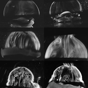
Dark-field slit microscopy was performed on fresh, unfixed, post-mortem human eyes that had undergone dissection to peel off the sclera, choroid, and retina. The vitreous body remains attached to the anterior segment which is seen below, while the posterior pole is above in these images. The top panel demonstrates the absence of internal vitreous structures that scatter light in youth (left image from an 11 year-old girl, right image from a 14 year-old boy. The middle panel demonstrates light scattering from linear, fibrous structures that have an antero-posterior orientation with insertions into the vitreous base peripherally and the posterior vitreous cortex, typical in middle age (left image from a 56 year-old and right image from a 59 year-old). The bottom panel illustrates advance fibrous liquefaction in old age (88-year-old subject). [From Sebag J, Niemeyer M, Koss M: Anomalous PVD and vitreoschisis. In: Vitreous – in Health & Disease (J. Sebag, ed.) Springer, New York, 2014, pg. 245; image © Springer Nature, reprinted with permission]
Condition/keywords: vitreous
-
 Vitreoschisis Membrane in Myopic Traction Maculopathy
May 27 2020 by Raja Rami P Reddy, MD FRCS FASRS
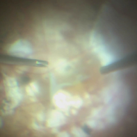
Triamcinolone enhanced visualization of vitreoschisis membrane and removal in a case of myopic traction maculopathy.
Imaging device: intra operative photograph, SONY camera
Condition/keywords: myopic traction maculopathy, vitreoschisis
-
 Vitreoschisis
Jan 26 2017 by Sara Sella
70-year-old male with high myopia -18D underwent successful surgery of pars plana vitrectomy +posterior hyaloid peel (vitreoschisis) +ELX + SF6.
Photographer: Sara Sella
Condition/keywords: high myopia, vitreoschisis
-
 Vitreoschisis
Jan 26 2017 by Sara Sella
70-year-old male with high myopia -18D underwent successful surgery of pars plana vitrectomy +posterior hyaloid peel (vitreoschisis) +ELX + SF6
Photographer: Sara Sella
Condition/keywords: high myopia, vitreoschisis
-
 Vitreoschisis
Jan 26 2017 by Sara Sella
70-year-old male with high myopia -18D underwent successful surgery of pars plana vitrectomy +posterior hyloid peel (vitreoschisis) +ELX + SF6 .
Photographer: Sara Sella
Condition/keywords: high myopia, vitreoschisis
-
 Vitreoschisis ( Spider-like)
May 31 2014 by Hamid Ahmadieh, MD
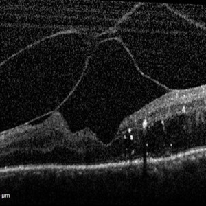
OCT image of the left eye of a 70-year-old woman with proliferative diabetic retinopathy associated with vitreoschisis and diabetic macular edema. The vitreal changes simulate the appearance of a spider on the retina !
Photographer: Nayereh Hadipour, Negah Eye Center, Tehran
Condition/keywords: diabetic macular edema, optical coherence tomography (OCT), vitreoschisis
 Loading…