 Initializing download.
-
 By J. Sebag, MD, FACS, FRCOphth, FARVO
VMR Institute for Vitreous Macula Retina - Uploaded on Sep 1, 2020.
- Last modified by Caroline Bozell on Sep 1, 2020.
- Rating
- Appears in
- Vitreous
- Condition/keywords
- pathology, vitreoschisis
- Description
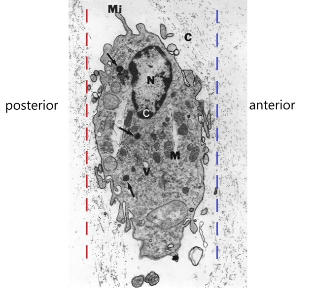
- Transmission electron microscopy of human hyalocyte in situ demonstrates embedding within the dense collagen matrix of the posterior vitreous cortex. The retina was to the left (“posterior”) and the anterior segment was to the right (“anterior”). The red dashed line indicates the level of vitreoschisis split that might occur posterior to the level of the hyalocyte monolayer, leaving a thin, hypocellular membrane attached to the macula. The dashed blue line indicates the level of vitreoschisis split that might occur anterior to the level of the hyalocyte monolayer, leaving a thick, hypercellular membrane attached to the macula. The former is more likely to present as macular hole, while the latter as macular pucker (see Figure 12). Mi = microvilli; black C = collagen of posterior vitreous cortex; N = lobulated nucleus typical of mononuclear phagocytes; white C = dense marginal chromatin in nucleus; M = mitochondria; V = vacuoles; arrows = dense granule (original magnification = 11,670) [Modified from Sebag J: Anomalous PVD – a unifying concept in vitreo-retinal diseases. Graefe’s Arch Clin Exp Ophthalmol 2004;242:690-8 and Sebag J, Niemeyer M, Koss M: Anomalous PVD and vitreoschisis. In: Vitreous – in Health & Disease (J. Sebag, ed.) Springer, New York, 2014, pg. 252]
---thumb.jpg/image-square;max$79,0.ImageHandler "Toxocariasis Cyst Gross Pathology Specimen")


---thumb.jpg/image-square;max$79,0.ImageHandler "Infectious Retinitis")

