Search results (68 results)
-
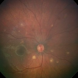
 Fractal Pattern of Chronic Serpiginous Choroiditis
Jun 17 2025 by Guilherme Sturzeneker, MD, MSc
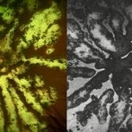
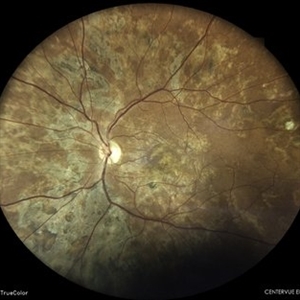
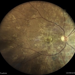
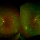
Ultra-widefield fundus photograph and autofluorescence of a 33-year-old woman with longstanding serpiginous choroiditis in the right eye. The image reveals centrifugal chorioretinal atrophy forming a dramatic fractal-like pattern, sparing the fovea. The patient is several years post-onset, with repeated negative workups, including for tuberculosis. Despite extensive lesions, the patient retains 20/20 vision in both eyes. Management included azathioprine monotherapy, as systemic steroids were contraindicated due to bipolar disorder.
Photographer: Andrea Almeida, IPEPO - Instituto da Visão
Imaging device: Optos Silverstone
Condition/keywords: autoimmune uveitis, azathioprine, chorioretinal atrophy, serpiginous choroiditis, ultra-wide field imaging
-
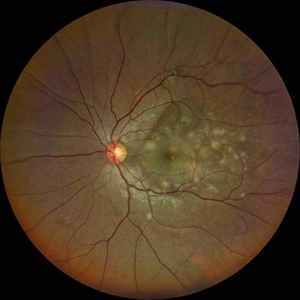
 Optic Disc Granuloma
May 7 2025 by Aayesha - Khanum, MBBS. D.N.B
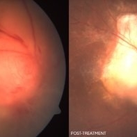
A 45-year-old male presented with diminished vision for one month. His Mantoux test was negative, but as steroids worsened the condition, quantiferon TB was advised and it returned positive. He was started on anti-tuberculosis treatment (ATT). Oral steroids were reintroduced after one week of ATT. Optic disc granulomas can arise from direct invasion of the optic nerve or may represent hypersensitivity reaction to tuberculous antigens. The pathogenesis involves infiltration of immune cells, leading to formation of a granulomatous structure that disrupts normal architecture and function of the optic disc. Steroids with ATT facilitated regression of granulomatous lesion.
Photographer: Ms. Krishna Jeyanthi
Imaging device: Zeiss Clarus 500
Condition/keywords: Tuberculosis
-
 Tuberculoma
Sep 23 2024 by DR Rohit Gupta
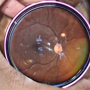
Fundus photograph of a 26 year old female suffering from pulmonary tuberculosis, on fundus examination a tuberculoma seen in her left eye.
Photographer: DR Rohit gupta
Imaging device: Samsung S21
Condition/keywords: tubercular choroidal granuloma
-
 Atypical Tubercular Occlusive Peripheral Retinal Vasculitis
Jun 21 2024 by Tejaswita Verma
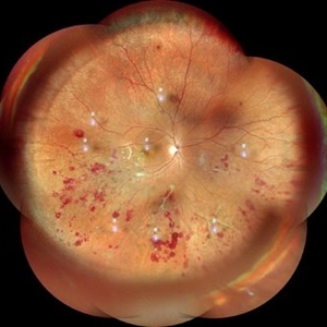
Follow up right eye fundus photograph of a 27 year old male with vision 6/12 , diagnosed with systemic tuberculosis(mediastinal lymphadenopathy on chest CT) on oral steroids, and started on ATT .We can see a parafoveal sub-ILM hemorrhage with vascular sheathing and hemorrhages in inferior and temporal quadrants . The patient was advised anti-VEGF intravitreal injection, later sectoral laser after resolution of inflammation
Photographer: DR. TEJASWITA VERMA
Imaging device: MIRANTE
Condition/keywords: obliterative peripheral vasculitis, ocular tuberculosis
-
 FFA in Atypical Tubercular Peripheral Occlusive Retinal Vasculitis
Jun 21 2024 by Tejaswita Verma
Right eye FFA montage of a 27 year male with peripheral occlusive tubercular vasculitis, showing CNP areas inferiorly and temporally, leakages and blocked fluorescence due to hemorrhages. The patient was advised intravitreal anti-VEGF injection and later sectoral laser once inflammation subsides.
Photographer: DR. TEJASWITA VERMA
Imaging device: MIRANTE
Condition/keywords: obliterative peripheral vasculitis, ocular tuberculosis
-
 Atypical Tubercular Peripheral Occlusive Retinal Vasculitis
Jun 21 2024 by Tejaswita Verma
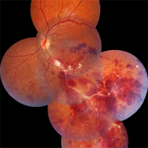
Fundus montage of the right eye of a 27 year old male with macula threatening occlusive vasculitis showing hemorrhages in inferior, temporal quadrant with vascular sheathing. The patient was Mantoux positive (20 mm induration) and IGRA (TB-GOLD)positive and started on oral steroids. The case was atypical due to no vitritis at presentation which is unusual of tuberculosis. Behcet's disease was ruled out as there was no panuveitis like picture.
Photographer: DR. TEJASWITA VERMA
Imaging device: MIRANTE
Condition/keywords: occlusive vasculitis, ocular tuberculosis
-
 Old Healed Serpiginous Like Choroiditis With Disc Pallor
Apr 8 2024 by Akansha Sharma
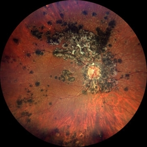
Color fundus photograph of a 40 year old male patient with old healed serpiginous like choroiditis with disc pallor.
Photographer: Dr. Akansha Sharma, Bharati Eye Hospital
Condition/keywords: Tuberculosis
-
 Old Healed Serpiginous Like Choroiditis
Apr 8 2024 by Akansha Sharma
Color fundus photograph of a 40 year old male patient with old healed serpiginous like choroiditis.
Photographer: Dr. Akansha Sharma, Bharati Eye Hospital
Condition/keywords: serpiginous like choroiditis, Tuberculosis
-
 Serpiginous-Choroiditis-Like
Mar 30 2024 by Karen Flores Guevara
Fundus photograph of a 26-year-old woman with a serpiginous-choroiditis-like presentation secundary to a tuberculosis activation disease, started with visual acuity and systemic syntomps
Photographer: Andrés Santiago Pérez-Giraldez, MD. Asociación para evitar la Ceguera en Mexico I.A.P. Mexico
Condition/keywords: ocular tuberculosis, serpiginous choroiditis, tuberculosis
-
 Choroidal-Tuberculoma
Mar 29 2024 by Carlos Emiliano Rodriguez Lopez
Multimodal image. 50-year-old female with choroidal neovascularization secondary to choroidal tuberculoma.
Photographer: Carlos Emiliano Rodríguez López. Instituto de Oftalmología FAP Conde de Valenciana, IAP.
Imaging device: Heidelberg Spectralis
Condition/keywords: Choroidal Neovascularization, Choroidal-tuberculoma, Tuberculosis
-
 Serpiginious Chorioretinitis
Sep 6 2023 by PUSHPANJALI BADOLE
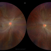
Fundus photograph of an 18 year-old male with bilateral choroiditis. Tuberculosis work-up revealed positive QuantiFERON Gold test and Mantoux test. Patient was started anti-tuberculosis treatment along with oral corticosteroids. Optos fundus photography shows extensive active plus healed lesions with pigmentary change in the midperipheral retina and periphery suggesting varied stage presentation of lesions. There are few hemorrhages in right eye superotemporal retina.
Photographer: Hitesh Rawlani, Isha Netralaya, Kalyan.
Condition/keywords: serpiginous choroiditis
-
 Frosted branch angitis
Jan 9 2023 by Keshavi Shah
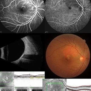
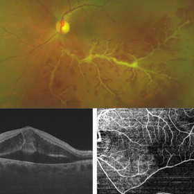
Tuberculosis associated Frosted branch angitis involving inferior temporal vessels (top) with intra- and sub-retinal exudation noted on OCT imaging (bottom left) and perfusion deficit noted on OCTA imaging (bottom right).
Photographer: Shrushti Sharma
Imaging device: Nikon Optos Daytona, DRI OCT Triton Plus
Condition/keywords: Frosted Branch Angitis
-
 Tuberculosis-related serpiginous-like choroiditis
Nov 22 2022 by Ricardo Leitão Guerra
True color BLFI of a 60-year-old male presenting chorioretinal scars from a tuberculosis-related serpiginous-like choroiditis.
Photographer: Ricardo Leitão Guerra
Imaging device: Zeiss Clarus 700
Condition/keywords: serpiginous choroiditis, tuberculosis
-
 Tubercular Retinal Vasculitis
Mar 31 2022 by Lucas Zago Ribeiro, MD
Fundus image of 22yr male, presenting with unilateral vision loss
Photographer: Lucas Zago Ribeiro, UNIFESP / EPM
Imaging device: Zeiss Visucam 524
Condition/keywords: ocular tuberculosis, tuberculosis, vasculitis
-
 Miliary Tuberculosis
Mar 17 2022 by Franco Benvenuto, MD
Fundus photograph of a 9-month-old baby with hemophagocytic syndrome secondary to Tuberculosis infection.
Photographer: Franco Benvenuto, Universidad de Buenos Aires, Argentina
Imaging device: RetCam
Condition/keywords: ocular tuberculosis
-
 Tubercular Multifocal Choroiditis
Aug 18 2021 by Priyanka Raj, MBBS, MS
Fundus photograph of a 28 year-old man with multifocal tubercular choroiditis.
Photographer: Priyanka Raj, Prakash Netra Kendr, Lucknow, India
Imaging device: Zeiss Clarus 500
Condition/keywords: choroiditis, tuberculosis
-
 Choroidal Tubeculoma
Feb 12 2021 by Sham Talati, DOMS
A 9-year-old male patient who is a known case of pulmonary tuberculosis presented with choroidal tubeculoma in his right eye.
Photographer: Dr. Sham Talati,Retina Foundation,Ahmedabad
Imaging device: Nidek Mirante
Condition/keywords: choroidal tuberculoma, ocular tuberculosis, tuberculosis
-
 Choroidal Tuberculoma Fluorescence Angiography Montage
Feb 12 2021 by Sham Talati, DOMS
A 9-year-old male patient who is a known case of pulmonary tuberculosis presented with choroidal tuberculoma in his right eye.
Photographer: Dr. Sham Talati,Retina Foundation,Ahmedabad
Imaging device: Nidek Mirante
Condition/keywords: choroidal tuberculoma, ocular tuberculosis, tuberculosis
-
 Active Vasculitis with Proliferative Retinopathy
Jan 30 2021 by Raja Rami P Reddy, MD FRCS FASRS
25-year-old boy with unilateral recent onset visual loss. Fundus shows areas of active vasculitis nasally and large neovascular complexes temporally and on the disc and early fibrous membrane formation. Fellow eye fundus is normal. Further investigations suggested tubercular etiology
Photographer: Raja Rami Reddy P
Imaging device: fundus camera
Condition/keywords: proliferative retinopathy, tuberculosis, vasculitis
-
 Ocular Tuberculosis
May 18 2020 by McGill University Health Centre
Ocular tuberculosis refers to necrotizing granulomatous uveitis caused by Mycobacterium tuberculosis infection. The mechanism of infection of ocular structures is via hematogenous dissemination of the bacteria during the primary infection. In this enucleation specimen, the anterior chamber and vitreous cavity are filled by caseous exudate. The iris is also replaced by a whitish material, and the retina is completely detached. Note the whitish thickening of the posterior choroid (arrow).
Condition/keywords: caseous exudate, enucleation, ocular tuberculosis, uveitis
-
 Choroidal Miliary Tubercles
Feb 4 2020 by Pierre-Henry Gabrielle, MD
Fundus photograph of an 8-year-old girl with bilateral choroidal miliary tubercles due to acute miliary tuberculosis.
Photographer: Pierre-Henry Gabrielle, Ophthalmology department, Dijon University Hospital
Imaging device: Optos
Condition/keywords: choroidal tuberculoma, color fundus photograph, tuberculosis
-
 Choroidal Granuloma
Apr 23 2019 by Purva Patwari
22-year-old male patient presented with blurring of vision in the right eye noticed since last one week. He was asymptomatic a week ago when he noticed the blurring in his right eye. On examination his vision was 6/6 in both eyes. Anterior segment was normal. Posterior segment was normal for the left eye. Right eye examination revealed a clear vitreous cavity with choroidal granulomas scattered throughout the fundus. The present picture shows choroidal granulomas with OCT segment passing through the parafoveal lesion showing subretinal fluid accumulation and corresponding thickening of the retinal layers. CT scan reveals heterogeneously enhancing lymph nodes showing conglomerationin the hilar region-possibility of tubercular etiology.
Photographer: Dr Purva Patwari, Patwari Retina Center
Imaging device: Zeiss Visu 500
Condition/keywords: choroidal granuloma, choroiditis, granulomatous choroiditis, tubercular choroidal granuloma, tuberculosis
-
 NVE in a Patient With Vasculitis
Nov 5 2018 by awaneesh m upadhyay, MBBS, DNB
FFA image of a 22-year-old male vasculitis patient with NVE.
Photographer: Hiteshwar Saikia
Condition/keywords: neovascularization elsewhere (NVE), tuberculosis, vasculitis
-
 TB Granuloma
Sep 21 2017 by Theodore Leng, MD, MS, FASRS
TB granuloma.
Condition/keywords: choroidal tuberculoma, tuberculosis
 Loading…