Search results (44 results)
-
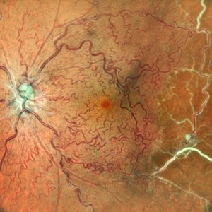
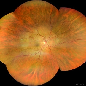
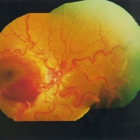
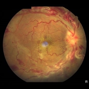 Central Retinal Vein Occlusion
Jun 21 2025 by Moazzam Parvez
Fundus photograph of a 56 year old male presenting with dilated tortuous vessels with adjoining Hard exudates and macular star.
Photographer: Moazzam Parvez , Netralayam , Kolkata
Imaging device: Topcon Maestro 2
Condition/keywords: CRVO with macular edema, hard exudates, macular star
-
 CRVO
CRVO
Jan 15 2025 by Virginia Gebhart
65 year old male with new central retinal vein occlusion with macular edema. Carotid ultrasound showed less than 50% stenosis bilateral. Dilated and tortuous vessels as well as cystoid macular edema and flame-shaped hemes in all 4 quadrants. Treated with IVA
Photographer: Virginia Gebhart, Retina Consultants of Carolina
Imaging device: Optos California
Condition/keywords: central retinal vein occlusion (CRVO), macular edema
-
 Diabetic Retinopathy
Nov 20 2024 by Korey Starkey
64 year old female being monitored for moderate-severe diabetic retinopathy.
Photographer: Korey Starkey
Condition/keywords: capillary nonperfusion, FA, FLUORESCEIN ANGIOGRAPHY, microaneurysms, nonproliferative diabetic retinopathy, Optos, OPTOS CALIFORNIA, tortuous vessels
-
 Sea Fan Neovascular Frond in Rare Case of Fanconi Anemia
May 18 2024 by Amol yuvraj ganvir
A 15-year-old female patient brought by her parents with defective vision in left eye for 15 days. The patient had a known case of Fanconi Anemia. Left eye tortuous vessels and new retinal vessels along the major temporal arcade, with intraretinal and subretinal hemorrhage covering the macula.
Photographer: Dr. Amol Ganvir
Condition/keywords: Fanconi Anemia, sea fan
-
 Congenital Retinal Vessel Tortuosity
Apr 2 2024 by Pablo Angel Garcia Uribe
Fundus photograph of a 29-year-old man with bilateral congenital retinal vessel tortuosity. This image shows the sinuous course of retinal arterioles and a shiny internal limiting membrane.
Photographer: Pablo Ángel García-Uribe, Clínica Oftalmológica Salauno, Mexico City
Imaging device: NIDEK OCT RS-330 Duo 2
Condition/keywords: abnormal retinal vessel, anomalous vessels, Retina, tortuous vessels
-
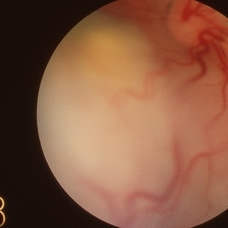
 Left Eye Arteriovenous Malformation, Vein Occlusion and Ruptured Macroaneurysm
Feb 9 2024 by Sandra R Montezuma, MD
47 year old female presented with acute changes in vision in the left eye, flashes of light and a new supero temporal scotoma. No history of trauma. She has history of retina bleeding in 1998 when she was pregnant and had pre-eclampsia. She was told had a retina scar. Her VA was 20/500. Fundus exam revealed an arteriovenous malformation along inferonasal vessels with prominent tortuous vessels. The optic nerve was hyperemic and there was peripapillary pre-retinal hemorrhage. There is a central macula scar and retina hemorrhage in the macula and mid periphery. In the nasal mid periphery, there is a ruptured macroaneurysm with hemorrhage in all layers of the retina. There are diffuse IRH. Her OCT revealed abnormal foveal contour with intraretinal fluid, Outer retinal atrophy and increased hyperreflectivity of the inner retina layers. The patient was treated with avastin injections with some improvement of the vision and resolution of the intraretinal fluid. Her MRI was normal.
Photographer: University of Minnnesota
Condition/keywords: arteriovenous malformation, macroaneurysm, vein occlusion
-
 Racemose angioma
Sep 21 2023 by Ben Serar
Fundus photograph showing markedly dilated and tortuous vessels in a case of racemose angioma.
Condition/keywords: Racemose angioma
-
 Racemose angioma
Sep 14 2023 by Ben Serar
Fundus photograph showing markedly dilated and tortuous vessels arising from the disc ,in a case of racemose angioma.
Condition/keywords: Racemose angioma
-
 Racemose angioma
Sep 14 2023 by Ben Serar
Fundus photograph showing markedly dilated and tortuous vessels in a case of racemose angioma.
Condition/keywords: Racemose angioma
-
 Tortuous Retinal Vessels
Sep 12 2023 by Ben Serar
Fundus photograph of RE showing tortuous retinal arteries and veins.
Condition/keywords: tortuous vessels
-
 Optociliary Shunts
Sep 12 2023 by Ben Serar
Fundus photograph showing Optociliary shunts with tortuous vessels.
Condition/keywords: Optociliary Shunts, tortuous vessels
-
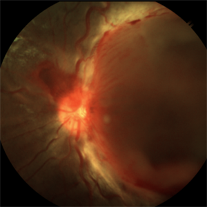
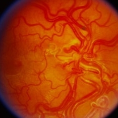
 OLD ISCHEMIC CRVO
Jun 11 2022 by Nivesh Gupta
Central fundus image showing tortuous vessels.
Photographer: DR. NIVESH GUPTA, RETINA FELLOW , RETINA FOUNDATION, AHMEDABAD
Imaging device: NIDEK MIRANTE
Condition/keywords: ischemic CRVO
-
 Impending-CRVO
Feb 20 2022 by Vishal Gupta, MBBS, MS
Impending CRVO in a 54 year old hypertensive female patient with Collaterals and Grade 2 Chronic Hypertensive retinopathy.
Photographer: Dr Shobhit Chawla, Prakash Netra Kendr, Lucknow, UP, INDIA
Imaging device: Zeiss Clarus 500
Condition/keywords: central retinal vein occlusion (CRVO), collaterals, dilated tortuous vessels, hypertensive retinopathy
-
 Retinal Capillary Hemangioma
Sep 9 2021 by Jesus Lozano, MD
60 year-old woman with a Peripheral RCH treated with laser photocoagulation.
Photographer: Yair Bet Yosef, Hadassah Medical Center. Israel
Imaging device: Optos Silverstone
Condition/keywords: abnormal retinal vessel, anomalous vessels, dilated tortuous vessels, hemangioma, retina
-
 Tortuous Retinal Vessels
Apr 29 2021 by Giselle DeOliveira
Infared photograph of 30-year-old male with tortuous retinal vessels .
Photographer: Giselle DeOliveira
Imaging device: Heidelberg Spectralis
Condition/keywords: tortuous vessels
-
 Tortuous Vessels in the Controlateral Eye in a Patient with Choroidal Hemangioma
Nov 11 2019 by Sophia El Hamichi, MD
A 51-year-old female with a history of treated choroidal hemangioma OD, presents with an interesting finding of vascular tortuosity of her good eye OS. VA 20/20 in this eye.
Photographer: Sophia El Hamichi, MD, Murray Ocular Oncology and Retina, Miami
Condition/keywords: choroidal hemangioma, Optos, tortuous vessels, vascular tortousity
-
 Retinoblastoma
Apr 2 2019 by Gary R. Cook, MD, FACS
Another view of the solid white exophytic retinoblastoma lesion with dilated and tortuous vessels visible on its surface in the left eye of a white female infant.
Condition/keywords: retinoblastoma
-
 Exophytic Retinoblastoma
Apr 2 2019 by Gary R. Cook, MD, FACS
Solid white-appearing exophytic retinoblastoma with dilated tortuous vessels visible on its surface in the left eye of a WF infant.
Condition/keywords: retinoblastoma
-
 Color Montage Picture
Jan 15 2019 by Aniruddha Maiti, MBBS DO DNB FRVS FICO MRCS FACS FASRS FRCOphth
Dilated tortuous vessels with intraretinal and subretinal hemorrhage at fovea.
Photographer: Sangeeta Mohanta
Imaging device: Heidelberg spectralis
Condition/keywords: color photo, dilated tortuous vessels, subretinal hemorrhage
-
 Color Montage Picture
Jan 15 2019 by Aniruddha Maiti, MBBS DO DNB FRVS FICO MRCS FACS FASRS FRCOphth
Dilated tortuous vessels with intraretinal and subretinal hemorrhage at fovea.
Photographer: Sangeeta Mohanta
Imaging device: Zeiss FF450 plus IR
Condition/keywords: dilated tortuous vessels, subretinal hemorrhage, Wyburn-Mason
-
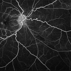
 Central Retinal Vein Occlusion With Macular Edema
Aug 29 2018 by Olivia Rainey
Ultra-widefield fluorescein angiogram of an 58-year-old male with a central retinal vein occlusion with macular edema affecting his right eye. Fluorescein showed delayed transit with late leakage.
Photographer: Olivia Rainey
Imaging device: Optos
Condition/keywords: central retinal vein occlusion (CRVO), FA late phase leakage, fluorescein angiogram (FA), Optos, tortuous vessels, ultra-wide field imaging
-
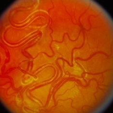
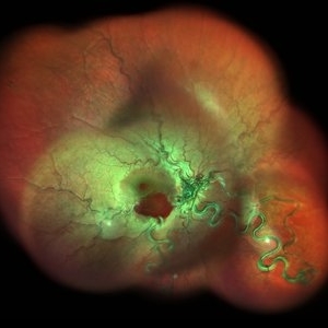
 Wyburn-Mason Syndrome (Racemose Hemangiomatosis)
Mar 30 2018 by Rameez N Hussain, MD
A 7-year-old Portuguese girl with unilateral retinal arteriovenous malformations composed of dilated, tortuous vessels with normal vision.
Photographer: Thambi Durai
Imaging device: TOPCON
Condition/keywords: arteriovenous malformation, racemose hemangioma, Wyburn-Mason
-
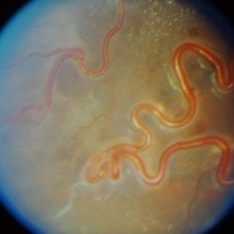
 Retinal Arteriovenous Malformations (Racemose Hemangiomatosis)
Mar 30 2018 by Rameez N Hussain, MD
A 7-year-old Portuguese girl with unilateral retinal arteriovenous malformations composed of dilated, tortuous vessels with normal vision.
Photographer: Thambi Durai, Consultant Optometrist, Orbit Health Care - Dr Agarwal's Eye Hospital, Maputo, Mozambique
Imaging device: TOPCON
Condition/keywords: racemose hemangioma, retinal arteriovenous malformations, Wyburn-Mason
-
 Retinal Arteriovenous Malformations (Racemose Hemangiomatosis)
Mar 30 2018 by Rameez N Hussain, MD
A 7-years-old Portuguese girl with unilateral retinal arteriovenous malformations composed of dilated, tortuous vessels with normal vision.
Photographer: Thambi Durai. Consultant Optometrist, Orbit Health Care - Dr Agarwal's Eye Hospital, Maputo, Mozambique
Imaging device: TOPCON
Condition/keywords: racemose hemangioma, retinal arteriovenous malformations, Wyburn-Mason
-
 Fluorescein Angiogram - Tortuous Vessels of DME Right Eye
Dec 10 2015 by James B. Soque, CRA, OCT-C, COA, FOPS
Early fluorescein angiogram of diabetic macular edema and tortuous vessels in the superior macula of the right eye.
Photographer: James B Soque, CRA, COA
Imaging device: Top[con TRC-50 DX with MERGE Winstation V 11.2.0
Condition/keywords: diabetes, diabetic macular edema, microaneurysms, microangiopathy, tortuous vessels
 Loading…