Search results (16 results)
-
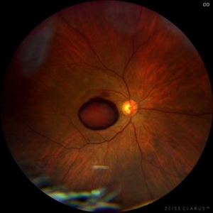
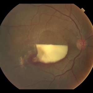
 Sub-Inner Limiting Membrane Hemorrhage
Mar 30 2024 by Karen Flores Guevara
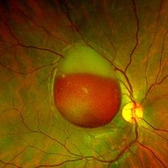
Fundus photograph of a 14-year-old-man with a Sub-inner limiting membrane hemorrhage.
Photographer: Diana Elizabeth Arellano AcostaPediatric Retina,Asociación para Evitar la Ceguera en México IAP. México
Condition/keywords: Sub-inner limiting membrane haemorrhage
-
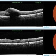
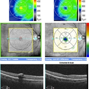
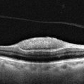 OCT of a sub-internal limiting membrane hemorrhage in Valsalva retinopathy
Jul 29 2022 by JORGE SOBERANES
Optical coherence tomography of a 70-year-old man with a sub-internal limiting membrane due to Valsalva retinopathy
Photographer: Jorge I. Soberanes, Asociación para Evitar la Ceguera en México.
Imaging device: PLEX Elite 9000, Zeiss
Condition/keywords: OCT, sub-inner limiting membrane hemorrhage, swept source, valsalva retinopathy
-
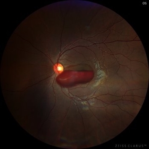
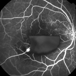
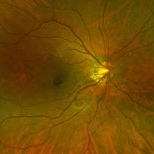
 Sub-internal limiting membrane hemorrhage in Valsalva retinopathy
Jul 29 2022 by JORGE SOBERANES
A fundus photography of a 70-year-old man with premacular hemorrhage (Sub-internal limiting membrane) due to Valsalva retinopathy
Photographer: Jorge I. Soberanes, Asociación para Evitar la Ceguera en México.
Imaging device: Zeiss Clarus 700
Condition/keywords: premacular hemorrhage, sub-inner limiting membrane hemorrhage, valsalva retinopathy
-
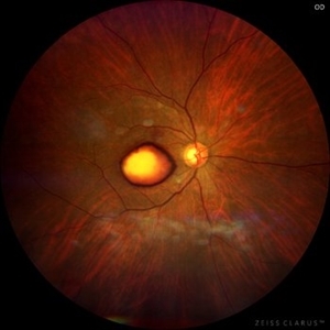 Dehemoglobinized sub-internal limiting membrane hemorrhage
Jul 29 2022 by JORGE SOBERANES
Fundus photograph of a 70-year-old man with Valsalva retinopathy manifested as premacular hemorrhage (sub-ILM) in dehemoglobinized process.
Photographer: Jorge I. Soberanes, Asociación para Evitar la Ceguera en México.
Imaging device: Zeiss Clarus 700
Condition/keywords: dehemoglobinized hemorrhage, sub-inner limiting membrane hemorrhage, valsalva retinopathy
-
 Sub-ILM Hemorrhage (Dehemoglobinized + Red) - Valsalva Retinopathy
Jun 12 2021 by RUSHIK PATEL
Fundus Image of 21-year-old boy with sub-ILM hemorrhage (dehemoglobinized + red ) following valsalva maneuver 10 days back.
Photographer: Rushik Patel, Netralaya Super Speciality Eye Hospital, Ahmedabad, Gujarat
Imaging device: Optos
Condition/keywords: sub-inner limiting membrane hemorrhage, valsalva retinopathy
-
 Sub-Internal Limiting Membrane Hemorrhage - Pre and Post YAG Laser
May 21 2021 by Anmol Naik
A 36-year-old male complained of central scotoma in the right eye after observing intense laser lights at a local festival celebration. His BCVA was 6/60. On examination, he had a sub-internal limiting membrane (ILM) hemorrhage, which was treated with focal frequency-doubled Nd:YAG laser. Two weeks later, the hemorrhage resolved completely with BCVA of 6/6.
Photographer: Anmol Naik, MS, Insight Institute of Ophthalmology, Pune, India.
Imaging device: Topcon 3D Maestro 1, integrated Fundus camera and OCT
Condition/keywords: focal laser, laser photocoagulation, sub-inner limiting membrane hemorrhage
-
 RAMA with Sub ILM Hemorrhage
Jan 31 2018 by John S. King, MD
73-year-old with well controlled diabetes and hypertension presented with a month onset of acute central scotoma; CF 5'; SUB-ILM vs subyaloid elevation
Photographer: Stacey
Imaging device: Cirrus
Condition/keywords: ruptured macroaneurysm, sub-inner limiting membrane hemorrhage
-
 RAMA with Sub ILM Hemorrhage
Jan 31 2018 by John S. King, MD
73 -year-old with well controlled diabetes and hypertension presented with a month onset of acute central scotoma; CF 5'; FA shows pooling in the aneurysm, blockage by dehemoglobinized heme, some diabetic changes and some IRMAs likely from old vein occlusion (s)
Photographer: Stacey
Imaging device: Cirrus
Condition/keywords: ruptured macroaneurysm, sub-inner limiting membrane hemorrhage
-
 RAMA with Sub ILM Hemorrhage
Jan 31 2018 by John S. King, MD
73-year-old with well controlled diabetes and hypertension presented with a month onset of acute central scotoma; CF 5'
Photographer: Stacey
Imaging device: Cirrus
Condition/keywords: ruptured macroaneurysm, sub-inner limiting membrane hemorrhage
-
 Sub-ILM Heme in patient with Roth Spots - Bacterial Endocarditis
Dec 3 2017 by John S. King, MD
29 yo wf denied ivdu p/c with acute scotoma. OCT shows sub-ILM heme in foveal region (left) 20/300 that resolved spontaneously a few weeks later, back to baseline acuity (right).
Imaging device: Cirrus
Condition/keywords: bacterial endocarditis, sub-inner limiting membrane hemorrhage, white centered retinal hemorrhage (Roth Spot)
-
 Roth Spots - Bacterial Endocarditis
Dec 3 2017 by John S. King, MD
Initial presentation; 29-year-old white female denied ivdu p/c with acute scotoma due to the sub-ILM foveal heme. She did have some roth spots in both eyes. There was a focal area of periphlebitis just superior to the fovea OD. Work up for roth spots and retinal vasculitis initiated. She did have a low grade fever that she attributed to a urinary tract infection being treated by her PCP.
Imaging device: Optos
Condition/keywords: sub-inner limiting membrane hemorrhage, white centered retinal hemorrhage (Roth Spot)
-
 Sub ILM Hemorrhage
Jul 29 2014 by Mallika Goyal, MD
OCT of the left eye of a 26-year-old lady who presented with sudden vision drop (VA 20/40) reveals sub-ILM haemorrhage. There was no history of trauma, valsalva maneuvre or other contributing factors. This heme cleared within 10 days following gas injection and prone positioning with visual recovery to 20/20.
Photographer: Mallika Goyal, MD, Apollo Health City, Jubilee Hills, Hyderabad-500033
Condition/keywords: sub-inner limiting membrane hemorrhage
-
 Sub ILM Hemorrhage
Jul 29 2014 by Mallika Goyal, MD
Fundus photograph of the left eye of a 26-year-old lady who presented with sudden vision drop (VA 20/40) reveals sub-ILM hemorrhage. There was no history of trauma, valsalva maneuvre or other contributing factors. This heme cleared within 10 days following gas injection and prone positioning with visual recovery to 20/20.
Photographer: Mallika Goyal, MD, Apollo Health City, Jubilee Hills, Hyderabad-500033
Condition/keywords: sub-inner limiting membrane hemorrhage
-
 Blood Under the Internal Limiting Membrane
May 29 2013 by Zofia Anna Nawrocka (vel Michalewska), MD, PhD
Scanning laser ophthalmoscopy and SD-OCT of a 25-year-old patient with sub- ILM hemorrhage.
Photographer: Slawomir Cisiecki, MD, PhD, Ophthalmic Clinic "Jasne Blonia", Lodz, Poland
Imaging device: Heidelberg Spectralis
Condition/keywords: sub-inner limiting membrane hemorrhage
-
---thumb.jpg/image-square;max$300,300.ImageHandler) Terson Syndrome
Terson Syndrome
Mar 9 2013 by Young-Gyun Kim, MD
OCT image of a 56-year-old woman with a sub-ILM hemorrhage. She has subarachnoid hemorrhage.
Photographer: Shin Ji-Young, Eulji university, Seoul
Imaging device: Heidelberg Spectralis
Condition/keywords: sub-inner limiting membrane hemorrhage, Terson's Syndrome
-
 Terson Syndrome
Mar 9 2013 by Young-Gyun Kim, MD
Fundus photograph of a 56-year-old woman with a sub-ILM hemorrhage. She has subarachnoid hemorrhage.
Photographer: Shin Ji-Young, Eulji university, Seoul
Imaging device: Topcon TRC 50 EX
Condition/keywords: sub-inner limiting membrane hemorrhage, Terson's Syndrome
 Loading…