Search results (57 results)
-
 Hamartoma of the Retina and Retinal Pigment Epithelium
Jan 5 2025 by César Adrián Gómez Valdivia, MD
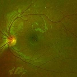
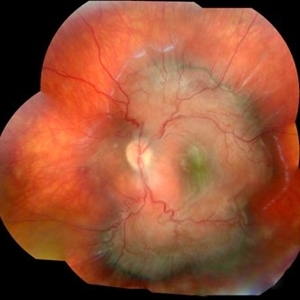
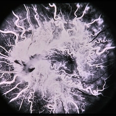
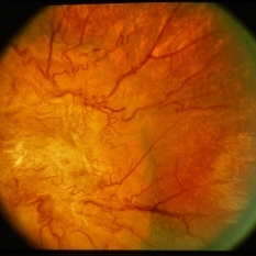
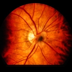
Hamartoma of the retina and retinal pigment epithelium found in a 10 year-old male patient with type 2 neurofibromatosis history. Overlaying fibrous proliferation can be appreciated. Findings were unilateral.
Photographer: @eyemissu2
Imaging device: TOPCON TRC-50DX
Condition/keywords: hamartoma, retinal pigment epithelium (RPE) hamartoma
-
 Hamartoma of the Retinal Pigment Epithelium
Apr 2 2024 by José Laércio Araújo Filho
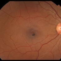
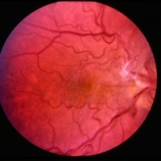
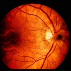
Fundus photograph of a 36-year-old man with a Hamartoma of the retinal pigment epithelium.
Photographer: José Laércio de Araújo Filho, Universidade de São Paulo, São Paulo
Imaging device: Optos Daytona P200T / A10600
Condition/keywords: retinal pigment epithelium (RPE) hamartoma
-
 Congenital Simple Hamartoma of the Retinal Pigment Epithelium
May 16 2022 by David C Sousa, MD PhD FRANZCO
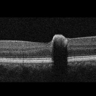
A 49-year-old man was referred after an incidental finding in the right eye macula. Best-corrected visual acuity was 20/25. Anterior segment examination was unremarkable. Fundoscopy revealed a juxta-foveal heavily pigmented well-demarcated slightly elevated lesion measuring 0.4 x 0.4 mm. No other changes were observed adjacent to the lesion or elsewhere in either eye. Optical coherence tomography revealed an area of retinal elevation with high optical reflectivity and posterior shadowing. The findings are consistent with congenital simple hamartoma of the retinal pigment epithelium. Given the benign, non-progressive and usually asymptomatic nature of this condition, most patients are diagnosed in adulthood.
Imaging device: Topcon Maestro2
Condition/keywords: retinal pigment epithelium (RPE) hamartoma
-
 Congenital Simple Hamartoma of the Retinal Pigment Epithelium
May 16 2022 by David C Sousa, MD PhD FRANZCO
A 49-year-old man was referred after an incidental finding in the right eye macula. Best-corrected visual acuity was 20/25. Anterior segment examination was unremarkable. Fundoscopy revealed a juxta-foveal heavily pigmented well-demarcated slightly elevated lesion measuring 0.4 x 0.4 mm. No other changes were observed adjacent to the lesion or elsewhere in either eye. Optical coherence tomography revealed an area of retinal elevation with high optical reflectivity and posterior shadowing. The findings are consistent with congenital simple hamartoma of the retinal pigment epithelium. Given the benign, non-progressive and usually asymptomatic nature of this condition, most patients are diagnosed in adulthood.
Imaging device: Topcon Maestro2
Condition/keywords: retinal pigment epithelium (RPE) hamartoma
-
 Combined Hamartoma of Retina and Retinal Pigment Epithelium
Apr 30 2021 by ARVIND JAIN M
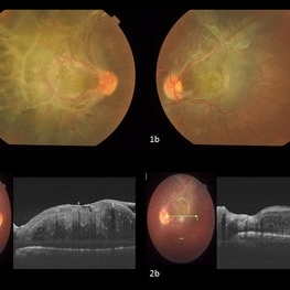
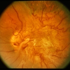
A 26-year-old gentlemen came with complains of defective vision in both eyes since childhood. His BCVA was right eye 5/60 and left eye 6/60. His anterior segment examination showed no abnormality with posterior segment examination showed both eyes (1a and 1b) greyish white elevated lesion involving the macula with thick fibrotic epiretinal membrane causing the macular drag temporally in right eye and supero-temporally in left eye. (2a and 2b) showing the thick ERM with the hamartoma of the retina and RPE.
Photographer: DR ARVIND JAIN, ARAVIND EYE HOSPITAL, COIMBATORE,INDIA
Condition/keywords: combined hamartoma, congenital hypertrophy of the retinal pigment epithelium (CHRPE), epiretinal membrane (ERM), retinal pigment epithelium (RPE) hamartoma
-
 Hamartoma of the Retina
May 29 2018 by JEFFERSON R SOUSA, Tecg.º (Biomedical Systems Technology)
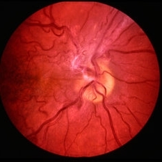
A 4-year-old male patient attended the clinic for evaluation. In the mapping examination and retina and retinography, important alterations were observed in the posterior pole of the left eye. This in turn was sent to perform the ocular ultrasonography examination, which together with the previous examinations, confirmed changes that suggested diagnosis of: COMBINED HAMARTOMA OF RETINA AND PIGMENTARY EPITHELIUM.
Photographer: JEFFERSON R SOUSA - Study Center and Ophthalmological Research Dr. Andre M V Gomes, Institute Dr. Suel Abujamra São Paulo-Brazil
Imaging device: Topcon TRC-50 DX, Imaginet 5.0, angle de 35º . Flash 36 / Mosaic with 9 images.
Condition/keywords: combined hamartoma, retinal pigment epithelium (RPE) hamartoma, tumor
-
 Combined Hamartoma of the Retina and Retinal Pigment Epithelium
Dec 22 2015 by P. Mahesh Shanmugam, MBBS, DO, FRCSEd, PhD, FAICO
A fundus photo of 11-year-old boy with CHRRPE, gliotic epiretinal membrane overlying deep greyish lesion. Retinal wrinkling and dragging of macula is seen.
Condition/keywords: combined hamartoma, retinal pigment epithelium (RPE) hamartoma
-
 Combined Hamartoma of the Retina and RPE
Jul 8 2015 by Emmanuel Chang, MD PhD FACS FASRS
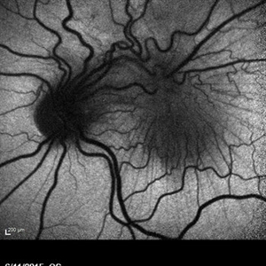
10-year-old with history of progressive severe distortion in the left eye over the past year.
Photographer: Retina and Vitreous of Texas
Imaging device: Heidelberg Autofluorescence
Condition/keywords: combined hamartoma, epiretinal membrane (ERM), retinal pigment epithelium (RPE) hamartoma
-
 Epiretinal Membrane/Macular Pucker With Combined Hamartoma of Retina and RPE
Jul 8 2015 by Emmanuel Chang, MD PhD FACS FASRS
10-year-old with history of progressive severe distortion in the left eye over the past year.
Photographer: Retina and Vitreous of Texas
Imaging device: Heidelberg Autofluorescence
Condition/keywords: combined hamartoma, epiretinal membrane (ERM), retinal pigment epithelium (RPE) hamartoma
-
 Combined Hamartoma of the Retina and RPE
Jul 8 2015 by Emmanuel Chang, MD PhD FACS FASRS
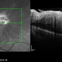
10-year-old with history of progressive severe distortion in the left eye over the past year.
Photographer: Retina and Vitreous of Texas
Imaging device: Heidelberg Spectralis
Condition/keywords: combined hamartoma, epiretinal membrane (ERM), retinal pigment epithelium (RPE) hamartoma
-
 Combined Hamartoma of the RPE
Dec 22 2014 by H. Michael Lambert, MD
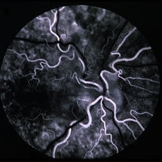
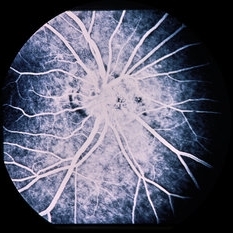
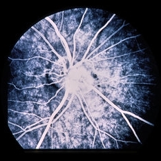
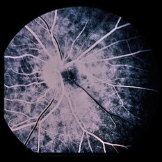
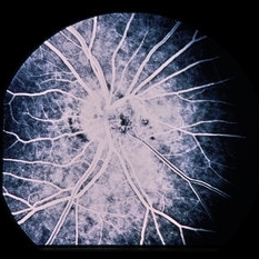
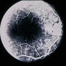
Fluorescein angiogram of second patient.
Condition/keywords: combined hamartoma, retinal pigment epithelium (RPE) hamartoma
-
 Combined Hamartoma of the RPE
Dec 22 2014 by H. Michael Lambert, MD
Fluorescein angiogram of second patient.
Condition/keywords: combined hamartoma, retinal pigment epithelium (RPE) hamartoma
-
 Combined Hamartoma of the RPE
Dec 22 2014 by H. Michael Lambert, MD
Fundus photo of second patient.
Condition/keywords: combined hamartoma, retinal pigment epithelium (RPE) hamartoma
-
 Combined Hamartoma of the RPE
Dec 22 2014 by H. Michael Lambert, MD
Fundus photo of first patient.
Condition/keywords: combined hamartoma, retinal pigment epithelium (RPE) hamartoma
-
 Combined Hamartoma of the RPE
Dec 22 2014 by H. Michael Lambert, MD
Fundus photo of first patient.
Condition/keywords: combined hamartoma, retinal pigment epithelium (RPE) hamartoma
-
 Combined Hamartoma of the RPE
Dec 22 2014 by H. Michael Lambert, MD
Fundus photo of first patient.
Condition/keywords: combined hamartoma, retinal pigment epithelium (RPE) hamartoma
-
 RPE Hamartoma
Dec 22 2014 by H. Michael Lambert, MD
Fundus photo; it looks like a melanocytoma.
Condition/keywords: retinal pigment epithelium (RPE) hamartoma
-
 RPE Hamartoma
Dec 22 2014 by H. Michael Lambert, MD
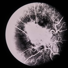
Fluorescein angiogram of the lesion.
Condition/keywords: retinal pigment epithelium (RPE) hamartoma
-
 RPE Hamartoma
Dec 22 2014 by H. Michael Lambert, MD
Fluorescein angiogram of the lesion.
Condition/keywords: retinal pigment epithelium (RPE) hamartoma
-
 RPE Hamartoma
Dec 22 2014 by H. Michael Lambert, MD
Fundus photo; it looks like a melanocytoma.
Condition/keywords: retinal pigment epithelium (RPE) hamartoma
-
 RPE Hamartoma
Dec 22 2014 by H. Michael Lambert, MD
Fluorescein angiogram of the lesion.
Condition/keywords: retinal pigment epithelium (RPE) hamartoma
-
 RPE Hamartoma
Dec 22 2014 by H. Michael Lambert, MD
Fluorescein angiogram of the lesion.
Condition/keywords: retinal pigment epithelium (RPE) hamartoma
-
 Combined Retinal & RPE Hamartoma
Jun 13 2014 by David Callanan, MD
66-year-old white female, combined retinal & RPE hamartoma.
Condition/keywords: combined hamartoma, retinal pigment epithelium (RPE) hamartoma
-
 Combined Retinal & RPE Hamartoma
Jun 13 2014 by David Callanan, MD
66-year-old white female, combined retinal & RPE hamartoma.
Condition/keywords: combined hamartoma, retinal pigment epithelium (RPE) hamartoma
-
 Combined Retinal & RPE Hamartoma
Jun 13 2014 by David Callanan, MD
66-year-old white female, combined retinal & RPE hamartoma.
Condition/keywords: combined hamartoma, retinal pigment epithelium (RPE) hamartoma
 Loading…