Search results (805 results)
-
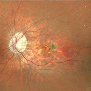
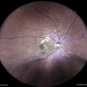
 Retinal Arteriovenous Malformation
Oct 7 2025 by Korey Starkey
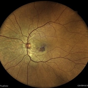
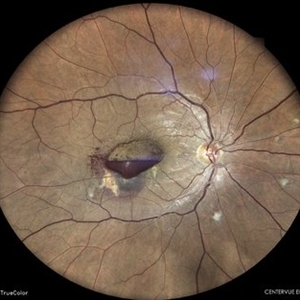
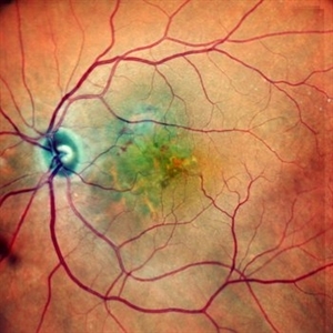
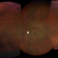
55 year-old patient presents with retinal arteriovenous malformation in the left eye and BRVO w/retinal neovascularization. Patient is asymptomatic. No edema or treatment necessary today, signs of old RVO with MAs along inferior arcade and dot heme.
Photographer: Korey Starkey
Imaging device: Topcon
Condition/keywords: branch retinal vein occlusion (BRVO), fundus photography, inferior arcade, microaneurysms, retinal arteriovenous malformations, retinal neovascularization, Topcon
-
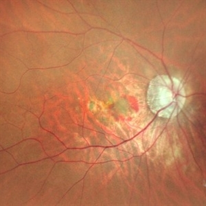
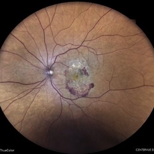
 Subretinal Neovascular Membrane
Aug 15 2025 by Akansha Sharma
Color fundus photograph of a 40 year old male with subretinal neovascular membrane.
Photographer: DR. AKANSHA SHARMA
Condition/keywords: choroidal neovascular membrane (CNVM), CNVM, SRNVM, subretinal neovascularization (SRNV), wet age-related macular degeneration (wet AMD)
-
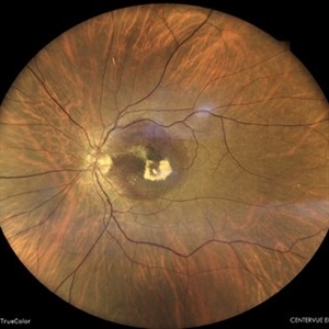
 Subretinal Neovascular Membrane
Aug 15 2025 by Akansha Sharma
Color fundus photograph of a 40 year old male with subretinal neovascular membrane.
Photographer: DR. AKANSHA SHARMA
Condition/keywords: choroidal neovascular membrane (CNVM), CNVM, Myopic CNVM, SRNVM, subretinal neovascularization (SRNV), Wet age related macular degeneration
-
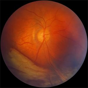 Traction in Progression
Aug 6 2025 by Claudio Brancato, MD
This image captures stage 4A Retinopathy of Prematurity, showing partial retinal detachment sparing the macula. The elevated retina and fibrous ridge indicate tractional forces secondary to extraretinal neovascularization. A striking representation of disease evolution, poised between reversibility and vision loss.
Photographer: Gregorio Lo Giudice, ARNAS Civico Hospital, Palermo, Italy
Imaging device: RETCAM 3 (enhanced via IA)
Condition/keywords: retinopathy of prematurity
-
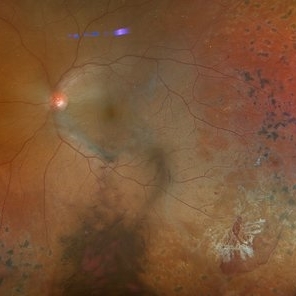 Actively Bleeding NVE
Apr 1 2025 by Jordyn Beckman
47 year old woman presented with actively bleeding NVE temporally on exam with complaints of foggy vision and floaters.
Photographer: Jordyn Beckman, Retina Consultants of Carolina, P.A.
Imaging device: Optos California
Condition/keywords: active bleeding, Elevated retinal neovascularization, vitreous hemorrhage
-
 Ischemic BRVO
Aug 26 2024 by Nassim Alejandro Abreu Arbaje, MD
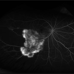
Montage of fluorescein angiography and color fundus photos of a 60 years old male with diabetic retinopathy complicated with extensive retinal neovascularization secondary to and ischemic BRVO.
Photographer: Nassim Abreu, Hospital Dr. Elías Santana
Condition/keywords: BRVO, diabetic retinopathy, Neovascularisation elsewhere (NVE)
-
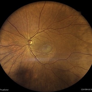
 Subretinal Neovascular Membrane
Jun 5 2024 by Akansha Sharma
Color fundus photograph of a 49 year old female with subretinal bleed suggestive of subretinal neovascular membrane.
Photographer: Dr. Akansha Sharma, Bharati Eye Hospital
Condition/keywords: choroidal neovascular membrane (CNVM), CNVM, SRNVM, subretinal neovascularization (SRNV), wet age-related macular degeneration (wet AMD)
-
 Subretinal Neovascular Membrane
Jun 5 2024 by Akansha Sharma
Color fundus photograph of a 94 year old female with subretinal bleed suggestive of subretinal neovascular membrane.
Photographer: Dr. Akansha Sharma, Bharati Eye Hospital
Condition/keywords: choroidal neovascular membrane (CNVM), CNVM, SRNVM, subretinal neovascularization (SRNV), wet age-related macular degeneration (wet AMD)
-
 Subretinal Neovascular Membrane
Apr 19 2024 by Akansha Sharma
Color fundus photograph of a 74 year old female with subretinal neovascular membrane with scarring below the fovea.
Photographer: Dr. Akansha Sharma, Bharati Eye Hospital
Condition/keywords: SRNVM, subretinal neovascularization (SRNV)
-
 Subretinal Neovascular Membrane with PED
Apr 17 2024 by Akansha Sharma
Color fundus photograph of a 72 year old male with ped along with subretinal bleed around it.
Photographer: Dr. Akansha Sharma, Bharati Eye Hospital
Condition/keywords: CNVM, PED, SRNVM, subretinal neovascularization (SRNV), wet age-related macular degeneration (wet AMD)
-
 Subretinal Neovascular Membrane
Mar 26 2024 by Akansha Sharma
Color fundus photograph of a 65 year old female patient with subretinal bleed suggestive of subretinal neovascular membrane.
Photographer: Dr. Akansha Sharma, Bharati Eye Hospital
Condition/keywords: CNVM, SRNVM, subretinal neovascularization (SRNV), wet age-related macular degeneration (wet AMD)
-
 Branch Retinal Vein Occlusion with Retinal Neovascularization
Mar 21 2024 by Isaac Agranoff
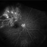
Fundus angiography photograph of a 63 year old male presenting with worsening blurry vision OD for 4 years with new transient floaters (vision 20/160 PH 20/60). Fluorescein angiography revealed significant capillary non-perfusion corresponding to the area, with peripheral vascular remodeling. Physician recommended anti-VEGF therapy and FA-guided supplemental PRP given the size of the NVE.
Photographer: Isaac Agranoff
Imaging device: Optos California
Condition/keywords: branch retinal vein occlusion (BRVO), EYLEA, FLUORESCEIN ANGIOGRAPHY, Neovascularisation elsewhere (NVE), Optos
-
 Sea fan neovascularization
Jul 19 2023 by Mariam Cernichiaro-Espinosa, MD
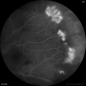
Sea fan neovascularization on a fluorescein angiography
Photographer: Mariam Cernichiaro-Espinosa, Asociación para Evitar la Ceguera en México, I.A.P. Mexico City, Mexico.
Imaging device: Zeiss Clarus
Condition/keywords: retinal neovascularization, sea fan
-
 Subretinal Neovascular Membrane
May 8 2023 by Akansha Sharma
Colour fundus photograph of a 81 year old female with subretinal neovascular memebrane
Photographer: Dr. Urmil Shah, Dr. Denish Patel, Dr. Akansha Sharma, Bharati Eye Clinic, Ahmedabad, Gujarat
Condition/keywords: CNVM, subretinal neovascularization (SRNV)
-
 Subhyaloid with Subretinal Bleed
May 8 2023 by Akansha Sharma
Colour fundus photograph of a 38 year old male with subhyaloid bleed with subretinal bleed
Photographer: Dr. Urmil Shah, Dr. Denish Patel, Dr. Akansha Sharma, Bharati Eye Clinic, Ahmedabad, Gujarat
Condition/keywords: SHH, subretinal neovascularization (SRNV)
-
 Retinal neovascularization
Feb 28 2023 by Nassim Alejandro Abreu Arbaje, MD
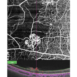
OCT and OCTa of a diabetic patient with severe PDR, showing the anatomical location and blood flow of neovessels
Photographer: Nassim Abreu, Centro de Oftalmologia y Glaucoma
Imaging device: Topcon Triton Plus
Condition/keywords: neovascularization (NV), OCT, OCT Angiography, PDR
-
 WET AGE RELATED MACULAR DEGENERATION
Nov 21 2022 by Akansha Sharma
COLOUR FUNDUS PHOTOGRAPH OF A 71 YEAR OLD MALE WITH SUBRETINAL BLEED IN A CASE OF WET AGE RELATED MACULAR DEGENERATION
Photographer: Dr. Akansha Sharma-Retina Foundation, Ahmedabad
Condition/keywords: CNVM, subretinal neovascularization (SRNV), wet age-related macular degeneration (wet AMD)
-
 SUB-RETINAL NEOVASCULAR MEMBRANE
Nov 21 2022 by Akansha Sharma
COLOUR FUNDUS PHOTOGRAPH OF A 57 YEAR OLD MALE WITH SUBRETINAL NEOVASCULAR MEMBRANE
Photographer: Dr. Akansha Sharma-Retina Foundation, Ahmedabad
Condition/keywords: choroidal neovascularization (CNV), CNVM, subretinal neovascularization (SRNV)
-
 SUB-RETINAL NEOVASCULAR MEMBRANE
Nov 21 2022 by Akansha Sharma
COLOUR FUNDUS PHOTO OF A 79 YEAR OLD MALE PATIENT WITH SUBRETINAL NEOVASCULAR MEMBRANE
Photographer: Dr. Akansha Sharma-Retina Foundation, Ahmedabad
Condition/keywords: choroidal neovascular membrane (CNVM), CNVM, subretinal neovascularization (SRNV)
-
 Central Retinal Vein Occlusion with Retinal Neovascularization
Jan 19 2022 by Olivia Rainey
Ultra-widefield fluorescein angiogram of a 56-year-old male with a Central Retinal Vein Occlusion with Retinal Neovascularization affecting his left eye. The patient presented on 1/19/2022 with scNLP vision in the left eye. The patient has good PRP, however areas of ischemia still remain untreated by laser. He also has severe neovascular glaucoma contributing to his poor vision.
Photographer: Olivia Rainey, OCT-C, COA
Imaging device: Optos California
Condition/keywords: central retinal vein occlusion (CRVO), FA early phase, fluorescein angiogram (FA), hemorrhage, ischemic CRVO, left eye, neovascular glaucoma, Optos, pan-retinal photocoagulation (PRP), retinal ischemia, retinal neovascularization, ultra-wide field imaging
-
 Branch Retinal Vein Occlusion with Retinal Neovascularization
Jun 9 2021 by Shelby Helton
Ultrawide fluorescein angiography of a 83-year-old female with a branch retinal vein occlusion with retinal neovascularization affecting her right eye.
Photographer: Shelby Helton, Retina Specialist Of Michigan
Imaging device: Optos California
Condition/keywords: branch retinal vein occlusion (BRVO)
-
 Retinal Angiomatous Proliferation
May 7 2021 by Dhaivat Shah
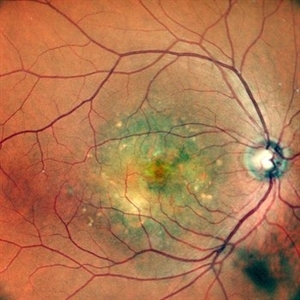
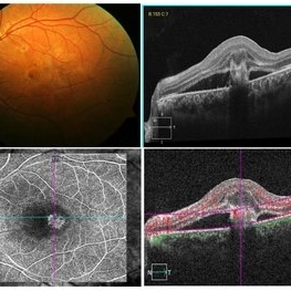
Retinal angiomatous proliferation (RAP) is a distinct variant of neovascular age-related degeneration (AMD) that usually initiates at the retina and progresses posteriorly into sub retinal space. In most recent study, it was suggested that angiogenesis may begin in the retina, choroid, or both, and introduced a new name for the process: Type 3 neovascularization. The frequency of RAP has been studied in many studies, with figures ranging from 10% to 21% of exudative AMD. Clinically, three stages were originally described as intraretinal neovascularization (IRN), subretinal neovascularization (SRN), choroidal neovascularization (CNV). RAP predominantly intraretinal hard exudates, and intra/pre retinal hemorrhages along with intraretinal edema, associated pigment epithelial detachment beneath it, at times retinochoroidal, retino-retinal anastomosis. Apart from conventional OCT, FFA and ICG, OCT-A has now been used primarily as a tool in the diagnosis RAP. Here we present imaging of a 30-year-old young male diagnosed as RAP stage 3 (Type 3 CNVM). Patient was started on intravitreal anti-VEGF monotherapy therapy.
Photographer: Choithram Netralaya Indore
Condition/keywords: retinal angiomatous proliferation (RAP)
-
 Ischemic BRVO With Retinal Neovascularization
Apr 26 2021 by Niloofar Piri, MD
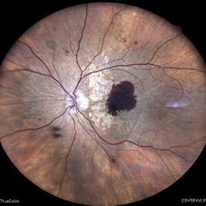
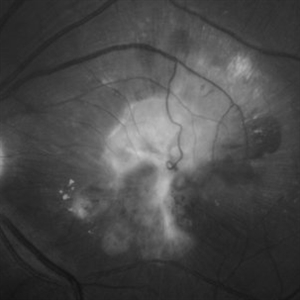
Wide field fundus photograph of the left eye demonstrating sclerotic superotemporal vein resulting from ischemic old BRVO with secondary retinal neovascularization. Note the unrelated atrophic hole within lattice degeneration in temporal periphery.
Photographer: Niloofar Piri, MD, St. louis University
Condition/keywords: branch retinal vein occlusion (BRVO), ischemia, non-perfused branch retinal vein occlusion (BRVO), retinal neovascularization
-
 Into de depth!
Mar 14 2021 by Luiz A Zago, PhD
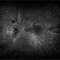
Diving anastomotic vein! Blood secondary to a sub retinal neovascular membrane in an old fibrotic scar. Colateral drainage anastomosis with the choroid
Photographer: Dr Luiz Alberto Zago Filho
Imaging device: Topcon 50IX
Condition/keywords: chorioretinal scar, retina vessels, subretinal hemorrhage, subretinal neovascularization (SRNV)
-
 Mixed Occlusion of Artery and Vein
Jan 6 2021 by Renata Garcia Franco, Md
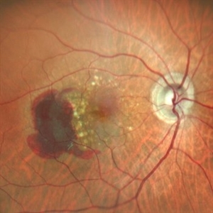
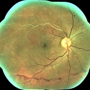
Male with a history of smoking, sudden low vision of the right eye, retinal neovascularization and inferior preretinal hemorrhage.
Photographer: Fatima Hernandez, Instituto de la Retina del Bajio SC
Imaging device: Zeiss
Condition/keywords: arterial occlusion
 Loading…