 Initializing download.
-
 By Dhaivat Shah
Sankara Nethralaya
Co-author(s): Dr Shams Tabrez, Choithram Netralaya Indore - Uploaded on May 7, 2021.
- Last modified by Caroline Bozell on May 7, 2021.
- Rating
- Appears in
- Imaging marvels
- Condition/keywords
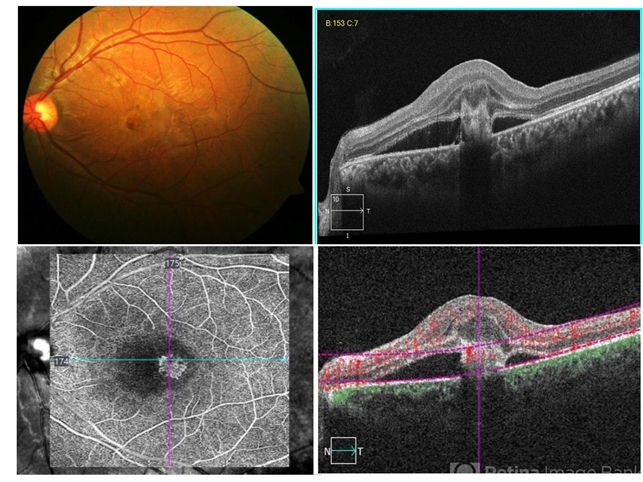
- retinal angiomatous proliferation (RAP)
- Photographer
- Choithram Netralaya Indore
- Imaging device
- Optical coherence tomography system
- Description
- Retinal angiomatous proliferation (RAP) is a distinct variant of neovascular age-related degeneration (AMD) that usually initiates at the retina and progresses posteriorly into sub retinal space. In most recent study, it was suggested that angiogenesis may begin in the retina, choroid, or both, and introduced a new name for the process: Type 3 neovascularization. The frequency of RAP has been studied in many studies, with figures ranging from 10% to 21% of exudative AMD. Clinically, three stages were originally described as intraretinal neovascularization (IRN), subretinal neovascularization (SRN), choroidal neovascularization (CNV). RAP predominantly intraretinal hard exudates, and intra/pre retinal hemorrhages along with intraretinal edema, associated pigment epithelial detachment beneath it, at times retinochoroidal, retino-retinal anastomosis. Apart from conventional OCT, FFA and ICG, OCT-A has now been used primarily as a tool in the diagnosis RAP. Here we present imaging of a 30-year-old young male diagnosed as RAP stage 3 (Type 3 CNVM). Patient was started on intravitreal anti-VEGF monotherapy therapy.


