Search results (82 results)
-
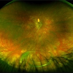
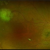
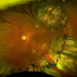 CHRPE
CHRPE
Mar 25 2025 by Toolie Winters
Ultra-wide field fundus photograph of a 78-year-old woman with extensive CHRPE lesions OS. Continued observation has been recommended at this time.
Photographer: Toolie Winters
Imaging device: Optos California
Condition/keywords: CHRPE, congenital hypertrophy of the retinal pigment epithelium (CHRPE), fundus photography, Optos, Optos California, pseudocolor, ultra-wide field imaging
-
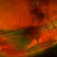
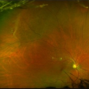
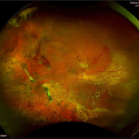 Peripheral Exudative Hemorrhagic Chorioretinopathy
Nov 19 2024 by Toolie Winters
Ultra-wide field fundus photograph of an 85-year-old woman with Peripheral Exudative Hemorrhagic Chorioretinopathy (PECHR) affecting the right eye. Patient presented with a blind spot centrally in the right eye which she first noticed 4 months prior to this image being taken. The patient states that in the month prior to this image, she began noticing bright lights flash across her vision 4-5x/day which last about 15 seconds. The flashes are either black with a blue ring around them or yellow, and their frequency has increased over time. The patient's vision at the time of this appointment was Dcc20/100+1 PHNI. This photo also shows diffuse hemorrhage, lipid, and an eccentric disciform lesion.
Photographer: Toolie Winters
Imaging device: Optos California
Condition/keywords: fundus photography, neovascular age-related macular degeneration (AMD), Optos, OPTOS CALIFORNIA, peripheral exudative hemorrhagic chorioretinopathy (PEHCR), pseudocolor, ultra-wide field imaging, wet age-related macular degeneration (wet AMD)
-
 Leber´s Congenital Amaurosis
Sep 6 2024 by Mauricio Bayram-Suverza, MD
13-year-old female patient with severe nyctalopia, photophobia, and reduced peripheral vision. CRB1-related Leber´s Congenital Amaurosis. The ultra-widefield pseudocolor image shows attenuated arterioles and diffuse nummular pigmentation with important atrophy.
Photographer: Mauricio Bayram-Suverza, Casey Eye Institute, OHSU.
Imaging device: Optos California
Condition/keywords: genetic testing, Leber's congenital amaurosis, nyctalopia, retinal dystrophy
-
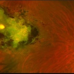
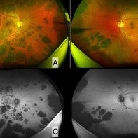 Bear Track Lesions in a Case of Congenital Hypertrophy of RPE
Sep 6 2024 by Giriraj Vibhute
Fundus photo and autofluorescence image in a 58-year-old woman A&B: Right and left eye ultrawidefield pseudocolor imaging in case of congenital hypertrophy of RPE. C&D: Fundus autofluorescence of right and left eye of the same patient. The patient/ family members did not have any history of colon cancer. Patient was advised colonoscopy and family members were screened.
Photographer: Giriraj Vibhute, dept of retina, M M Joshi eye institute, Hubli. India
Imaging device: Optos daytona
Condition/keywords: bear tracks, congenital hypertrophy of the retinal pigment epithelium (CHRPE)
-
 A Fleet of Boat-Shaped Hemorrhages
Aug 1 2024 by James P Dossett, MD
Pseudocolor fundus photograph of the left eye of a 54-year-old diabetic man presenting with bilateral vision loss. Examination revealed 20/200 vision OS with extensive preretinal and vitreous hemorrhage, marked diffuse neovascularization, macular edema and hard exudates.
Photographer: Beth Smith, West Virginia University Eye Institute
Condition/keywords: proliferative diabetic retinopathy (PDR)
-
 Subretinal Gas After Pneumatic Retinopexy
Mar 6 2024 by James P Dossett, MD
Pseudocolor fundus photograph of a 68-year-old man who presented with a macula-on rhegmatogenous retinal detachment with a single horseshoe tear at 12 o'clock. Pneumatic retinopexy was performed with cryopexy to the retinal break. He returned to clinic three days later and the entire SF6 gas bubble was noted to have migrated to the subretinal space through the retinal break. Pars plana vitrectomy was performed that day with retinal reattachment and improvement in vision to 20/40 now 6 months postoperatively.
Imaging device: Optos
Condition/keywords: pneumatic retinopexy, subretinal gas bubble
-
 Roth Spots
Mar 5 2024 by James P Dossett, MD
Pseudocolor fundus photograph of the right eye of a 56-year-old man who presented for evaluation of floaters noted to have bilateral Roth spots on dilated fundus exam. WBC count was obtained and was >300k. Bone marrow biopsy was performed and was consistent with chronic myelogenous leukemia. He was started on dasatinib and hydroxycarbamide. 1 month later the hemorrhages had improved significantly.
Imaging device: Optos
Condition/keywords: Roth spots
-
 Toxoplasmosis Chorioretinitis
Mar 2 2024 by James P Dossett, MD
Pseudocolor fundus photograph of the right eye of a 34-year-old man with retinitis along the inferotemporal arcade with associated subretinal fluid and overlying vitritis. Aqueous paracentesis was performed and PCR was positive for Toxoplasma gondii. He was administered intravitreal clindamycin.
Imaging device: Optos
Condition/keywords: posterior uveitis, toxoplasmosis chorioretinitis
-
 Vitreoretinal Traction with Adjacent Tear and Vitreous Hemorrhage
Oct 3 2023 by Alexis Singstock
Ultra-widefield fundus photograph of a 76 year old woman with vitreoretinal traction, an adjacent retinal tear and vitreous hemorrhage affecting the left eye. Patient was referred for retinal detachment and vitreous hemorrhage. Patient reports waking up the day prior to their appointment with "a lot of lines coming down the front, like swirling dirt in the left eye". Patient's vision was counting fingers at 1 ft. Dr. Joseph Boss noticed a horseshoe tear inferior to traction on exam and with the help of ultra-widefield imaging. Dr. Boss performed laser retinopexy to tear and impending tear at site of traction. Patient is scheduled for pars plana vitrectomy for dense vitreous hemorrhage.
Photographer: Alexis Singstock
Imaging device: Optos California
Condition/keywords: acute posterior vitreous detachment, fundus photography, left eye, Optos, OPTOS CALIFORNIA, pseudocolor, ULTRA WIDE FIELD, vitreoretinal traction, vitreous hemorrhage
-
 Dislocated Lens
Apr 26 2023 by Chloe Hanifan
Ultra wide field fundus photograph of a 41-year-old male with a dislocated lens affecting his right eye. IOL noted inferior vitreous base and vitrectomy surgery for removal of IOL was recommended. Patient has history of retinitis pigmentosa as well. Patient's vision at the time of presentation was counting fingers at 2 feet.
Photographer: Chloe Hanifan
Imaging device: Optos California
Condition/keywords: dislocated lens, fundus photography, Optos, pseudocolor, retinitis pigmentosa, ULTRA WIDE FIELD
-
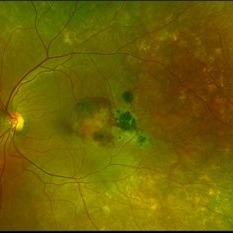 Subretinal Hemorrhage with Chorioretinal Macular Scars
Sep 28 2022 by Denica Rodriguez
Ultra-widefield pseudocolor fundus photograph of a 59 year old female with Subretinal Hemorrhage with Chorioretinal Macular Scars affecting her left eye. The physician presumes the etiology is CNV from adjacent scarring/choroidal rupture. Patient has history of ocular trauma with cricket ball at age 10-12 years old. She suspects that she might have suffered choroidal rupture, which has resulted in secondary CNV and hemorrhage that we are seeing today. She recommends treatment with Eylea sample injection in a series of 4 at a 4-5 week interval. The patient's vision at the time of her appointment was Dcc20/40-2 PHNI.
Photographer: Denica Rodriguez, COA
Imaging device: Optos California
Condition/keywords: antiVEGF therapy, chorioretinal scar, choroidal neovascular membrane (CNVM), fundus photography, left eye, macular scar, Optos, peripheral drusen, pseudocolor, secondary CNV, subretinal hemorrhage, ULTRA WIDE FIELD, ultra-wide field imaging
-
 Diabetic Traction Detachment of Retina
Sep 28 2022 by Chloe Hanifan
Ultra-widefield pseudo color fundus photograph of a 50-year-old female with a Diabetic Traction Detachment of Retina affecting her left eye. The patient was unable to proceed with surgery due to other health issues in May of 2022, when she presented in the office in September of 2022, a guarded prognosis given chronicity and associated ischemia. The patient was LP at the time of the September appointment.
Photographer: Chloe Hanifan
Imaging device: Optos California
Condition/keywords: Diabetes, diabetic traction detachment, fundus photography, left eye, neovascularization (NV), Optos, proliferative diabetic retinopathy (PDR), pseudocolor, ULTRA WIDE FIELD
-
 Proliferative Diabetic Retinopathy with Vitreous Hemorrhage
Sep 28 2022 by Chloe Hanifan
Ultra-widefield pseudo color fundus photograph of a 50-year-old female with Proliferative Diabetic Retinopathy with Vitreous Hemorrhage. The patient's new vitreous hemorrhage symptoms prompted September urgent visit. AntiVEGF was postponed due to chronic health complications with multiple strokes and PRP is recommended in the near future. Her vision was sc20/200-1 PH20/100-2 at the time of her September appointment.
Photographer: Chloe Hanifan
Imaging device: Optos California
Condition/keywords: diabetes, fundus photography, neovascularization, Optos, proliferative diabetic retinopathy (PDR), pseudocolor, right eye, ULTRA WIDE FIELD, vitreous hemorrhage
-
 Diabetic Traction Detachment of Retina
Sep 28 2022 by Chloe Hanifan
Ultra-widefield pseudo color fundus photograph of a 50-year-old female with a Diabetic Traction Detachment of Retina affecting her left eye. The patient was unable to proceed with surgery due to other health issues in May of 2022, when she presented in the office in September of 2022, a guarded prognosis given chronicity and associated ischemia. The patient was LP at the time of the September appointment.
Photographer: Chloe Hanifan
Imaging device: Optos California
Condition/keywords: diabetes, diabetic traction detachment, fundus photography, left eye, neovascularization (NV), Optos, proliferative diabetic retinopathy (PDR), pseudocolor, ULTRA WIDE FIELD
-
 Total retinal Detachment multiple holes
Sep 26 2022 by Denica Rodriguez
60 year old Male presented with two week old Macula off Retinal detachment with multiple tears.
Photographer: Denica Rodriguez
Imaging device: Optos California
Condition/keywords: color fundus photograph, color photo, macula-off, optos, pseudocolor, Retinal detachment, retinal holes, retinal tear, Retinal tear with detachment, superior arcade, superior field, superior retina, total retinal detachment
-
 Methotrexate Bubble following Intravitreal Injection for PVR
Sep 21 2022 by Zach Seim
Ultra-widefield fundus photograph of an 81 year old female with a Methotrexate bubble following an Intravitreal Injection for Proliferative Vitreoretinopathy. Patient has been presenting to the office for two week interval Methotrexate injections in her left eye. The image was taken prior to her eighth injection which revealed a residual Methotrexate bubble in her inferior retinal image. This patient was seeing "lots" of floaters, as well as having visual acuity of cc20/400 cc20/200 PH.
Photographer: Zach Seim
Imaging device: OPTOS California
Condition/keywords: bubble, fundus photograph, fundus photography, intravitreal injection, left eye, methotrexate, nasal retina, Optos, proliferative vitreoretinopathy (PVR), pseudocolor, ultra-wide field imaging
-
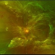
 Choroideremia
Sep 21 2022 by Zach Seim
Ultra-widefield fundus photo of a 74 year old male presenting with severe vision loss beginning at age 55. Patient sought a second opinion with our office and was diagnosed with Choroideremia. Patient denies hearing loss, heart problems, balance issues, polydactyly, kidney problems, and dental problems. Patient reports that nobody in the family had blindness. Choroideremia is an X-linked chorioretinal dystrophy characterized by the diffuse, progressive degeneration of the retinal pigment epithelium (RPE), photoreceptors and choriocapillaris. It is caused by a mutation in the CHM gene.
Photographer: Zach Seim
Imaging device: Optos California
Condition/keywords: choroideremia, hereditary choroidal atrophy, hereditary retinal dystrophy, left eye, light perception, low vision, Optos, pseudocolor, ultra-wide field imaging
-
 Choroideremia
Sep 21 2022 by Zach Seim
Ultra-widefield fundus photo of a 74 year old male presenting with severe vision loss beginning at age 55. Patient sought a second opinion with our office and was diagnosed with Choroideremia. Patient denies hearing loss, heart problems, balance issues, polydactyly, kidney problems, and dental problems. Patient reports that nobody in the family had blindness. Choroideremia is an X-linked chorioretinal dystrophy characterized by the diffuse, progressive degeneration of the retinal pigment epithelium (RPE), photoreceptors and choriocapillaris. It is caused by a mutation in the CHM gene.
Photographer: Zach Seim
Imaging device: Optos California
Condition/keywords: choroideremia, hereditary choroidal atrophy, hereditary retinal dystrophy, Optos, pseudocolor, ultra-wide field imaging
-
 Choroidal Detachment
Jan 17 2022 by Logan ryzenga
Left ultra-wide field photograph of an 81-year old female with a choroidal detachment affecting her left eye. Patient had a stent placed November, 2021 and following the procedure she complains of variable blurred vision and severe constricted visual fields. She presented at our office with flashes a month prior but without pain or floaters.
Photographer: Logan Ryzenga
Imaging device: Optos California
Condition/keywords: choroidal detachment, fundus photograph, left eye, Optos, pseudocolor, superior retina, ultra-wide field imaging
-
 Familial Exudative Vitreoretinopathy
Aug 18 2021 by Samuel Dada
Ultra-widefield optos image of a 40-year with Familial Exudative Vitreoretinopathy, affecting his left eye. Patient born at 38 weeks. No NICU time. Has had genetic testing to determine cause of blindness. Physician suspects FEVR and will carry out further testing. Patient uses a 200x or 600x magnifying lens to view and focus on objects at a distance. Patient's vision on initial visit was 20/70.
Photographer: Samuel Dada
Imaging device: Optos California
Condition/keywords: dysplastic excavation, familial exudative vitreoretinopathy (FEVR), fundus photograph, left eye, Optos, pseudocolor, ultra-wide field imaging
-
 Pre-op Anti-VEGF and Regression of Vascularity Within Fibrovascular Proliferation in Proliferative Diabetic Retinopathy
May 8 2021 by Kushal S Delhiwala, MBBS, MS, FMRF,FICO, FAICO
Fundus photograph of 47-year-old phakic female with right eye proliferative diabetic retinopathy associated tractional retinal detachment involving macula. Vitrectomy was planned with pre-op anti-VEGF before 4 days. Pseudocolor fundus image (A) shows extensive triangular vascular proliferation (FVP) around disc and macula (white arrows). Fundus image (B) shows significant FVP regression following anti-VEGF.
Photographer: Kushal Delhiwala, Netralaya superspeciality eye hospital, Ahmedabad, Gujarat,India
Imaging device: Optos Daytona
Condition/keywords: anti-VEGF, fibrovascular proliferation, intravitreal bevacizumab, pre-op, proliferative diabetic retinopathy (PDR), tractional retinal detachment
-
 Proliferative Sickle Cell Retinopathy
Jan 29 2021 by Olivia Rainey
Ultra-widefield fundus photograph of a 24-year-old female with proliferative sickle cell retinopathy affecting her right eye. He performed scatter PRP OD on 12/2/2020 to nonperfusion in temporal far periphery. The patient's 12/2020 Hb electrophoresis came back showing Hb SC (rather than sickle cell trait). Patient was born at full term, but she reports that her mother used drugs while pregnant with the patient. The patient also mentioned that her niece has full sickle cell disease and her grandmother, mother, and sibling all have condition on the sickle cell spectrum.
Photographer: Olivia Rainey, OCT-C, COA
Imaging device: Optos California
Condition/keywords: fundus photograph, laser photocoagulation, neovascularization (NV), neovascularization elsewhere (NVE), Optos, pseudocolor, sea fan, sickle cell retinopathy
-
 Retinal Cavernous Hemangioma
Oct 21 2020 by Olivia Rainey
Ultra-widefield image of a 31-year-old male presenting with a Retinal Cavernous Hemangioma affecting his left eye. Patient was 18-years-old when he was diagnosed with a retinal cavernous hemangioma. He has had a few episodes of vitreous hemorrhages since then. His vision was 20/20-1 in both eyes.
Photographer: Olivia Rainey, OCT-C, COA
Imaging device: Optos California
Condition/keywords: cavernous hemangioma of the retina, color fundus photograph, fundus photograph, left eye, Optos, pseudocolor, ultra-wide field imaging
-
 Bullous Retinoschisis with Outer Retinal Holes
Jun 15 2020 by Olivia Rainey
Ultra-widefield pseudocolor fundus photograph of a 56-year-old female with bullous retinoschisis with outer retinal holes affecting her right eye. The physician noted superotemporal retinoschisis in her monoculcar functioning eye. There was no demarcation line and no inner or outer layer breaks at her first appointment in February of 2020. On 6/15/20 she had a new onset outer holes and SRF tracking inferiorly. The physician recommended observation, however if this continues to progress we have discussed indications for barrier laser.
Photographer: Olivia Rainey, OCT-C, COA
Imaging device: Optos California
Condition/keywords: bullous retinoschisis, Optos, outer layer breaks, outer layer hole, pseudocolor, subretinal fluid, superior retina, ultra-wide field imaging
 Loading…