Search results (82 results)
-
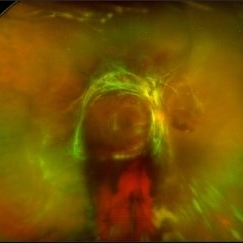
 Bilateral Lymphoma Metastasis after Resolution with IVM
Sep 19 2018 by Olivia Rainey
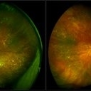
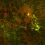
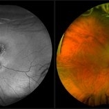
Ultra-wide field, pseudocolor fundus images of an 86-year-old female treated with intravitreal methothrexate as a management of subretinal infiltrate in the macula of the right eye, as a manifestation of leukemia. Her last intravitreal methotrexate injection was 5/1/18.
Photographer: Olivia Rainey
Imaging device: Optos
Condition/keywords: bilateral, leukemia, methotrexate, Optos, pseudocolor, ultra-wide field imaging, uveitis
-
 Blunt Ocular Trauma Due to Firework Injury
Jun 9 2020 by Brittany Rota
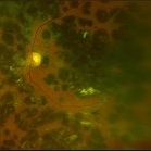
Ultra- widefield pseudocolor image of an 18-year-old male with blunt ocular trauma in the right eye due to a firework injury. The patient presented with commotio retinae (sclopteria), an acute vitreous hemorrhage, choroidal rupture, and a subretinal hemorrhage. The referring physician performed surgery on the lateral rectus muscle which was macerated but not severed, and several orbital fibrous foreign bodies were removed from the posterior orbit. The globe was intact. There is no evidence of retinal tear in the region of sclopetaria; however, there is complete necrosis of the temporal peripheral choroid and retina. The vitreous hemorrhage was slowly clearing on his exam 6-9-2020. The patient is developing subretinal fibrosis. The physician is concerned about the choroidal rupture that is visible through the submacular hemorrhage. There is one rupture that appears to course directly under the fovea. The physician states that if this is the case, his vision most likely will be 20/200 or worse. His vision was hand motion in all fields except nasally, which he was unable to see hand motion at his visit on 6-9-2020.
Photographer: Brittany Rota
Imaging device: Optos California
Condition/keywords: blunt trauma, choroidal rupture, commotio retinae, fibrosis, firework injury, fundus photograph, hand motion, necrotizing retina, Optos, pseudocolor, subretinal hemorrhage, vitreous hemorrhage
-
 Branch Retinal Vein Occlusion
Sep 11 2018 by Olivia Rainey
Ultra-wide field pseudocolor montage of an 84-year-old female with a branch retinal vein occlusion affecting her left eye. Patient recently had a PPV for a epiretinal membrane in her left eye and shortly after developed an occlusion.
Photographer: Olivia Rainey
Imaging device: Optos
Condition/keywords: branch retinal artery occlusion (BRAO), hemorrhage, left eye, montage, Optos, pseudocolor, ultra-wide field imaging
-
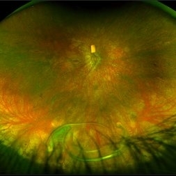
 Bullous Retinoschisis with Outer Retinal Holes
Jun 15 2020 by Olivia Rainey
Ultra-widefield pseudocolor fundus photograph of a 56-year-old female with bullous retinoschisis with outer retinal holes affecting her right eye. The physician noted superotemporal retinoschisis in her monoculcar functioning eye. There was no demarcation line and no inner or outer layer breaks at her first appointment in February of 2020. On 6/15/20 she had a new onset outer holes and SRF tracking inferiorly. The physician recommended observation, however if this continues to progress we have discussed indications for barrier laser.
Photographer: Olivia Rainey, OCT-C, COA
Imaging device: Optos California
Condition/keywords: bullous retinoschisis, Optos, outer layer breaks, outer layer hole, pseudocolor, subretinal fluid, superior retina, ultra-wide field imaging
-
 Central Retinal Vein Occlusion
Jul 13 2018 by Olivia Rainey
Ultra-wide field, pseudocolor montage of a patient presenting with a central retinal vein occlusion, as well as, an inferior chorioretinal scar in their right eye.
Photographer: Olivia Rainey
Imaging device: Optos
Condition/keywords: central retinal vein occlusion (CRVO), chorioretinal scar, montage, Optos, pseudocolor, ultra-wide field imaging
-
 Choroidal Detachment
Jan 17 2022 by Logan ryzenga
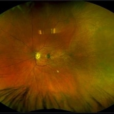
Left ultra-wide field photograph of an 81-year old female with a choroidal detachment affecting her left eye. Patient had a stent placed November, 2021 and following the procedure she complains of variable blurred vision and severe constricted visual fields. She presented at our office with flashes a month prior but without pain or floaters.
Photographer: Logan Ryzenga
Imaging device: Optos California
Condition/keywords: choroidal detachment, fundus photograph, left eye, Optos, pseudocolor, superior retina, ultra-wide field imaging
-
 Choroideremia
Sep 21 2022 by Zach Seim
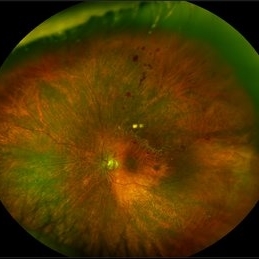
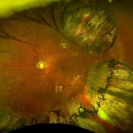
Ultra-widefield fundus photo of a 74 year old male presenting with severe vision loss beginning at age 55. Patient sought a second opinion with our office and was diagnosed with Choroideremia. Patient denies hearing loss, heart problems, balance issues, polydactyly, kidney problems, and dental problems. Patient reports that nobody in the family had blindness. Choroideremia is an X-linked chorioretinal dystrophy characterized by the diffuse, progressive degeneration of the retinal pigment epithelium (RPE), photoreceptors and choriocapillaris. It is caused by a mutation in the CHM gene.
Photographer: Zach Seim
Imaging device: Optos California
Condition/keywords: choroideremia, hereditary choroidal atrophy, hereditary retinal dystrophy, Optos, pseudocolor, ultra-wide field imaging
-
 Choroideremia
Sep 21 2022 by Zach Seim
Ultra-widefield fundus photo of a 74 year old male presenting with severe vision loss beginning at age 55. Patient sought a second opinion with our office and was diagnosed with Choroideremia. Patient denies hearing loss, heart problems, balance issues, polydactyly, kidney problems, and dental problems. Patient reports that nobody in the family had blindness. Choroideremia is an X-linked chorioretinal dystrophy characterized by the diffuse, progressive degeneration of the retinal pigment epithelium (RPE), photoreceptors and choriocapillaris. It is caused by a mutation in the CHM gene.
Photographer: Zach Seim
Imaging device: Optos California
Condition/keywords: choroideremia, hereditary choroidal atrophy, hereditary retinal dystrophy, left eye, light perception, low vision, Optos, pseudocolor, ultra-wide field imaging
-
 CHRPE
CHRPE
Mar 25 2025 by Toolie Winters
Ultra-wide field fundus photograph of a 78-year-old woman with extensive CHRPE lesions OS. Continued observation has been recommended at this time.
Photographer: Toolie Winters
Imaging device: Optos California
Condition/keywords: CHRPE, congenital hypertrophy of the retinal pigment epithelium (CHRPE), fundus photography, Optos, Optos California, pseudocolor, ultra-wide field imaging
-
 Congenital Hypertrophy of the Retinal Pigment Epithelium
Nov 11 2019 by Jessica Norkus
Bilateral Optos ultra wide field imaging of a 31-year-old female patient with CHRPE lesions. Lesions in OD were suspicious of Gardner Syndrome due to familial history of cancerous polyps in colon. Patient underwent colonoscopy and was deemed clear.
Photographer: Jessica Norkus, COA, Retina Specialists of Michigan
Imaging device: Optos Ultra Wide Field Camera
Condition/keywords: bear tracks, bilateral, color fundus photograph, color photo, congenital hypertrophy of the retinal pigment epithelium (CHRPE), fundus autofluorescence (FAF), fundus photograph, lacunae, macula, optic disc, Optos, pseudocolor, ultra-wide field imaging
-
 Diabetic Traction Detachment of Retina
Sep 28 2022 by Chloe Hanifan
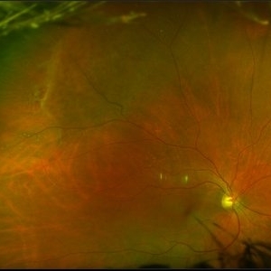
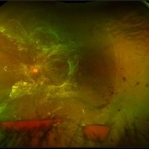
Ultra-widefield pseudo color fundus photograph of a 50-year-old female with a Diabetic Traction Detachment of Retina affecting her left eye. The patient was unable to proceed with surgery due to other health issues in May of 2022, when she presented in the office in September of 2022, a guarded prognosis given chronicity and associated ischemia. The patient was LP at the time of the September appointment.
Photographer: Chloe Hanifan
Imaging device: Optos California
Condition/keywords: diabetes, diabetic traction detachment, fundus photography, left eye, neovascularization (NV), Optos, proliferative diabetic retinopathy (PDR), pseudocolor, ULTRA WIDE FIELD
-
 Diabetic Traction Detachment of Retina
Sep 28 2022 by Chloe Hanifan
Ultra-widefield pseudo color fundus photograph of a 50-year-old female with a Diabetic Traction Detachment of Retina affecting her left eye. The patient was unable to proceed with surgery due to other health issues in May of 2022, when she presented in the office in September of 2022, a guarded prognosis given chronicity and associated ischemia. The patient was LP at the time of the September appointment.
Photographer: Chloe Hanifan
Imaging device: Optos California
Condition/keywords: Diabetes, diabetic traction detachment, fundus photography, left eye, neovascularization (NV), Optos, proliferative diabetic retinopathy (PDR), pseudocolor, ULTRA WIDE FIELD
-
 Diabetic Tractional Retinal Detachment
Jan 23 2019 by Olivia Rainey
Ultra-wide field pseudocolor image of an 43-year-old female with a diabetic tractional retinal detachment and a vitreous hemorrhage affecting her right eye.
Photographer: Olivia Rainey
Imaging device: Optos
Condition/keywords: diabetes, diabetic traction detachment, Optos, pan-retinal photocoagulation (PRP), proliferative diabetic retinopathy (PDR), pseudocolor, ultra-wide field imaging, vitreous hemorrhage
-
 Dislocated Lens
Apr 26 2023 by Chloe Hanifan
Ultra wide field fundus photograph of a 41-year-old male with a dislocated lens affecting his right eye. IOL noted inferior vitreous base and vitrectomy surgery for removal of IOL was recommended. Patient has history of retinitis pigmentosa as well. Patient's vision at the time of presentation was counting fingers at 2 feet.
Photographer: Chloe Hanifan
Imaging device: Optos California
Condition/keywords: dislocated lens, fundus photography, Optos, pseudocolor, retinitis pigmentosa, ULTRA WIDE FIELD
-
 Endogenous Endophthalmitis With Suspected Systemic Candidiasis
Jan 9 2018 by Olivia Rainey
Ultra-wide field Optos pseudocolor image of an 45-year-old male presenting with endogenous endophthalmitis affecting his left eye. Candidiasis was at high considerations due to intravenous drug abuse and recent history of dental abscess. Patient developed a subretinal infiltrate resulting in central scotoma and responded well to anti-fungal treatment.
Photographer: Olivia Rainey
Imaging device: Optos California
Condition/keywords: candida endophthalmitis, drug abuse, endogenous endophthalmitis, left eye, Optos, pseudocolor, retinal infiltrates, scotoma, ultra-wide field imaging
-
 Familial Exudative Vitreoretinopathy
Aug 18 2021 by Samuel Dada
Ultra-widefield optos image of a 40-year with Familial Exudative Vitreoretinopathy, affecting his left eye. Patient born at 38 weeks. No NICU time. Has had genetic testing to determine cause of blindness. Physician suspects FEVR and will carry out further testing. Patient uses a 200x or 600x magnifying lens to view and focus on objects at a distance. Patient's vision on initial visit was 20/70.
Photographer: Samuel Dada
Imaging device: Optos California
Condition/keywords: dysplastic excavation, familial exudative vitreoretinopathy (FEVR), fundus photograph, left eye, Optos, pseudocolor, ultra-wide field imaging
-
 Heavy focal laser pseudocolor photograph - OD
Jul 20 2018 by Hosam Attia, MD
65-year-old, African American, woman with inactive PDR, S/P multiple PRP/ heavy focal OU, now receiving simultaneous Ozurdex/ Eylea injection OS, on regular basis w/ long standing poor vision 20/200-20/400 OS, since 2016 - patient was and currently being treated by another physician.
Imaging device: Optos California
Condition/keywords: focal laser, proliferative diabetic retinopathy (PDR), pseudocolor
-
 Heavy Focal Laser Pseudocolor Photograph - OS
Jul 20 2018 by Hosam Attia, MD
65-year-old, African American, woman with inactive PDR, S/P multiple PRP/ heavy focal OU, now receiving simultaneous Ozurdex/ Eylea injection OS, on regular basis w/ long standing poor vision 20/200-20/400 OS, since 2016 - patient was and currently being treated by another physician.
Condition/keywords: focal laser, proliferative diabetic retinopathy (PDR), pseudocolor
-
 Macula Off Retinal Detachment with CNV
Nov 11 2019 by Olivia Rainey
Ultra-wide field pseudocolor photograph of a 42-year-old female with a long-standing, macula-off retinal detachment affecting her left eye. Patient was unaware of vision loss until testing her visual acuity and she denied seeing flashing lights. Patient decided to proceed with scleral buckling. The CNV is potentially secondary the retinal detachment, but may be myopic related or idiopathic. The CNV appears fibrotic and inactive. The patient was warned that this will absolutely limit how much vision she recovers once the retina is reattached.
Photographer: Olivia Rainey
Imaging device: Optos California
Condition/keywords: choroidal neovascularization (CNV), left eye, montage, Optos, pseudocolor, retinal detachment of the macula, ultra-wide field imaging
-
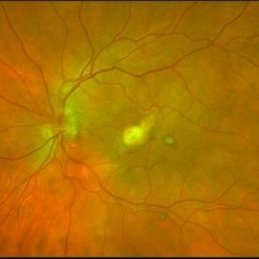
 Macular Degeneration with Significant Drusen
Jul 10 2018 by Karen Panzegrau
Zoomed-in ultra-wide field images of a 77-year-old female with macular degeneration with significant drusen.
Photographer: Karen Panzegrau
Imaging device: Optos
Condition/keywords: age-related macular degeneration (AMD), bilateral, drusen, fundus photograph, pseudocolor
-
 Macular Pattern Dystrophy Associated with MELAS
Dec 19 2019 by Olivia Rainey
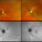
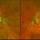
Bilateral wide field pseudocolor images of a 54-year-old female with macular pattern dystrophy associated with MELAS. The patient is positive for m.3243A>G in MT-TL1. She had stroke in her 40s, hearing loss in her 30s, and has early onset diabetes. MyRetinaTracker shows VUS in RP1L1. Mutation in RP1L1 have been describe in other families with occult macular dystrophy. Farnsworth D15 is showing mild tritan abnormality, which is most commonly seen with acquired maculopathies. 12/17/19 patient's Optos and OCT show mild progression of atrophy.
Photographer: Olivia Rainey
Imaging device: Optos California
Condition/keywords: advanced geographic atrophy, bilateral, fundus photograph, MELAS, Optos, pattern macular dystrophy, pseudocolor, wide angle imaging
-
 Malignant Melanoma
Sep 11 2018 by Olivia Rainey
Ultra-wide field autofluorescence and pseudocolor montage of a 57-year-old male s/p I-125 brachytherapy for malignant melanoma affecting his right eye. The patient’s radiation retinopathy has resulted in retinal vascular occlusive disease and optic nerve edema.
Photographer: Olivia Rainey
Imaging device: Optos
Condition/keywords: branch retinal vein occlusion (BRVO), fundus autofluorescence (FAF), I-125 brachytherapy, malignant melanoma, montage, Optos, pseudocolor, radiation retinopathy, ultra-wide field imaging
-
 Methotrexate Bubble following Intravitreal Injection for PVR
Sep 21 2022 by Zach Seim
Ultra-widefield fundus photograph of an 81 year old female with a Methotrexate bubble following an Intravitreal Injection for Proliferative Vitreoretinopathy. Patient has been presenting to the office for two week interval Methotrexate injections in her left eye. The image was taken prior to her eighth injection which revealed a residual Methotrexate bubble in her inferior retinal image. This patient was seeing "lots" of floaters, as well as having visual acuity of cc20/400 cc20/200 PH.
Photographer: Zach Seim
Imaging device: OPTOS California
Condition/keywords: bubble, fundus photograph, fundus photography, intravitreal injection, left eye, methotrexate, nasal retina, Optos, proliferative vitreoretinopathy (PVR), pseudocolor, ultra-wide field imaging
-
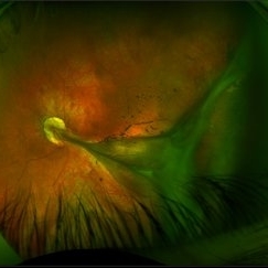
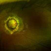 Morning Glory Syndrome
Jun 19 2019 by Olivia Rainey
Ultra-wide field pseudocolor image of an 10-year-old girl with Morning Glory Syndrome affecting her left eye. Patient is able to count fingers at 4 feet.
Photographer: Olivia Rainey
Imaging device: Optos
Condition/keywords: left eye, Morning Glory Syndrome, Optos, pseudocolor, ultra-wide field imaging
-
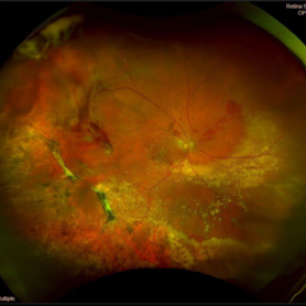 Peripheral Exudative Hemorrhagic Chorioretinopathy
Nov 19 2024 by Toolie Winters
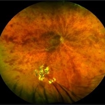
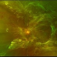
Ultra-wide field fundus photograph of an 85-year-old woman with Peripheral Exudative Hemorrhagic Chorioretinopathy (PECHR) affecting the right eye. Patient presented with a blind spot centrally in the right eye which she first noticed 4 months prior to this image being taken. The patient states that in the month prior to this image, she began noticing bright lights flash across her vision 4-5x/day which last about 15 seconds. The flashes are either black with a blue ring around them or yellow, and their frequency has increased over time. The patient's vision at the time of this appointment was Dcc20/100+1 PHNI. This photo also shows diffuse hemorrhage, lipid, and an eccentric disciform lesion.
Photographer: Toolie Winters
Imaging device: Optos California
Condition/keywords: fundus photography, neovascular age-related macular degeneration (AMD), Optos, OPTOS CALIFORNIA, peripheral exudative hemorrhagic chorioretinopathy (PEHCR), pseudocolor, ultra-wide field imaging, wet age-related macular degeneration (wet AMD)
 Loading…