Search results (10 results)
-
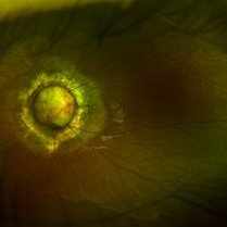
 Dislocated Lens
Apr 26 2023 by Chloe Hanifan
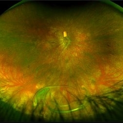
Ultra wide field fundus photograph of a 41-year-old male with a dislocated lens affecting his right eye. IOL noted inferior vitreous base and vitrectomy surgery for removal of IOL was recommended. Patient has history of retinitis pigmentosa as well. Patient's vision at the time of presentation was counting fingers at 2 feet.
Photographer: Chloe Hanifan
Imaging device: Optos California
Condition/keywords: dislocated lens, fundus photography, Optos, pseudocolor, retinitis pigmentosa, ULTRA WIDE FIELD
-
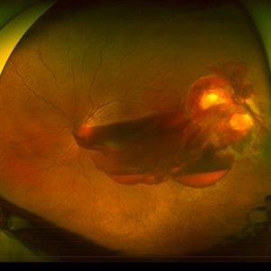
 Total retinal Detachment multiple holes
Sep 26 2022 by Denica Rodriguez
60 year old Male presented with two week old Macula off Retinal detachment with multiple tears.
Photographer: Denica Rodriguez
Imaging device: Optos California
Condition/keywords: color fundus photograph, color photo, macula-off, optos, pseudocolor, Retinal detachment, retinal holes, retinal tear, Retinal tear with detachment, superior arcade, superior field, superior retina, total retinal detachment
-
 Methotrexate Bubble following Intravitreal Injection for PVR
Sep 21 2022 by Zach Seim
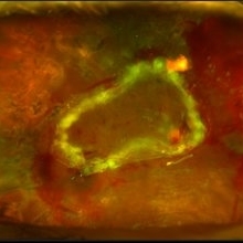
Ultra-widefield fundus photograph of an 81 year old female with a Methotrexate bubble following an Intravitreal Injection for Proliferative Vitreoretinopathy. Patient has been presenting to the office for two week interval Methotrexate injections in her left eye. The image was taken prior to her eighth injection which revealed a residual Methotrexate bubble in her inferior retinal image. This patient was seeing "lots" of floaters, as well as having visual acuity of cc20/400 cc20/200 PH.
Photographer: Zach Seim
Imaging device: OPTOS California
Condition/keywords: bubble, fundus photograph, fundus photography, intravitreal injection, left eye, methotrexate, nasal retina, Optos, proliferative vitreoretinopathy (PVR), pseudocolor, ultra-wide field imaging
-
 Choroidal Detachment
Jan 17 2022 by Logan ryzenga
Left ultra-wide field photograph of an 81-year old female with a choroidal detachment affecting her left eye. Patient had a stent placed November, 2021 and following the procedure she complains of variable blurred vision and severe constricted visual fields. She presented at our office with flashes a month prior but without pain or floaters.
Photographer: Logan Ryzenga
Imaging device: Optos California
Condition/keywords: choroidal detachment, fundus photograph, left eye, Optos, pseudocolor, superior retina, ultra-wide field imaging
-
 Blunt Ocular Trauma Due to Firework Injury
Jun 9 2020 by Brittany Rota
Ultra- widefield pseudocolor image of an 18-year-old male with blunt ocular trauma in the right eye due to a firework injury. The patient presented with commotio retinae (sclopteria), an acute vitreous hemorrhage, choroidal rupture, and a subretinal hemorrhage. The referring physician performed surgery on the lateral rectus muscle which was macerated but not severed, and several orbital fibrous foreign bodies were removed from the posterior orbit. The globe was intact. There is no evidence of retinal tear in the region of sclopetaria; however, there is complete necrosis of the temporal peripheral choroid and retina. The vitreous hemorrhage was slowly clearing on his exam 6-9-2020. The patient is developing subretinal fibrosis. The physician is concerned about the choroidal rupture that is visible through the submacular hemorrhage. There is one rupture that appears to course directly under the fovea. The physician states that if this is the case, his vision most likely will be 20/200 or worse. His vision was hand motion in all fields except nasally, which he was unable to see hand motion at his visit on 6-9-2020.
Photographer: Brittany Rota
Imaging device: Optos California
Condition/keywords: blunt trauma, choroidal rupture, commotio retinae, fibrosis, firework injury, fundus photograph, hand motion, necrotizing retina, Optos, pseudocolor, subretinal hemorrhage, vitreous hemorrhage
-
 Morning Glory Syndrome
Jan 6 2020 by Olivia Rainey
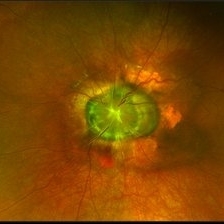
Ultra-wide field pseudocolor image of a 23-month-old male with morning glory syndrome affecting his left eye. Patient presented with esotropia affecting his left eye and strabismic amblyopia affecting both eyes. He could fix and follow on exam and his medical history was unremarkable.
Photographer: Olivia Rainey
Imaging device: Optos California
Condition/keywords: esotropia, left eye, macular, Morning Glory Syndrome, Optos, strabismic amblyopia, ultra-wide field imaging
-
 Vascular Sheathing
Dec 19 2019 by Lauren Schuler
Ultra-wide field pseudocolor fundus photograph of a 68-year-old female with vascular sheathing affecting her right eye. This was noted on initial exam on 1/15/16, at patient's first appointment. This remains unchanged and patient is asymptomatic at this time.
Photographer: Lauren Schuler
Imaging device: Optos California
Condition/keywords: fundus photograph, ghost vessels, pseudocolor, vascular sheathing of retina
-
 Retinal Detachment with PVR (s/ SPR, PPV, MPV, 360 Retinectomy, PFO, PI, FAx, SO)
Aug 22 2019 by Merrick Avila
Ultra-wide field pseudocolor fundus photograph of a 64-year-old female with a treated retinal detachment with proliferative vitreoretinopathy. Patient has a history of complex retinal detachments that have been treated multiple times. On exam 8-22-19, there were large macular holes with LP vision. There was a long discussion about guarded nature of her condition and goals or trial for repair including globe sparing prevention of phthisis.
Photographer: Merrick Avila
Imaging device: Optos
Condition/keywords: diabetic retinopathy, hemorrhage, Optos, proliferative vitreoretinopathy (PVR), retinectomy, silicone oil
-
 Morning Glory Syndrome
Jun 19 2019 by Olivia Rainey
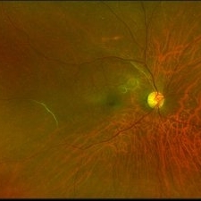
Ultra-wide field pseudocolor image of an 10-year-old girl with Morning Glory Syndrome affecting her left eye. Patient is able to count fingers at 4 feet.
Photographer: Olivia Rainey
Imaging device: Optos
Condition/keywords: left eye, Morning Glory Syndrome, Optos, pseudocolor, ultra-wide field imaging
-
 Penetrating Trauma with Retinal Detachment
Apr 30 2019 by Olivia Rainey
Ultra-wide field pseudocolor image of a 39-year-old female with penetrating trauma resulting in a retinal detachment with an intraretinal hemorrhage affecting the left eye. Patient was struck with a champagne glass in October of 2018, which lacerated the eyelid and globe. Patient was "seeing red" when she first came to the office and after multiple surgeries she was seeing 20/20 at her last check in April 2019.
Photographer: Olivia Rainey
Imaging device: Optos
Condition/keywords: hemorrhage, left eye, Optos, penetrating trauma, ruptured globe, ultra-wide field imaging
 Loading…