Search results (98 results)
-
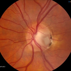
 Optic Nerve Pit
Feb 21 2024 by Virginia Gebhart
65 year old female with optic nerve pit. Asymptomatic, continued observation.
Photographer: Virginia Gebhart
Imaging device: Topcon TRC 50DX
Condition/keywords: congenital optic nerve pit, Optic nerve pit
-
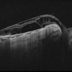
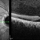
 Optic Disc Pit OCT
Aug 1 2023 by Aditya S Kelkar, MS, FRCS, FASRS,FRCOphth
Optical Coherence Tomography of an 21 year old male with a Optic Disc Pit.
Photographer: Dr. Ajinkya Rawale. National institute of Ophthalmology, Pune, India.
Imaging device: Zeiss Plex
Condition/keywords: congenital optic nerve pit
-
 Optic Disc Pit Associated with Multilayered Retinoschisis
Apr 26 2023 by Shaleen Arora
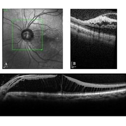
46-year-old female with an optic nerve pit in the left eye (A). OCT reveals retinoschisis involving multiple retinal layers due to intraretinal fluid tracking from the nerve pit (B). Muller cell processes maintain the architecture of individual retinal layers in the region of retinoschisis (C).
Photographer: George Washington University, Department of Ophthalmology
Condition/keywords: maculopathy, optic disc pit, optic pit
-
Surgery for optic nerve pit
Oct 24 2022 by Manish Nagpal, MD, FRCS (UK), FASRS
This video showcases the steps of surgery for optic nerve pit associated with sensory fluid under fovea, fovea sparing ilm peel is carried out followed by endolaser to the temporal edges of the disc margins followed by air fluid exchange and gas.
Photographer: Manish Nagpal
Condition/keywords: endolaser, ONP, optic nerve pit, video, vitrectomy
-
 Optic Disc Pit
Nov 8 2021 by Michael Grinton
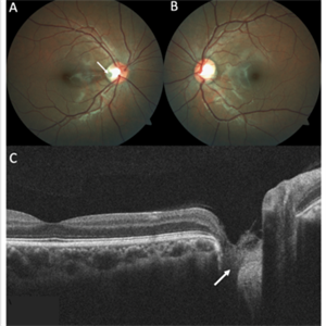
Optic disc pits are rare congenital abnormalities of the optic nerve head. Colour fundus image of an asymptomatic 18-year old male shows an optic disc pit in the right eye (A, white arrow); a small, grey, oval shaped excavation in the temporal segment of the optic disc. These pits are usually unilateral (B shows normal colour fundus of left eye) and asymptomatic. Imaging with optical coherence tomography (C) shows the optic disc pit in cross section (white arrow) and normal macular structure. In some patients with the condition, fluid can accumulate underneath the macular (serous macular detachment).
Condition/keywords: Optic disc pit, Optic nerve pit, Optic pit
-
 Optic Nerve Pit Right Eye
Feb 15 2021 by Kim Barrett
A 14-year-old male presented with vision loss and VF defect. Patient was treated for presumed amblyopia with patching since age 4. He has had neurologic care for post traumatic skull fracture and brain bleed in 2012. IOP's WNL. OD is without retinoschisis or subretinal fluid. Patient is at risk of serous detachment. Current VA OD 20/200+1 PH 20/80.
Photographer: Kim Barrett C.O.A. Retina Specialist of Michigan, Grand Rapids, MI
Imaging device: Optos California
Condition/keywords: amblyopia, hemifield, Humphrey visual field, nerve, optic nerve pit, visual field defect
-
 Optic Nerve Pit Left Eye
Feb 15 2021 by Kim Barrett
A 14-year-old male presented with vision loss and VF defect. Patient was treated for presumed amblyopia with patching since age 4. He has had neurologic care for post traumatic skull fracture and brain bleed in 2012. Patient has a superior hemifield defect OS on HVF. IOP's WNL. There are vessels emanating from the optic pit OS. Patient is at risk of serous detachment. Current VA 20/20-2+2
Photographer: Kim Barrett C.O.A. Retina Specialist of Michigan, Grand Rapids, MI
Imaging device: Optos California
Condition/keywords: amblyopia, hemifield, Humphrey visual field, nerve, optic nerve pit, visual field defect
-
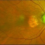
 Optic Disc Pit Associated with Optic Disc Coloboma and Retinochoroidal Coloboma
Jul 22 2020 by Deepak Bhojwani, MS
Fundus photograph of a 32-year-old male showing large optic disc pit in a colobomatous optic nerve head along with isolated inferior retino-choroidal coloboma. (A rare / coincidental occurrence of multiple congenital anomalies of optic disc and retina)
Photographer: DEEPAK BHOJWANI
Condition/keywords: coloboma of choroid, coloboma of optic disc, congenital optic nerve pit
-
 Colobomatous Optic Disc Maculopathy
Feb 13 2020 by Yoshihiro Yonekawa, MD, FASRS
EDI-OCT of a teenage girl with submacular fluid from a colobomatous optic disc. Note the subtle tracking of the subretinal fluid into the disc.
Photographer: Netanya Lerner, COA, Wills Eye Hospital/Mid Atlantic Retina
Imaging device: Topcon
Condition/keywords: chorioretinal coloboma, coloboma of optic disc, congenital optic nerve pit, subretinal fluid
-
 Colobomatous Optic Disc Maculopathy
Feb 13 2020 by Yoshihiro Yonekawa, MD, FASRS
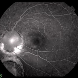
Fluorescein angiography, late frame, of a teenage girl with submacular fluid from a colobomatous optic disc. The camera is focused is on the elevated macula, and the disc is subtly defocused.
Photographer: Netanya Lerner, COA, Wills Eye Hospital/Mid Atlantic Retina
Imaging device: Topcon
Condition/keywords: chorioretinal coloboma, coloboma of optic disc, congenital optic nerve pit, subretinal fluid
-
 Colobomatous Optic Disc Maculopathy
Feb 13 2020 by Yoshihiro Yonekawa, MD, FASRS
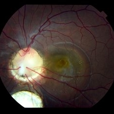
Beautifully focused fundus photograph of a teenage girl with submacular fluid from a colobomatous optic disc.
Photographer: Netanya Lerner, COA, Wills Eye Hospital/Mid Atlantic Retina
Imaging device: Topcon
Condition/keywords: chorioretinal coloboma, coloboma of optic disc, congenital optic nerve pit, subretinal fluid
-
 Slide 11-7
Feb 26 2019 by Lancaster Course in Ophthalmology
Optic nerve pit. This defect may lead to macular edema. (Courtesy of H. G. Scheie, M.D.}
Condition/keywords: optic nerve pit
-
 Coloboma
Sep 7 2018 by John S. King, MD
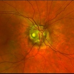
11-year-old white female with bilateral optic nerve and retinochoroidal colobomas and an optic nerve pit in the right eye looking almost like pseudoduplication of the optic nerve. She is currently 20/30 OD and 20/20 OS. She has a history of laser by Dr. Zocchi about 10 years ago for a low lying, macula involving, serous retinal detachment, and has responded well.
Photographer: Stacey Coleman
Imaging device: Topcon
Condition/keywords: chorioretinal coloboma, inferior optic nerve coloboma, optic disc pit
-
 Retinoschisis and Subretinal Fluid in Optic Disc Pit Related Maculopathy
Aug 20 2018 by DIEGO A BUESO PONCE, MD
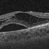
OCT B-scan of a 19-year-old female with congenital optic disc pit maculopathy.
Photographer: Diego Bueso Ponce, Clinica Unidad Laser, Barranquilla Colombia
Imaging device: Topcon DRI OCT Triton, Swept source OCT
Condition/keywords: B scan ultrasound, congenital optic nerve pit, retinoschisis
-
 Optic Disc Pit Maculopathy
Aug 20 2018 by DIEGO A BUESO PONCE, MD
Fundus photograph of an 19-year-old female with congenital optic disc pit and associated maculopathy with subretinal fluid and retinoschisis.
Photographer: Diego Bueso Ponce, Clinica Unidad Laser, Barranquilla Colombia
Imaging device: Topcon DRI OCT Triton, Swept source OCT
Condition/keywords: congenital optic nerve pit, maculopathy, optic disc pit, retinoschisis
-
 Optic Nerve Pit OD - OCT
Aug 6 2018 by Hosam Attia, MD
65-year-old white male, presented for a second opinion for possible cataract extraction OD. BCVA: OD: 20/70 OS: 20/60 WRx: OD: -3.75 +1.50 x 5 OS: -1.75 +1.50 x 178 SLE: +2 NS OD>OS DFE: OD: Nasal macular GA, connected by milder track of RPE changes to an optic nerve pit OD (no fluid seen clinically) OS: enlarged C/D w/ no pits, macular RPE change w/ No heme, CME/ SRF OCT: OD: Peri-papillary cystoid changes & outer retinal atrophy (corresponding to the area of GA on the pseudocolor photo) w/ No SRF (mimicking PP CNVM), connected to the optic disc pit by shallow sinus/ tract. OS: Drusenoid RPE changes, No cystoid changes/ SRF
Imaging device: Zeiss Cirrus -5000
Condition/keywords: congenital optic nerve pit
-
 Optic Nerve Pit OD - Pseudocolor Photo
Aug 6 2018 by Hosam Attia, MD
65-year-old white male, presented for a second opinion for possible cataract extraction OD. BCVA: OD: 20/70 OS: 20/60 WRx: OD: -3.75 +1.50 x 5 OS: -1.75 +1.50 x 178 SLE: +2 NS OD>OS DFE: OD: Nasal macular GA, connected by milder track of RPE changes to an optic nerve pit OD (no fluid seen clinically) OS: enlarged C/D w/ no pits, macular RPE change w/ No heme, CME/ SRF OCT: OD: Peri-papillary cystoid changes & outer retinal atrophy (corresponding to the area of GA on the pseudocolor photo) w/ No SRF (mimicking PP CNVM), connected to the optic disc pit by shallow sinus/ tract. OS: Drusenoid RPE changes, No cystoid changes/ SRF
Imaging device: Optos California
Condition/keywords: congenital optic nerve pit
-
 Optic Nerve Pit
Aug 4 2018 by Nilesh K Kanjani, MD
A female patient aged 40 years came with progressive dimness of vision.
Photographer: Dr Nilesh Kanjani
Condition/keywords: optic nerve pit
-
 Optic Nerve Pit
Aug 2 2018 by Nilesh K Kanjani, MD
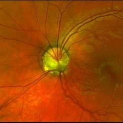
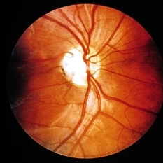
Fundus photograph of 45-year-old woman with complain of gradual progressive dimness of vision in LE.
Photographer: Dr Nilesh Kanjani
Condition/keywords: optic nerve pit
-
 Partial Optic Disc Avulsion with Optic Disc Pit
Jul 1 2018 by John S. King, MD
16-year-old with acute loss of vision after blunt finger injury to eye while playing football. This photo is three weeks post-injury. Vision HM. Retinal striae with subhyaloid heme. Decreased retinal whitening. Peripapillary heme clearing, and temporal optic disc avulsion with optic disc pit can be seen.
Photographer: Maisee Yang
Imaging device: Topcon
Condition/keywords: epiretinal membrane (ERM), optic nerve head avulsion, optic nerve pit, traumatic optic neuropathy
-
 Optic Nerve Pit With Sub-Retinal Fluid
Sep 17 2015 by Jason S. Calhoun
Young female with blurred vision in the left eye. Fundus photograph shows optic nerve pit adjacent to the macula where there is sub retinal fluid visible.
Photographer: Jason Calhoun, Mayo Clinic, Department of Ophthalmology
Imaging device: TOPCON-TRC50EX
Condition/keywords: congenital optic nerve pit
-
 Optic Nerve Pit with Sub-Retinal Fluid
Sep 17 2015 by Jason S. Calhoun
Young female with blurred vision in the left eye. Fundus photograph shows optic nerve pit adjacent to the macula where there is sub retinal fluid visible.
Photographer: Jason Calhoun, Mayo Clinic, Department of Ophthalmology
Imaging device: TOPCON-TRC50EX
Condition/keywords: congenital optic nerve pit
-
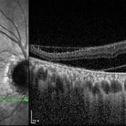 OCT Image of Optic Nerve Pit and Retinal Detachment
Aug 24 2015 by Young Hee Yoon, MD, PhD
OCT image of a 44-year-old man. There was optic nerve pit and associated foveal detachment. His best-corrected visual acuity was count finger in 30cm.
Photographer: Jung Im Cho, Asan Medical Center
Imaging device: Spectralis OCT
Condition/keywords: optic nerve pit
-
 OCT Image of Optic Nerve Pit and Retinal Detachment
Aug 24 2015 by Young Hee Yoon, MD, PhD
SD-OCT image of a 44-year-old man. There was optic nerve pit and associated foveal detachment. His best-corrected visual acuity was count finger in 30cm.
Photographer: Jung Im Cho, Asan Medical Center
Imaging device: Spectralis OCT
Condition/keywords: optic nerve pit
-
 OCT Image of Optic Nerve Pit and Retinal Detachment
Aug 24 2015 by Young Hee Yoon, MD, PhD
OCT image of a 44-year-old man. There was optic nerve pit and associated foveal detachment. His best-corrected visual acuity was count finger in 30cm.
Photographer: Jung Im Cho, Asan Medical Center
Imaging device: Spectralis OCT
Condition/keywords: optic nerve pit
 Loading…