Search results (42 results)
-
 IOL Dislocation
Sep 29 2025 by Carmen R. Negrin-Martin, MD
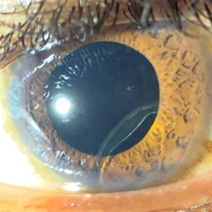
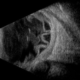
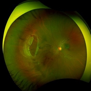
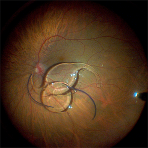
Male patient with a history of ocular trauma at 12 years of age. Underwent cataract surgery as an adult and now reports decreased vision of a few days’ duration. Referred to the retina service due to IOL dislocation. Visual acuity with +9.00: 20/40. Biomicroscopy: clear cornea, formed anterior chamber. The IOL haptic is observed hooked to the iris, with the optic in the posterior segment behind the iris. Posterior segment without alterations.
Photographer: Dr. Vizcaino MD
Imaging device: iPhone 16 pro Max
Condition/keywords: IOL dislocation
-
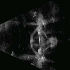
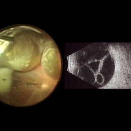
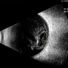 Ocular B-scan Ultrasound (Longitudinal Scan M6, gain 100 dB)
Jun 26 2025 by Hector Gabriel Moreno Solano, MD, MHA
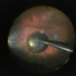
B-scan ultrasound was performed in longitudinal section M6 with a gain of 100 dB. A hyperechoic structure with posterior acoustic shadowing is observed, consistent with lens luxation and condensed vitreous bands adjacent to the lens. The dislocated lens measures approximately 9.54 mm x 4.62 mm. The study was conducted following blunt ocular trauma caused by a golf ball. The remaining vitreous cavity appears anechoic, with no evidence of retinal detachment or other structural abnormalities in this section.
Photographer: Hector Gabriel Moreno Solano, Instituto Mexicano de Oftalmología “IMO I.A.P”
Imaging device: Quantel Medical
Condition/keywords: B scan ultrasound, lens luxation, ocular trauma
-
 Fluorescein Angiography in Choroidal Rupture
Jun 26 2025 by Hector Gabriel Moreno Solano, MD, MHA
Fluorescein angiography of the right eye reveals three linear hypofluorescent lesions with progressive staining at the edges, consistent with choroidal ruptures. These lesions are temporally located in the posterior pole, with one of them situated near the fovea but without direct foveal involvement. The pattern is suggestive of previous blunt ocular trauma.
Photographer: Héctor Gabriel Moreno Solano, Instituto Mexicano de Oftalmología “IMO I.A.P”
Imaging device: CLARUS
Condition/keywords: Choroidal Rupture, fluorescein angiogram (FA)
-
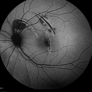
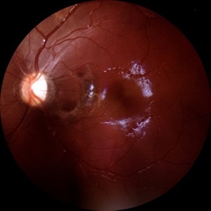 Choroidal Rupture
Jun 4 2025 by Paulina Araujo
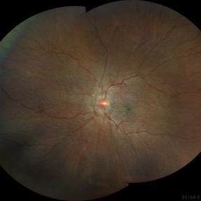
The 55-degree central fundus photograph of the left eye reveals a choroidal rupture in the nasal parafoveal area secondary to blunt ocular trauma.
Photographer: Paulina D.Araujo Martínez, Asociación para Evitar la Ceguera en México I.A.P., Hospital Dr Luis Sánchez Bulnes.
Condition/keywords: choroidal rupture
-
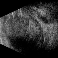 Scleral Rupture
May 9 2025 by Gustavo Uriel Fonseca Aguirre
This B-mode longitudinal ultrasound scan reveals dense vitreous hemorrhage, subretinal fluid, annular choroidal detachment, and scleral wall discontinuity with adjacent scleral folds. These findings indicate severe ocular trauma with probable scleral rupture and multi-compartment involvement.
Photographer: Gustavo U. Fonseca Aguirre, Hospital Conde de Valenciana, Ciudad de México
Condition/keywords: ocular trauma, scleral rupture
-
 Traumatic Retinal Detachment
May 5 2025 by Gustavo Uriel Fonseca Aguirre
This B-mode longitudinal ultrasound scan over the macular area reveals vitreous hemorrhage, retinal detachment with folding, peripheral annular choroidal detachment, and sub-Tenon's fluid in the setting of blunt ocular trauma. The findings indicate severe posterior segment disruption with multi-compartment involvement.
Photographer: Gustavo U. Fonseca Aguirre, Hospital Conde de Valenciana, Ciudad de México
Condition/keywords: blunt trauma, Retinal Detachment
-
 Traumatic Posterior Capsular Rupture
Apr 9 2025 by Gustavo Uriel Fonseca Aguirre
Immersion B-mode ultrasound in a patient with blunt ocular trauma demonstrates an isolated posterior lens capsule rupture accompanied by phacodonesis.
Photographer: Gustavo U. Fonseca Aguirre, Hospital Conde de Valenciana, Ciudad de México
Condition/keywords: blunt trauma, Posterior Capsular Rupture
-
 Choroidal Rupture
Apr 7 2025 by Ramses Rosales-Diaz
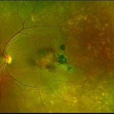
Autofluorescence image of a 39-year-old female patient who sustained blunt ocular trauma resulting in three choroidal ruptures.
Photographer: Ramses Rosales-Diaz, Asociación Para Evitar la Ceguera en México I.A.P., Mexico City
Imaging device: Heidelberg Spectralis
Condition/keywords: blunt trauma, Choroidal Rupture
-
 Giant Tear
Oct 28 2024 by Andreas Paulo Di Luciano Rojas, MD
Giant retinal tear secondary to trauma.
Photographer: Andreas Di-Luciano, MD
Imaging device: Optos
Condition/keywords: giant retinal tear, ocular trauma, proliferative vitreoretinopathy (PVR), retinectomy, Trauma
-
 Expulsion of Retina
Oct 23 2024 by Gustavo Uriel Fonseca Aguirre
Male patient with a history of penetrating keratopathy presents due to blunt ocular trauma. A disruption of the continuity at the interface between the donor and recipient corneas is observed, with expulsion of the lens and retina. Vision is limited to light perception.
Photographer: Lizeth Jiménez Santana, Fundación Hospital Nuestra Señora de la Luz, Ciudad de México
Condition/keywords: ocular trauma, penetrating keratoplasty
-
 Large Retinal Tear from a Shuttlecock Injury
Oct 11 2024 by Ahmad B. Tarabishy, MD
27 year old woman presenting with floaters and a shadow in her temporal visual field OS. Approximately one week earlier, she was injured in her left eye by a shuttlecock while playing badminton. Fundus exam reveals mild vitreous hemorrhage and a large retinal tear with a small cuff of surrounding SRF. This image was taken immediately following treatment with barrier laser retinopexy.
Photographer: Angela Rico, M.D.
Imaging device: Optos
Condition/keywords: blunt trauma, ocular trauma, retinal tear
-
 Large Retinal Tear from a Shuttlecock Injury
Oct 11 2024 by Ahmad B. Tarabishy, MD
27 year old woman presenting with floaters and a shadow in her temporal visual field OS. Approximately one week earlier, she was injured in her left eye by a shuttlecock while playing badminton. Fundus exam reveals mild vitreous hemorrhage and a large retinal tear with a small cuff of surrounding SRF.
Photographer: Angela Rico, M.D.
Imaging device: Optos
Condition/keywords: blunt trauma, ocular trauma, retinal tear
-
 Optic Nerve Head Avulsion
Sep 24 2024 by Gustavo Uriel Fonseca Aguirre
A 14-year-old male with a history of blunt ocular trauma in the right eye presented partial avulsion of the optic nerve head and submacular hemorrhage that was managed with neumatic displacement.
Photographer: Gustavo U. Fonseca Aguirre, Fundación Hospital Nuestra Señora de la Luz, Ciudad de México
Condition/keywords: optic nerve head avulsion
-
 Submacular Hemorrhage
Sep 24 2024 by Gustavo Uriel Fonseca Aguirre
24-year-old patient, submacular hemorrhage is observed in the left eye secondary to blunt ocular trauma.
Photographer: Gustavo U. Fonseca Aguirre, Fundación Hospital Nuestra Señora de la Luz, Ciudad de México
Condition/keywords: submacular hemorrhage
-
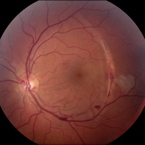
 Berlin’s Edema
Aug 10 2024 by Sachit Mahajan, MBBS MS
Fundus photograph of 10 year old boy, with a history of blunt trauma to left eye with cricket ball in school, showing Berlin’s Edema at posterior pole.
Photographer: Prattoy, Dr Shroff’s Charity Eye Hospital, New Delhi
Imaging device: Mirante, Nidek
Condition/keywords: Berlin's edema, blunt trauma, ocular trauma
-
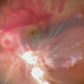 Retinal Detachment With Vitreous Hemorrhage With Sub Retinal Bleed With PVR Changes
Jul 4 2024 by Anjana Mirajkar, MS Ophthalmology
Intra operative image showing us sub retinal bleed at the macular area with superior vitreous hemorrhage with retinal detachment with inferior PVR changes likely in a case of trauma.
Photographer: Dr. Anjana Mirajkar -Retina Foundation, Ahmedabad
Condition/keywords: ocular trauma, retinal detachment with PVR, SUB RETINAL HEMORRHAGE, vitreous hemorrhage
-
 Proliferative Vitreoretinopathy
Jun 9 2024 by Marcelo Zas, MD PhD
We present a case of a 20-year-old patient who underwent surgery for congenital cataract when he was born and 20 years after he developed a retinal detachment with proliferative vitreoretinopathy. Proliferative vitreoretinopathy (PVR), a major complication of rhegmatogenous retinal detachment (RRD), is an abnormal process whereby proliferative, contractile cellular membranes form in the vitreous and on both sides of the retina, resulting in tractional retinal detachment with fixed retinal folds. PVR arises in an estimated 5-10% of RRD cases, and therefore represents a major complication of retinal detachment. The best treatment of PVR is its prevention. Clinical factors associated with increased risk of PVR include: • Chronic RRD • 2 o more horseshoe retinal tears and RRD exposing three-disc diameters or more of RPE • RD associated with giant retinal • RD associated with choroidal detachment • Ocular Trauma • RRD associated with vitreous hemorrhage • Aphakia and RRD • Failure of previous surgery or multiple retinal surgeries • Aggressive retinitis, etc.
Photographer: Luciano Scorsetti MD
Condition/keywords: proliferative vitreoretinopathy (PVR)
-
 Choroidal-rupture
Jan 2 2024 by Tahsin Khundkar, MD
37-year-old male with blunt ocular trauma presented with a choroidal rupture, pre -retinal and sub-retinal heme, and a heart shaped patch of commotio retinae.
Photographer: Jeffrey Zeigler, Concord Eye Center
Imaging device: Topcon
Condition/keywords: Choroidal Rupture, commotio retinae, Trauma
-
 Macrocysts in Kickboxer
Nov 17 2023 by Bradley T. Smith, MD, FASRS
Intraoperative photo and preoperative b scan of chronic retinal detachment with macrocysts in a kickboxer
Condition/keywords: B scan ultrasound, chronic retinal detachment, ocular trauma, pars plana vitrectomy (PPV), retinal macrocyst
-
 Fraternal Twins
May 22 2023 by Gustavo M. Hüning, MD, MBA, FASRS
Intrasurgical photograph using a non-contact system and 3D visualization system of a 65-year-old woman who suffered an ocular trauma.
Photographer: Gustavo M. Hüning, Hüning Clínica do Olhar, Santa Maria - Brazil
Imaging device: Alcon Luxor combined with Alcon nGenuity
Condition/keywords: dislocated intraocular lens (IOL), implant, pars plana vitrectomy (PPV)
-
 PPV retained cataract
Apr 19 2023 by Denica Rodriguez
A 46-year-old male with hypermature dense cataract. Patient got a piece of metal in his eye when he was 5 years old and was not able to see since. Patient was having cataract surgery and phacodonesis was present. The lens dropped to the back of the eye. Patient had to have another surgery to do vitrectomy. The lens removal was done with a fragmatome handpiece.
Photographer: Denica Rodriguez COA, ST
Imaging device: Zeiss Microscope with resight
Condition/keywords: cataract, dropped nucleus, fragmatome, lens capsule, ocular trauma, pars plana vitrectomy (PPV), retained lens fragments, Retina, retina surgery, traumatic cataract
-
 Hypotony maculopathy
Feb 23 2023 by Kamal Kishore, MD, MBBS
Ultrawide field fundus photograph of a 62-year-old male with hypotony following blunt ocular trauma
Photographer: Kim Grabill, COA, Illinois Retinal and Eye Associates, Peoria, IL, USA
Imaging device: Zeiss Clarus
Condition/keywords: hypotony maculopathy
-
 Subretinal Hemorrhage with Chorioretinal Macular Scars
Sep 28 2022 by Denica Rodriguez
Ultra-widefield pseudocolor fundus photograph of a 59 year old female with Subretinal Hemorrhage with Chorioretinal Macular Scars affecting her left eye. The physician presumes the etiology is CNV from adjacent scarring/choroidal rupture. Patient has history of ocular trauma with cricket ball at age 10-12 years old. She suspects that she might have suffered choroidal rupture, which has resulted in secondary CNV and hemorrhage that we are seeing today. She recommends treatment with Eylea sample injection in a series of 4 at a 4-5 week interval. The patient's vision at the time of her appointment was Dcc20/40-2 PHNI.
Photographer: Denica Rodriguez, COA
Imaging device: Optos California
Condition/keywords: antiVEGF therapy, chorioretinal scar, choroidal neovascular membrane (CNVM), fundus photography, left eye, macular scar, Optos, peripheral drusen, pseudocolor, secondary CNV, subretinal hemorrhage, ULTRA WIDE FIELD, ultra-wide field imaging
-
 Silicone oil in traumatic aniridia
Apr 19 2022 by Thais Bastos
A 27-year-old patient who developed aniridia, aphakia and retinal detachment after ocular trauma in the left eye. She underwent vitrectomy with silicone oil. Photo of the anterior segment 3 months after surgery showing a double meniscus made of silicone oil. Note red reflex, the retina is totally attached.
Photographer: Thaís Azeredo Bastos, CBCO Hospital de Olhos, Goiânia - Brazil
Imaging device: Zeiss Clarus 700
Condition/keywords: aniridia, ocular trauma, silicone oil
 Loading…