Search results (75 results)
-
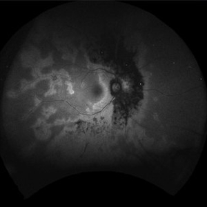
 Acute Zonal Occult Outer Retinopathy (AZOOR) FA, Fluorescein Angiography, Peripheral Vasculitis
Jan 19 2022 by James B. Soque, CRA, OCT-C, COA, FOPS
Acute Zonal Occult Outer Retinopathy (AZOOR). Peripheral Vasculitis OD. Fluorescein angiography showing vasculitis in the far right periphery 8-10 o'clock. 46-year-old white male, VA CC 20/16, 20/12.5, has had recurrent vasculitis for 11 years. No treatment.
Photographer: James Soque, CRA, OCT-C, COA, FOPS, Island Retina, Shirley, NY
Imaging device: Optos California
Condition/keywords: acute zonal occult outer retinopathy (AZOOR), FA early phase, fluorescein angiogram (FA), Peripheral Vasculitis, ultra-wide field imaging
-
 Acute Zonal Occult Outer Retinopathy (AZOOR) FA, Ultra Wide-Field Fluorescein Angiogram Early
Jan 19 2022 by James B. Soque, CRA, OCT-C, COA, FOPS
Acute Zonal Occult Outer Retinopathy, FA, Fluorescein Angiography, OD. 46-year-old white male, VA CC 10/16, 20/12.5, has had recurrent vasculitis for 11 years. No treatment.
Photographer: James Soque, CRA, OCT-C, COA, FOPS, Island Retina, Shirley, NY
Imaging device: Optos California
Condition/keywords: acute zonal occult outer retinopathy (AZOOR), FA EARLY, fluorescein angiogram (FA), ultra-wide field imaging
-
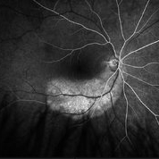
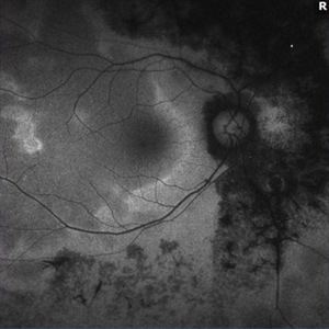
 Acute Zonal Occult Outer Retinopathy, (AZOOR) FAF, Fundus Autofluorescence
Jan 19 2022 by James B. Soque, CRA, OCT-C, COA, FOPS
Acute Zonal Occult Outer Retinopathy, FAF, Fundus Auto Fluorescence, OD. 46-year-old white male, VA CC 10/16, 20/12.5, has had recurrent vasculitis for 11 years. No treatment.
Photographer: James Soque, CRA, OCT-C, COA, FOPS, Island Retina, Shirley, NY
Imaging device: Optos California
Condition/keywords: acute zonal occult outer retinopathy (AZOOR), fundus autofluorescence (FAF), ultra-wide field imaging
-
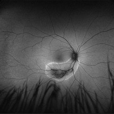
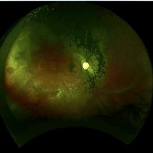
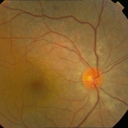
 Acute Zonal Occult Outer Retinopathy (AZOOR)
Jan 19 2022 by James B. Soque, CRA, OCT-C, COA, FOPS
Acute Zonal Occult Outer Retinopathy, OD. Color fundus, Ultra-wide field photograph. 46-year-old white male, VA CC 10/16, 20/12.5, has had recurrent vasculitis for 11 years. No treatment.
Photographer: James Soque, CRA, OCT-C, COA, FOPS, Island Retina, Shirley, NY
Imaging device: Optos California
Condition/keywords: acute zonal occult outer retinopathy (AZOOR), color wide field, optos, ultra-wide field imaging
-
 Rare Bilateral Choroidal Metastasis from Occult Primary Lung Cancer
May 5 2021 by Deependra Vikram Singh, MD FASRS
Fundus photographs and OCT scans of a 73-year-old non-smoker Indian male who presented to our retina clinic in 2013 with blurred vision in left eye for past 2 weeks. BCVA was 20/20 in right eye and 20/40 in left eye. Slit lamp exam was unremarkable for both eyes with no cells in aqueous or anterior vitreous. Fundus examination revealed creamy yellow choroidal lesions in both eyes. Lesion in right eye was one disc diameter (DD) in size and was located close to fovea (Fig-1a). Lesion in the left eye was bigger with a size of 2 DD located superior to fovea (Fig-1b). OCT scan for left eye revealed neurosensory detachment involving fovea (Fig-1c). Fundus fluorescein angiography was inconclusive for right eye and showed late hyper fluorescence the choroidal lesion in left eye. Patient underwent detailed systemic work up for malignancy that revealed primary lung non-small cell carcinoma. He had widespread metastasis affecting liver and brain. Palliative chemotherapy and radiotherapy were initiated 4 weeks after he presented to us. The choroidal lesions show progression on fundus picture and OCT scans done at 4 weeks follow up after initial presentation (Fig – 1d, e, f). The lesions in both eyes show regression at 4 weeks and 12 weeks follow up after initiation of therapy. Unfortunately, patient succumbed at 13 weeks follow up due to disease progression. The case demonstrates rare bilateral choroidal metastasis from primary lung cancer and also highlights that lesions can be asymptomatic till they develop neurosensory detachment as evident from asymptomatic lesion in right eye despite proximity to fovea and symptomatic lesion in left eye with NSD.
Photographer: Deependra Vikram Singh, Eye-Q Superspecialty Eye Hospitals, Gurugram
Imaging device: Topcon
Condition/keywords: choroidal mass, choroidal metastasis
-
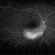
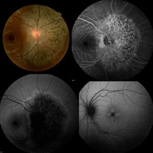
 Acute Zonal Occult Outer Retinopathy
Dec 16 2020 by Robert C Wann, MD
Fundus autofluorescence of a 28-year-old female with AZOOR.
Photographer: Retina Consultants of Alabama
Imaging device: Optos
Condition/keywords: acute zonal occult outer retinopathy (AZOOR)
-
 Acute Zonal Occult Outer Retinopathy
Dec 16 2020 by Robert C Wann, MD
Fundus autofluorescence of a 28-year-old female with AZOOR.
Photographer: Retina Consultants of Alabama
Imaging device: Optos
Condition/keywords: acute zonal occult outer retinopathy (AZOOR)
-
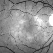
 Acute Zonal Occult Outer Retinopathy
Dec 16 2020 by Robert C Wann, MD
Fundus photo of a 28-year-old female with AZOOR.
Photographer: Retina Consultants of Alabama
Imaging device: Optos
Condition/keywords: acute zonal occult outer retinopathy (AZOOR)
-
 Unilateral AZOOR
May 4 2020 by Iuri Golubev, MD
Color, FA and FAF photos of a 17-year-old female w/h/o unilateral AZOOR OD for 5 years. FAF OS demonstrates an intact fundus.
Condition/keywords: acute zonal occult outer retinopathy (AZOOR)
-
 Macular Pattern Dystrophy Associated with MELAS
Dec 19 2019 by Olivia Rainey
Bilateral wide field fundus autofluorescence images of a 54-year-old female with macular pattern dystrophy associated with MELAS. The patient is positive for m.3243A>G in MT-TL1. She had stroke in her 40s, hearing loss in her 30s, and has early onset diabetes. MyRetinaTracker shows VUS in RP1L1. Mutation in RP1L1 have been describe in other families with occult macular dystrophy. Farnsworth D15 is showing mild tritan abnormality, which is most commonly seen with acquired maculopathies. 12/17/19 patient's Optos and OCT show mild progression of atrophy.
Photographer: Olivia Rainey
Imaging device: Optos California
Condition/keywords: advanced geographic atrophy, bilateral, fundus autofluorescence (FAF), MELAS, Optos, pattern macular dystrophy, wide angle imaging
-
 Macular Pattern Dystrophy Associated with MELAS
Dec 19 2019 by Olivia Rainey
Bilateral wide field pseudocolor images of a 54-year-old female with macular pattern dystrophy associated with MELAS. The patient is positive for m.3243A>G in MT-TL1. She had stroke in her 40s, hearing loss in her 30s, and has early onset diabetes. MyRetinaTracker shows VUS in RP1L1. Mutation in RP1L1 have been describe in other families with occult macular dystrophy. Farnsworth D15 is showing mild tritan abnormality, which is most commonly seen with acquired maculopathies. 12/17/19 patient's Optos and OCT show mild progression of atrophy.
Photographer: Olivia Rainey
Imaging device: Optos California
Condition/keywords: advanced geographic atrophy, bilateral, fundus photograph, MELAS, Optos, pattern macular dystrophy, pseudocolor, wide angle imaging
-
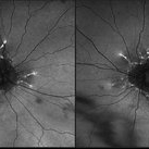
 FA ICG AZOOR
Oct 14 2017 by Navneet Mehrotra, DNB
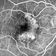
Fundus autofluorescence OS showing peripapillary hypoautofluorescence surrounded by an area of hyperautofluorescence with well demarcated margins suggestive of AZOOR.
Photographer: Ashish jain, Retina Foundation, Ahmedabad
Imaging device: Heidelberg spectralis
Condition/keywords: acute zonal occult outer retinopathy (AZOOR)
-
 FA ICG AZOOR
Oct 14 2017 by Navneet Mehrotra, DNB
fundus autofluorescence OD showing peripapillary hypoautofluorescence surrounded by an area of hyperautofluorescence with well demarcated margins suggestive of AZOOR.
Photographer: Ashish jain, Retina Foundation, Ahmedabad
Imaging device: Heidelberg spectralis
Condition/keywords: acute zonal occult outer retinopathy (AZOOR)
-
 Occult ARMD, Color Fundus Photograph
May 17 2016 by James B. Soque, CRA, OCT-C, COA, FOPS
86-year-old white female after anti veg-f therapy. Color fundus photo of occult SRN with pigment mottling of the right eye.
Photographer: James B Soque, CRA, OCT-C, COA, Island Retina, Shirley, NY
Imaging device: Topcon TRC 50 EX, OIS 5 MP Camera, MERGE software
Condition/keywords: after treatment, color fundus photograph, occult, occult choroidal neovascularization (CNV), subretinal neovascularization (SRNV), wet age-related macular degeneration (wet AMD)
-
 AZOOR
AZOOR
Mar 19 2015 by Niloofar Piri, MD
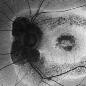
#2 : Fundus autofluorescence OS in the same patient demonstrates more severe changes ; peripapillary hypoAF and concentric rings of hyper and hypo AF in posterior pole
Imaging device: Heidelberg Spectralis
Condition/keywords: acute zonal occult outer retinopathy (AZOOR)
-
 AZOOR
AZOOR
Mar 19 2015 by Niloofar Piri, MD
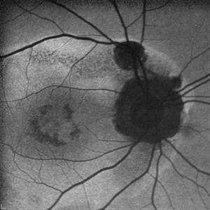
#1: Fundus autofluorescence OD in a patient with AZOOR demonstrates characteristic peripapillary hypoAF as well as concentric rings of hypo and hyper AF in posterior pole .
Imaging device: Heidelberg Spectralis
Condition/keywords: acute zonal occult outer retinopathy (AZOOR)
-
 Occult Wet AMD Fluorescein Angiogram
Jul 25 2014 by James B. Soque, CRA, OCT-C, COA, FOPS
FA OD image, early phase, 32 seconds, of 82-year-old white female with occult disease OD, anti-VEGF therapy since 2012, and retains 20/63 vision.
Photographer: James Soque, CRA COA
Imaging device: Topcon TRC 50 DX, OIS 5 MP Camera, MERGE software
Condition/keywords: wet age-related macular degeneration (wet AMD)
-
 Occult Wet ARMD, Red Free Image
Jul 25 2014 by James B. Soque, CRA, OCT-C, COA, FOPS
Red free image, OD of 82-year-old white female with occult disease, anti-VEGF therapy since 2012, and retains 20/63 vision.
Photographer: James Soque, CRA COA
Imaging device: Topcon TRC 50 DX, OIS 5 MP Camera, MERGE software
Condition/keywords: red-free, wet age-related macular degeneration (wet AMD)
-
 Occult Wet ARMD, Fundus Color Photograph
Jul 25 2014 by James B. Soque, CRA, OCT-C, COA, FOPS
FC image OD, of 82-year-old white female with occult disease OD, anti-VEGF therapy since 2012, and retains 20/63 vision.
Photographer: James Soque, CRA COA
Imaging device: Topcon 50 DX, OIS 5 MP Camera, MERGE software
Condition/keywords: color photo, fundus photograph, wet age-related macular degeneration (wet AMD)
-
 AZOOR vs. AAOOR
Mar 19 2014 by Ali Tavallali, MD, FASRS
FAF of a 47-year-old female with 20/20 VA of both eyes, note the progression of demarcation line after 4 months
Photographer: Neda Sheibani, Dr. Khodadoust Eye Hospital, Shiraz, Iran
Condition/keywords: acute zonal occult outer retinopathy (AZOOR)
-
 AZOOR vs. AAOOR
Mar 19 2014 by Ali Tavallali, MD, FASRS
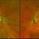
Color fundus photograph of a 47-year-old female with 20/20 VA of both eyes, note the progression of demarcation line after 4 months
Photographer: Neda Sheibani, Dr. Khodadoust Eye Hospital, Shiraz, Iran
Condition/keywords: acute zonal occult outer retinopathy (AZOOR)
-
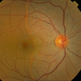 AZOOR vs. AAOOR
Mar 19 2014 by Ali Tavallali, MD, FASRS
Color fundus photograph of a 47-year-old female with 20/20 VA of both eyes, note the demarcation line
Photographer: Neda Sheibani, Dr. Khodadoust Eye Hospital, Shiraz, Iran
Condition/keywords: acute zonal occult outer retinopathy (AZOOR)
-
 Autofluorescence 10-14-13 AZOOR
Dec 14 2013 by Robert T. Wendel, MD
AF image
Imaging device: Heidelberg
Condition/keywords: acute zonal occult outer retinopathy (AZOOR)
-
 Autofluorescence 12-5-13 AZOOR
Dec 14 2013 by Robert T. Wendel, MD
AF 12-5-16
Condition/keywords: acute zonal occult outer retinopathy (AZOOR), autofluorescence imaging
-
 Autofluorescence 10-14-13 AZOOR
Dec 14 2013 by Robert T. Wendel, MD
Autofluorescence 10-14-13 AZOOR
Condition/keywords: acute zonal occult outer retinopathy (AZOOR), autofluorescence imaging
 Loading…