Search results (75 results)
-
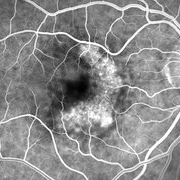
 Pigment Epithelial Detachment late FA with small occult CNV
Jul 6 2012 by Tarek S. Hassan, MD, FASRS
72-year-old man with VA loss and metamorphopsia of 2 months duration. PED found, testing done to rule out CNV. Very suspicious for CNV in superonasal fovea/parafovea.
Condition/keywords: choroidal neovascularization (CNV), pigment epithelial detachment (PED)
-
 WET Age Related Macular Degeneration (WET AMD)
Sep 8 2012 by Ratimir Lazic, MD, PhD
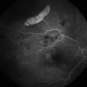
FAG image of a 65 - year- old male. In early venous phase hyperfluorescence due to feeling of occult CNV can be observed
Photographer: Ratimir Lazic, PhD MD
Imaging device: Zeis Visucam Lite 2
Condition/keywords: fundus photograph
-
 AZOOR 10-10-13
Oct 11 2013 by Robert T. Wendel, MD
Acute zonal occult outer retinopathy (AZOOR).
Condition/keywords: acute zonal occult outer retinopathy (AZOOR)
-
 RPE rip macular OCT
Dec 23 2012 by Alex P. Hunyor, MD
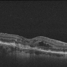
80-year-old female with subfoveal occult CNV and large extrafoveal PED which underwent spontaneous RPE rip. OCT shows subfoveal CNV and intraretinal cystic edema
Condition/keywords: pigment epithelial detachment (PED), retinal pigment epithelium (RPE) tear
-
 AZOOR
AZOOR
Mar 19 2015 by Niloofar Piri, MD
#1: Fundus autofluorescence OD in a patient with AZOOR demonstrates characteristic peripapillary hypoAF as well as concentric rings of hypo and hyper AF in posterior pole .
Imaging device: Heidelberg Spectralis
Condition/keywords: acute zonal occult outer retinopathy (AZOOR)
-
 Wet Age Related Macular Degeneration (WET AMD)
Sep 8 2012 by Ratimir Lazic, MD, PhD
FAG image of a 65 - year - old male. In the late venous phase further leakage from the occult CNV membrane can be noted
Photographer: Ratimir Lazic, PhD MD
Imaging device: Zeis Visucam Lite 2
Condition/keywords: fundus photograph
-
 AZOOR-OCT
Oct 11 2013 by Robert T. Wendel, MD
Acute zonal occult outer retinopathy (AZOOR).
Condition/keywords: acute zonal occult outer retinopathy (AZOOR)
-
 Age Related Macular Degeneration - Occult CNV
May 3 2013 by Suber S. Huang, MD, MBA, FASRS
Age related macular degeneration - occult CNV
Imaging device: Retina Diseases Imaging Analysis Reading Center
Condition/keywords: choroidal neovascularization (CNV), choroidal neovascularization occult
-
 Autofluorescence 10-14-13 AZOOR
Dec 14 2013 by Robert T. Wendel, MD
Autofluorescence 10-14-13 AZOOR
Condition/keywords: acute zonal occult outer retinopathy (AZOOR), autofluorescence imaging
-
 AZOOR
AZOOR
Mar 19 2015 by Niloofar Piri, MD
#2 : Fundus autofluorescence OS in the same patient demonstrates more severe changes ; peripapillary hypoAF and concentric rings of hyper and hypo AF in posterior pole
Imaging device: Heidelberg Spectralis
Condition/keywords: acute zonal occult outer retinopathy (AZOOR)
-
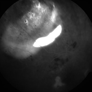
 Extrafoveal PED with RPE rip AF
Dec 23 2012 by Alex P. Hunyor, MD
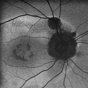
80-year-old female with subfoveal occult CNV and large extrafoveal PED which underwent spontaneous RPE rip. Autofluorescence image shows hypoautofluorescence in crescentic area of absent RPE due to rip, and also RPE atrophy adjacent to fovea. Intervening small areas of hypoautofluorescence are due to subretinal haemorrhage.
Condition/keywords: pigment epithelial detachment (PED), retinal pigment epithelium (RPE) tear
-
 AZOOR-OCT8
Oct 11 2013 by Robert T. Wendel, MD
Acute zonal occult outer retinopathy (AZOOR).
Condition/keywords: acute zonal occult outer retinopathy (AZOOR)
-
 AZOOR Color 10-14-2013
Dec 14 2013 by Robert T. Wendel, MD
AZOOR
Condition/keywords: acute zonal occult outer retinopathy (AZOOR)
-
 Occult Wet AMD Fluorescein Angiogram
Jul 25 2014 by James B. Soque, CRA, OCT-C, COA, FOPS
FA OD image, early phase, 32 seconds, of 82-year-old white female with occult disease OD, anti-VEGF therapy since 2012, and retains 20/63 vision.
Photographer: James Soque, CRA COA
Imaging device: Topcon TRC 50 DX, OIS 5 MP Camera, MERGE software
Condition/keywords: wet age-related macular degeneration (wet AMD)
-
 Extrafoveal PED with RPE rip FA1
Dec 23 2012 by Alex P. Hunyor, MD
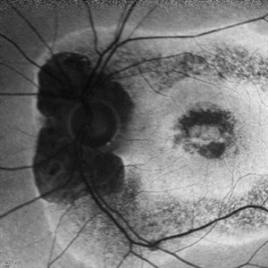
80-year-old female with subfoveal occult CNV and large extrafoveal PED which underwent spontaneous RPE rip. Early phase FA showing intense hyperfluorescence in the area of acute absence of RPE.
Condition/keywords: pigment epithelial detachment (PED), retinal pigment epithelium (RPE) tear
-
 AZOOR vs. AAOOR
Mar 19 2014 by Ali Tavallali, MD, FASRS
FAF of a 47-year-old female with 20/20 VA of both eyes, note the progression of demarcation line after 4 months
Photographer: Neda Sheibani, Dr. Khodadoust Eye Hospital, Shiraz, Iran
Condition/keywords: acute zonal occult outer retinopathy (AZOOR)
-
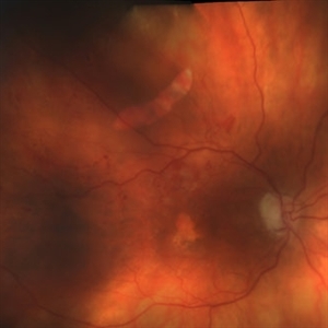 Extrafoveal PED with RPE rip colour photo
Dec 23 2012 by Alex P. Hunyor, MD
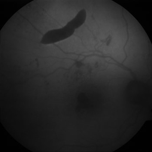
80-year-old female with subfoveal occult CNV and large extrafoveal PED which underwent spontaneous RPE rip.
Condition/keywords: pigment epithelial detachment (PED), retinal pigment epithelium (RPE) tear
-
---thumb.JPG/image-square;max$300,300.ImageHandler) Co-existing CNVM and CSCR
Co-existing CNVM and CSCR
Dec 10 2012 by Mallika Goyal, MD
Right eye of a 54-year-old gentleman shows an occult CNVM with CSCR. FFA and OCT confirmed the diagnoses. There has been no change after treatment with avastin and focal photocoagulation for the extrafoveal RPE leak. PDT has been performed, and result will be uploaded later.
Photographer: Mallika Goyal, MD, Apollo Health City, Hyderabad, India
Condition/keywords: central serous chorioretinopathy (CSCR)
-
 AZOOR OCT 10-10-13
Dec 14 2013 by Robert T. Wendel, MD
AZOOR OCT 10-10-13
Condition/keywords: acute zonal occult outer retinopathy (AZOOR), optical coherence tomography (OCT)
-
 Occult ARMD, Color Fundus Photograph
May 17 2016 by James B. Soque, CRA, OCT-C, COA, FOPS
86-year-old white female after anti veg-f therapy. Color fundus photo of occult SRN with pigment mottling of the right eye.
Photographer: James B Soque, CRA, OCT-C, COA, Island Retina, Shirley, NY
Imaging device: Topcon TRC 50 EX, OIS 5 MP Camera, MERGE software
Condition/keywords: after treatment, color fundus photograph, occult, occult choroidal neovascularization (CNV), subretinal neovascularization (SRNV), wet age-related macular degeneration (wet AMD)
-
 Autofluorescence 10-14-13 AZOOR
Dec 14 2013 by Robert T. Wendel, MD
Autofluorescence 10-14-13 AZOOR
Condition/keywords: acute zonal occult outer retinopathy (AZOOR), autofluorescence imaging
-
 Extrafoveal PED with RPE rip FA4
Dec 23 2012 by Alex P. Hunyor, MD
80-year-old female with subfoveal occult CNV and large extrafoveal PED which underwent spontaneous RPE rip. FA shows intense hyperfluorescence in area of absent RPE, progressive filling of extrafoveal PED, and hyperfluorescence in macula from atrophy and occult CNV.
Condition/keywords: pigment epithelial detachment (PED), retinal pigment epithelium (RPE) tear
-
 Autofluorescence 12-5-13 AZOOR
Dec 14 2013 by Robert T. Wendel, MD
Autofluorescence 12-5-13 AZOOR
Condition/keywords: acute zonal occult outer retinopathy (AZOOR), autofluorescence imaging
-
 AZOOR-OCT4
Oct 11 2013 by Robert T. Wendel, MD
Acute zonal occult outer retinopathy (AZOOR).
Condition/keywords: acute zonal occult outer retinopathy (AZOOR)
-
 AZOOR OCT2 10-10-13
Dec 14 2013 by Robert T. Wendel, MD
AZOOR OCT2 10-10-13
Condition/keywords: acute zonal occult outer retinopathy (AZOOR), optical coherence tomography (OCT)
 Loading…