Search results (146 results)
-
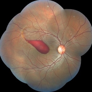
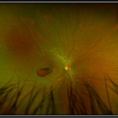 Premacular Hemorrhage Secondary to Recreational Laser Exposure
Aug 22 2025 by Carlos Valdez Prado
A 40-year-old male with accidental exposure to a recreational laser during a party presents with sudden visual loss following the laser exposure. On examination, a premacular hemorrhage with a double-ring sign is observed in the macular region.
Photographer: Dr. Carlos Antonio Valdez Prado, Hospital Militar de Especialidades Oftalmologicas
Imaging device: Optos
Condition/keywords: Hemorrhage, premacular hemorrhage, Retina, subhyaloid hemorrhage, sublimitante
-
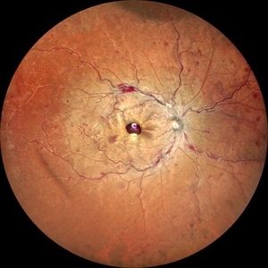 Ultra-Wide Field Image of Central Retinal Vein Occlusion with Foveal Hemorrhage
Apr 17 2025 by Malvika Singh
Ultra- wide field fundus photograph of a 41 year-old male, with a central retinal vein occlusion and a foveal sub-internal limiting membrane hemorrhage.
Photographer: Dr Malvika Singh, Retina Foundation, Ahmedabad, India
Imaging device: Mirante SLO/OCT
Condition/keywords: central retinal vein occlusion (CRVO), macular hemorrhage, Ultra-wide field retinal imaging
-
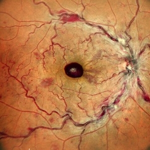 Central Retinal Vein Occlusion with Foveal Hemorrhage
Apr 17 2025 by Malvika Singh
Fundus photograph of a 41 year-old, male, with a central retinal vein occlusion and a foveal sub-internal limiting membrane hemorrhage.
Photographer: Dr Malvika Singh, Retina Foundation, Ahmedabad, India
Imaging device: Mirante SLO/OCT
Condition/keywords: central retinal vein occlusion (CRVO), macular hemorrhage
-
 Sub-Retinal Blood Air and TPA
Jan 31 2025 by Thirumalesh Mochi Basavaraj, MD
Intra Operative View of a 76 year old gentleman with Submacular bleed treated with Sub Retinal TPA, Ranibizumab and air, one can appreciate at multiple levels
Photographer: Thirumalesh Mochi Basavaraj
Condition/keywords: submacular hemorrhage, tissue plasminogen activator (tPA)
-
 Optic Nerve Head Avulsion
Sep 24 2024 by Gustavo Uriel Fonseca Aguirre
A 14-year-old male with a history of blunt ocular trauma in the right eye presented partial avulsion of the optic nerve head and submacular hemorrhage that was managed with neumatic displacement.
Photographer: Gustavo U. Fonseca Aguirre, Fundación Hospital Nuestra Señora de la Luz, Ciudad de México
Condition/keywords: optic nerve head avulsion
-
 Submacular Hemorrhage
Sep 24 2024 by Gustavo Uriel Fonseca Aguirre
24-year-old patient, submacular hemorrhage is observed in the left eye secondary to blunt ocular trauma.
Photographer: Gustavo U. Fonseca Aguirre, Fundación Hospital Nuestra Señora de la Luz, Ciudad de México
Condition/keywords: submacular hemorrhage
-
 Nd: Yag Hyaloidotomy for Dense Subhyaloid Hemorrhage Caused by Diabetic Retinopathy
Aug 1 2024 by Yodpong Chantarasorn, MD
A 52-year-old female declined surgical procedures and opted for Nd:Yag Hyaloidotomy to treat a dense subhyaloid hemorrhage from proliferative diabetic retinopathy. She experienced a full recovery from the hemorrhage, while PRP laser treatment was successfully completed.
Photographer: Yodpong Chantarasorn
Condition/keywords: NdYAG laser, premacular hemorrhage, YAG HYALOIDOTOMY
-
 Submacular Hemorrhage
Jun 12 2024 by Anand Temkar
Intraoperative still of a 58 year old male with submacular hemorrhage (LE).
Photographer: Dr.Anand Temkar- Retina Foundation, Ahmedabad
Condition/keywords: submacular hemorrhage, subretinal hemorrhage
-
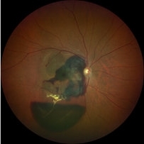
 Sub Macular Fibrosis in the Setting of Old Sub Macular Hemorrhage
Feb 15 2024 by Sayena . Jabbehdari, MD, MPH, MBA
Pseudo color fundus photo of 78 years old male with history of sub macular hemorrhage in the setting of wet age-related macular degeneration. You can appreciate the tear shaped appearance of blood due to gravity. The OCT of the macula depicts the huge (>950mm) sub retinal fibrosis.
Photographer: Sayena Jabbehdari MD MPH , University of Arkansas in Little Rock
Imaging device: Clarus
Condition/keywords: macular neovascular disease, retina, submacular hemorrhage, Wet age related macular degeneration
-
 Sub-Macular Haemorrhage
Apr 15 2023 by Veer Singh, MS, FVRS, FMRF, FICO (Retina)
Sub-Retinal haemorrhage secondary to PCV in an elderly gentleman.
Photographer: Dr. Veer Singh
Condition/keywords: polypoidal choroidal vasculopathy (PCV), submacular hemorrhage
-
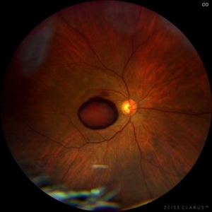 Sub-internal limiting membrane hemorrhage in Valsalva retinopathy
Jul 29 2022 by JORGE SOBERANES
A fundus photography of a 70-year-old man with premacular hemorrhage (Sub-internal limiting membrane) due to Valsalva retinopathy
Photographer: Jorge I. Soberanes, Asociación para Evitar la Ceguera en México.
Imaging device: Zeiss Clarus 700
Condition/keywords: premacular hemorrhage, sub-inner limiting membrane hemorrhage, valsalva retinopathy
-
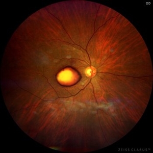 Dehemoglobinized sub-internal limiting membrane hemorrhage
Jul 29 2022 by JORGE SOBERANES
Fundus photograph of a 70-year-old man with Valsalva retinopathy manifested as premacular hemorrhage (sub-ILM) in dehemoglobinized process.
Photographer: Jorge I. Soberanes, Asociación para Evitar la Ceguera en México.
Imaging device: Zeiss Clarus 700
Condition/keywords: dehemoglobinized hemorrhage, sub-inner limiting membrane hemorrhage, valsalva retinopathy
-
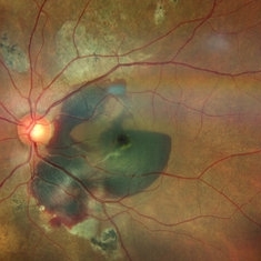
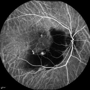
 Permacular fold in Terson's syndrome
Jun 1 2022 by Deependra Vikram Singh, MD FASRS
Fundus Fluorescein Angiography picture of a 36-year-old male with chronic liver disease who has undergone 25G vitrectomy for Vitreous and sub-ILM haemorrhage from Terson's syndrome.
Photographer: Deependra Vikram Singh, Eye-Q Superspeciality Eye Hospitals, Gurugram, India
Imaging device: ZEISS
Condition/keywords: macular fold, sub internal limiting membrane haemorrhage, submacular hemorrhage, Terson's Syndrome
-
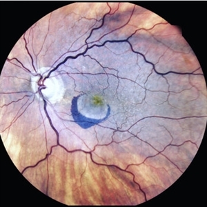
 Perimacular Fold in Terson's Syndrome
Jun 1 2022 by Deependra Vikram Singh, MD FASRS
Fundus photograph of a 36-year-old man with chronic liver disease who had undergone 25G vitrectomy for vitreous and sub-ILM haemorrhage resulting from Terson's syndrome. Image was clicked in 2016 and shows striking perimacular fold. Patient had vitreous haemorrhage in both eyes with left eye not requiring vitrectomy.
Photographer: Deependra Vikram Singh, Eye-Q Superspeciality Eye Hospitals, Gurugram India
Imaging device: Zeiss VISUCAM 500
Condition/keywords: macular fold, sub internal limiting membrane haemorrhage, submacular hemorrhage, Terson
-
 Submacular Hemorrhage PCV
May 6 2022 by Shobhit Chawla, M.S.
Submacular hemorrhage in a 38 years old female patient cause polyp bleed in PCV.
Photographer: Shobhit Chawla
Imaging device: Zeiss Clarus 500
Condition/keywords: polypoidal choroidal vasculopathy (PCV), submacular hemorrhage
-
 Post Subretinal tpa , viterectomy and gas
May 6 2022 by Shobhit Chawla, M.S.
SUBMACULAR HAEMORRHAGE IN A 38YEAR OLD LADY PATIENT CAUSE POLYP BLEED IN PCV. Following viterectomy , subretinal tpa . gas and aflibercept injection. 7 day post operative image.
Photographer: Shobhit Chawla
Imaging device: Zeiss Clarus 500
Condition/keywords: aflibercept, intravitreal gas bubble, submacular hemorrhage, tissue plasminogen activator (tPA), vitrectomy
-
 Macular Hemorrhage Secondary to Anemic Retinopathy
Apr 18 2022 by Deepak Bhojwani, MS
Fundus image of a young 28 year old patient who has been diagnosed as 'PRIMARY BONE MARROW APLASIA' by hematologist showing large macular hemorrhage (sub -ILM Heme mound). Few Roth spots were also seen in midperiphery suggesting 'ANEMIC RETINOPATHY'.
Photographer: DEEPAK BHOJWANI
Condition/keywords: anaemic retinopathy, BONE MARROW APLASIA
-
 Post Aflibercept Sub-Retinal Hemorrhage
Sep 29 2021 by Hilton Arcoverde Medeiros, PhD
Fundus photograph of a 92-year-old man with sub macular hemorrhage 2 days after Aflibercept vitreo injection.
Photographer: Hilton Medeiros, Hospital de olhos João Eugênio, Brasília Brazil
Condition/keywords: aflibercept, dry age-related macular degeneration (dry AMD)
-
 Sub-Macular Hemorrhage Indocyanine Green Angiography
Jul 5 2021 by Fang Helen Mi
Indocyanine green angiography revealed three polypoidal choroidal vasculopathy lesions, and the patient underwent photodynamic therapy the following week. His vision improved to 20/30.
Condition/keywords: indocyanine green (ICG) angiography, polypoidal choroidal vasculopathy (PCV), submacular hemorrhage
-
 Submacular Hemorrhage after C3F8 Pneumatic Displacement
Jul 5 2021 by Fang Helen Mi
The patient underwent urgent intravitreal injection of recombinant tissue plasminogen activator and pneumatic displacement with 100% C3F8 gas. The following day, the sub-macular haemorrhage was largely displaced away from the fovea inferiorly and nasally, with the gas bubble seen superiorly.
Imaging device: Clarus Camera
Condition/keywords: gas pneumatic displacement, polypoidal choroidal vasculopathy (PCV), submacular hemorrhage
-
 Submacular Hemorrhage in a Patient on Rivaroxaban
Jul 5 2021 by Fang Helen Mi
A 69-year-old gentleman with a history of atrial fibrillation on Factor Xa inhibitor (rivaroxaban) presented to the Ophthalmology service with acute blurring of vision in the right eye one day after intravitreal aflibercept injection. Fundus examination revealed a massive foveal-involving sub-macular hemorrhage with associated serosanguineous retinal detachment and hemorrhagic retinal pigment epithelial detachment. This multi-layer retinal hemorrhage was further confirmed with optical coherence tomography.
Imaging device: Clarus Camera
Condition/keywords: polypoidal choroidal vasculopathy (PCV), Rivaroxaban, submacular hemorrhage
-
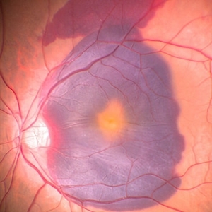
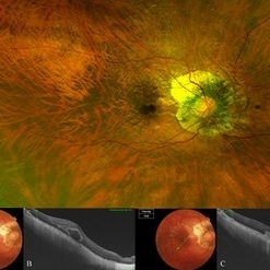 Bow-Tie Macular Hemorrhage With Cyst- Atypical Presentation of Myopic Choroidal Neovascularization
Mar 26 2021 by RUSHIK PATEL
The image of right eye of 51-year-old lady with high myopia show " Bow-Tie" macular hemorrhage (A). Optical coherence tomography (B) scan passing through hemorrhage showed intraretinal cystic lesion. During the course of intravitreal anti-VEGF injection treatment, the lesion converted into typical myopic choroidal neovascularization (C).
Photographer: Rushik Patel, Netralaya Super Speciality Eye Hospital
Condition/keywords: cyst, macular hemorrhage, myopic choroidal neovascularization (CNV)
-
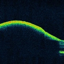 OCT Showing Premacular Hemorrhage
Nov 26 2020 by Priya Rasipuram Chandrasekaran, MBBS, DO, DNB, FRCS
A 26-year-old male with no h/o trauma or underlying systemic disease presented with the complaint of central scotoma in the right eye since 1 month and fundus examination showed preretinal hemorrhage in the supero-temporal quadrant extending into the macular area and OCT macula showing premacular hemorrhage.
Condition/keywords: premacular hemorrhage
-
 Preretinal Hemorrhage Extending into the Macula
Nov 26 2020 by Priya Rasipuram Chandrasekaran, MBBS, DO, DNB, FRCS
A 26-year-old male with no h/o trauma or underlying systemic disease presented with the complaint of central scotoma in the right eye since 1 month and fundus examination showed preretinal hemorrhage in the supero-temporal quadrant extending into the macular area and OCT macula showing premacular hemorrhage.
Condition/keywords: preretinal hemorrhage
-
 The Cliff Hanger-Subhyaloid Hemorrhage Over Macula in Proliferative BRVO
Sep 26 2020 by Kushal S Delhiwala, MBBS, MS, FMRF,FICO, FAICO
Fundus photograph of 52-year-old hypertensive female having fresh subhyaloid hemorrhage of 2 days history hanging over macula secondary to Proliferative superotemporal branch retinal vein occlusion.
Photographer: KUSHAL DELHIWALA, Netralaya superspeciality eye hospital, Ahmedabad
Imaging device: Optos Daytona
Condition/keywords: branch retinal vein occlusion (BRVO), premacular hemorrhage, subhyaloid hemorrhage
 Loading…