Search results (43 results)
-
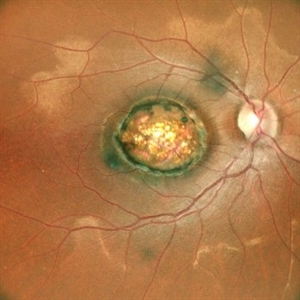
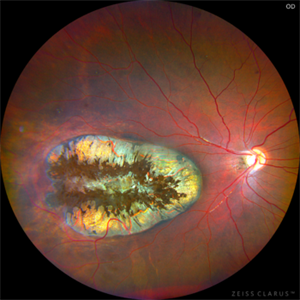 Presumed Congenital Toxoplasmosis Macular Coloboma
Aug 16 2025 by Vishal Agrawal, MD, FRCS,FACS,FASRS
7-year-old boy presented with esotropia in OD with light perception positive. Fundus reveals a large macular coloboma occupying nearly the entire macula. OCT scan shows complete atrophy and disorganization of the overlying RPE and neurosensory retina. A much smaller lesion was observed in OS with BCVA 20/40.
Photographer: Dr Ayushi Gupta
Imaging device: Clarus 700
Condition/keywords: Coloboma, congenital toxoplasmosis
-
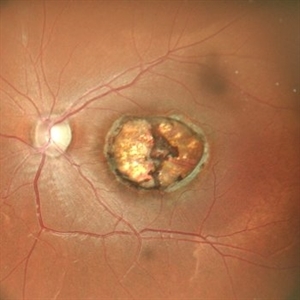
 Macular Coloboma
Jun 5 2025 by César Adrián Gómez Valdivia, MD
Macular Coloboma found in a 28 year-old male patient, visual acuity was 20/60. Resulting due to fusion failure of the optic fissure, colobomas are commonly found in the infero-nasal quadrant. If the retina is involved, it is reduced to glial tissue with no underlying RPE or choroid. This appears as an area of whitening often with pigment deposition at the junction of the coloboma and normal retina. Findings were bilateral.
Photographer: @eyemissu2
Imaging device: TOPCON TRC-50DX
Condition/keywords: coloboma
-
 Signet Ring in the Eye
Sep 28 2024 by Tejaswita Verma
Infrared fundus image of the LE of a 32 year-old male showing macular coloboma.
Photographer: DR. TEJASWITA VERMA
Imaging device: MIRANTE
Condition/keywords: macular coloboma
-
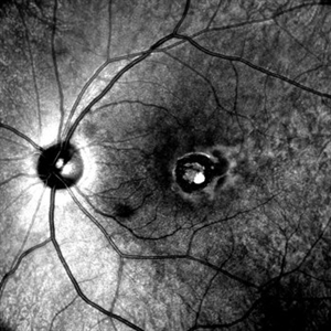
 OCT in Case of Macular Coloboma (LE)
Sep 18 2024 by Anand Temkar
A 24 year old male came with chief complaint of diminution of vision in both eyes since childhood. Vision in both eyes was 6/24. IOP in RE was 12 and LE was 14 mm of Hg. On fundus examination periphery was within normal limits and central fundus revealed this picture. The serology testing such as serum IgM, IgG for toxoplasma and cytomegalovirus was negative. I have also uploaded LE color photo and BE OCT of this patient.
Photographer: Dr.Anand Temkar- Retina Foundation, Ahmedabad
Imaging device: Mirante
Condition/keywords: Coloboma
-
 OCT in Case of Macular Coloboma (RE)
Sep 18 2024 by Anand Temkar
A 24 year old male came with chief complaint of diminution of vision in both eyes since childhood. Vision in both eyes was 6/24. IOP in RE was 12 and LE was 14 mm of Hg. On fundus examination periphery was within normal limits and central fundus revealed this picture. The serology testing such as serum IgM, IgG for toxoplasma and cytomegalovirus was negative. I have also uploaded LE color photo and BE OCT of this patient.
Photographer: Dr.Anand Temkar- Retina Foundation, Ahmedabad
Imaging device: Mirante
Condition/keywords: coloboma
-
 Macular Coloboma (LE)
Sep 18 2024 by Anand Temkar
A 24 year old male came with chief complaint of diminution of vision in both eyes since childhood. Vision in both eyes was 6/24. IOP in RE was 12 and LE was 14 mm of Hg. On fundus examination periphery was within normal limits and central fundus revealed this picture. The serology testing such as serum IgM, IgG for toxoplasma and cytomegalovirus was negative. I have also uploaded LE color photo and BE OCT of this patient.
Photographer: Dr.Anand Temkar- Retina Foundation, Ahmedabad
Imaging device: Mirante
Condition/keywords: macular coloboma
-
 Macular Coloboma (RE)
Sep 18 2024 by Anand Temkar
A 24 year old male came with chief complaint of diminution of vision in both eyes since childhood. Vision in both eyes was 6/24. IOP in RE was 12 and LE was 14 mm of Hg. On fundus examination periphery was within normal limits and central fundus revealed this picture. The serology testing such as serum IgM, IgG for toxoplasma and cytomegalovirus was negative. I have also uploaded LE color photo and BE OCT of this patient.
Photographer: Dr.Anand Temkar- Retina Foundation, Ahmedabad
Imaging device: Mirante
Condition/keywords: coloboma of macula
-
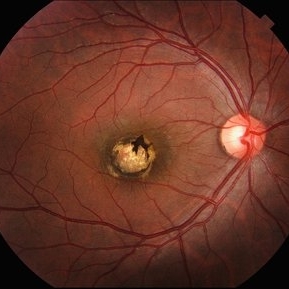
 Macular Coloboma
Jul 17 2024 by Anubhav Chauhan
This is fundus photograph of a 30 year male depicting a Macular coloboma in the right eye. The patient had a sharply defined large, yellowish white, coarsely pigmented, atrophic, round crater like defect at the macula. Spectral domain optical coherence tomography confirmed our diagnosis. The serology testing such as serum IgM, IgG for toxoplasma and cytomegalovirus was negative. His systemic examination was normal.
Photographer: Dr Anubhav Chauhan, Department of Ophthalmology, Shri Lal Bahadur Shastri Government Medical College, Nerchowk, District Mandi, Himachal Pradesh, India
Imaging device: Zeiss
Condition/keywords: macula, rare
-
 Macular Coloboma
Sep 26 2023 by Ben Serar
Fundus photograph of LE showing coloboma involving the macula.
Condition/keywords: macular coloboma
-
 MACULAR COLOBOMA
Oct 15 2022 by Akansha Sharma
COLOUR FUNDUS PHOTOGRAPH OF A 32 YEAR OLD MALE WITH MACULAR COLOBOMA
Photographer: Dr. Akansha Sharma-Retina Foundation, Ahmedabad
Condition/keywords: coloboma of macula
-
 MACULAR COLOBOMA
Oct 15 2022 by Akansha Sharma
COLOUR FUNDUS PHOTOGRAPH OF A 32 YEAR OLD MALE WITH MACULAR COLOBOMA
Photographer: Dr. Akansha Sharma-Retina Foundation, Ahmedabad
Condition/keywords: coloboma of macula
-
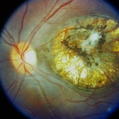
 Macular Colobomata
Jun 21 2022 by Sukanya Mondal, MBBS, MS, FICO, MRCSEd
Left eye fundus photograph of a 19-year-old girl, having low vision in the same eye since birth, showing well demarcated macular excavation and underlying scleral baring with hyper and hypopigmentated areas.
Photographer: Dr Sukanya Mondal, National Institute of Ophthalmology, Pune. India
Imaging device: Zeiss Clarus 500
Condition/keywords: macular coloboma
-
 Unilateral Macular Coloboma
Jul 29 2021 by Mihir Trivedi
Fundus examination of a 35-year-old man with focal areas of altered retinal pigment epithelium and subretinal yellowish lesion in the foveal area in the right eye. Left eye showed a punched out circumscribed lesion in the center of the macula with thin foveal roof suggestive probably of the internal limiting membrane. Macular coloboma is characterized by a sharply defined, oval or rounded, usually unilateral, atrophic lesions of varying size presenting rudimentary or absent retina, choroid and sclera located at the macula leading to decreased vision in the central area of the fundus. It can be associated with retinal dystrophy in the fellow eye, as was the case in our patient.
Photographer: Priyanshi Kambodi, RNC Eye Hospital, Valsad
Condition/keywords: macular coloboma
-
 Macular Colobomas in Congenital Zika Syndrome
Sep 26 2020 by Swati Agarwal-Sinha, MD, FASRS
Color fundus picture of the right (OD) and left (OS) eye of 3-day-old female infant with congenital Zika syndrome with bilateral macular colobomatous like chorioretinal atrophy, attenuated vessels, pigmentary changes, and optic disc pallor.
Photographer: Swati Agarwal-Sinha, MD
Condition/keywords: fundus photograph, macular coloboma, zika
-
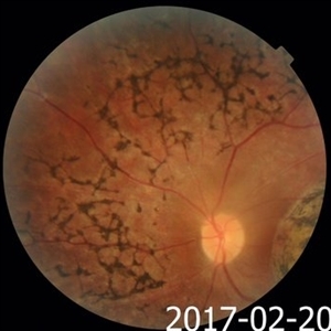
 Bilateral Macular Colobomata With Temporal Dragging of Optic Disc
Apr 24 2020 by Dinesh Rungta, MBBS, DNB
Optos ultra-widefield retinal image of a 7-year-old male child showing bilateral macular colobomata with temporal dragging of optic disc.
Photographer: Dr Shivam Madan, Giridhar Eye Institute, Kerala, India
Imaging device: Optos UWF Daytona Plus
Condition/keywords: bilateral macular colobomata, temporal dragging of optic disc
-
 Macular Coloboma With Macular Dystrophy
Jan 24 2020 by Deepak Bhojwani, MS
Fundus image of a 22-year-old gentlemen with poor vision since childhood with nystagmus. Fundus photo showing macular coloboma which is rarely seen in macular dystrophies.
Photographer: DEEPAK BHOJWANI
Condition/keywords: coloboma of macula, macular dystrophy
-
 Macular Coloboma With Macular Dystrophy
Jan 24 2020 by Deepak Bhojwani, MS
Fundus image of a 22-year-old gentlemen with poor vision since childhood with nystagmus. fundus photo showing macular coloboma which is rarely seen in macular dystrophies.
Photographer: DEEPAK BHOJWANI
Condition/keywords: macular dystrophy
-
 Macular Coloboma and Pigmentary Retinopathy
Feb 25 2017 by Hamid Ahmadieh, MD
Color fundus photograph of the right eye of a 25-year-old woman with the history of low vision since childhood. Bilateral macular colobomata and pigmentary retinopathy similar to Leber's congenital amaurosis are present.
Photographer: Shabnam Poureh, Negah Eye Center, Tehran, Iran
Condition/keywords: bilateral pigmentary retinopathy, color fundus photograph, macular coloboma, pigmentary retinal dystrophy
-
 Macular Coloboma and Pigmentary Retinopathy
Feb 25 2017 by Hamid Ahmadieh, MD
Color fundus photograph of the right eye of a 25-year-old woman with the history of low vision since childhood. Bilateral macular colobomata and pigmentary retinopathy similar to Leber's congenital amaurosis are present.
Photographer: Shabnam Poureh, Negah Eye Center, Tehran, Iran
Condition/keywords: bilateral pigmentary retinopathy, color fundus photograph, macular coloboma
-
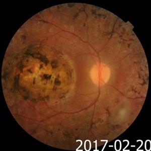
 Macular Coloboma and Pigmentary Retinopathy
Feb 25 2017 by Hamid Ahmadieh, MD
Color fundus photograph of the right eye of a 25-year-old woman with the history of low vision since childhood. Bilateral macular colobomata and pigmentary retinopathy similar to Leber's congenital amaurosis are present.
Photographer: Shabnam Poureh, Negah Eye Center, Tehran, Iran
Condition/keywords: color fundus photograph, pigmentary retinal dystrophy
-
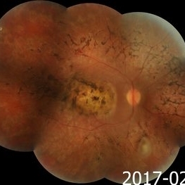 Macular Coloboma and Pigmentary Retinopathy
Feb 25 2017 by Hamid Ahmadieh, MD
Merged color fundus photograph of the right eye of a 25-year-old woman with the history of low vision since childhood. Bilateral macular colobomata and pigmentary retinopathy similar to Leber's congenital amaurosis are present.
Photographer: Shabnam Poureh, Negah Eye Center, Tehran, Iran
Condition/keywords: bilateral pigmentary retinopathy, color fundus photograph, macular coloboma, pigmentary retinal dystrophy
-
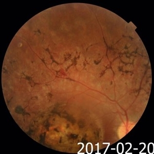
 Macular Coloboma and Pigmentary Retinopathy
Feb 25 2017 by Hamid Ahmadieh, MD
Color fundus photograph of the left eye of a 25-year-old woman with the history of low vision since childhood. Bilateral macular colobomata and pigmentary retinopathy similar to Leber's congenital amaurosis are present.
Photographer: Shabnam Poureh, Negah Eye Center, Tehran, Iran
Condition/keywords: bilateral pigmentary retinopathy, color fundus photograph, macular coloboma
-
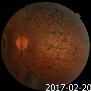
 Macular Coloboma and Pigmentary Retinopathy
Feb 25 2017 by Hamid Ahmadieh, MD
Color fundus photograph of the left eye of a 25-year-old woman with the history of low vision since childhood. Bilateral macular colobomata and pigmentary retinopathy similar to Leber's congenital amaurosis are present.
Photographer: Shabnam Poureh, Negah Eye Center, Tehran, Iran
Condition/keywords: bilateral pigmentary retinopathy, color fundus photograph, macular coloboma
-
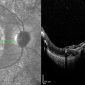 Macular Coloboma and Pigmentary Retinopathy
Feb 25 2017 by Hamid Ahmadieh, MD
Infrared and OCT images of the right eye of a 25-year-old woman with bilateral macular colobomata and pigmentary retinopathy similar to Leber's congenital amaurosis.
Photographer: Shabnam Poureh, Negah Eye Center, Tehran, Iran
Condition/keywords: infrared image, macular coloboma, optical coherence tomography (OCT)
-
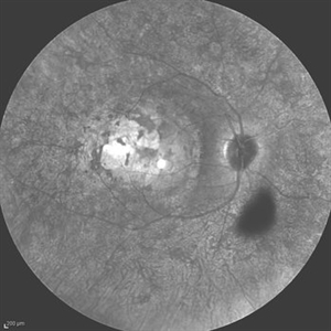 Leber's Congenital Amaurosis
Feb 25 2017 by Hamid Ahmadieh, MD
Infrared image of the right eye of a 25-year-old woman with bilateral macular colobomata and pigmentary retinopathy similar to Leber's congenital amaurosis.
Photographer: Shabnam Poureh, Negah Eye Center, Tehran, Iran
Condition/keywords: bilateral pigmentary retinopathy, infrared image, macular coloboma
 Loading…