Search results (136 results)
-
 T-Cell Lymphoma
Jul 3 2025 by Virginia Gebhart
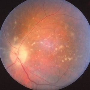
78 year old male s/p vitreous biopsy for T-Cell lymphoma. Pt presented with peripheral blot hemorrhages and numerous white subretinal infiltrates. Retinal pallor and thickening temporally. History of cutaneous T-cell lymphoma. PPV/vitreous biopsy performed to find differential diagnosis. Silicone oil was placed for 6 weeks, then removed and exchanged with a gas bubble. Hematology pathologist and Emory reviewed path report and agrees it is consistent with T-cell lymphoma. Pt received intravitreal Methotrexate and will be scheduled for weekly treatments. BCVA CF
Photographer: Virginia Gebhart, Retina Consultants of Carolina
Imaging device: Optos California
Condition/keywords: biopsy, gas bubble, lymphoma
-
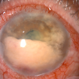
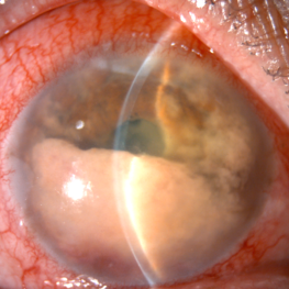
 Rosai-Dorfman Disease
Dec 4 2024 by Virginia Gebhart
72 year old female with temporal limbal lesion that extends onto the cornea from 10:00 - 8:00 encroaching on visual axis. Possible lymphomatous process. Will refer to Emory.
Photographer: Dr Chris Bergstrom MD, OD
Condition/keywords: corneal scars and opacities, Rosai-Dorfman Disease, Subconjunctival mass
-
 Benign Uveal Lymphoid Hyperplasia
Jan 24 2024 by Michell Goyal
Fundus photograph of woman with benign uveal lymphoid hyperplasia. The patient had no symptoms and tested 20/20 vision.
Condition/keywords: benign uveal lymphoid hyperplasia, lymphoid hyperplasia, Uveal Lymphoma
-
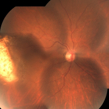 Primary intraocular lymphoma
Jul 21 2023 by AMIT NENE
Fundus photograph of 54year old female with primary intraocular lymphoma showing 'Leopard skin pigmentation'
Photographer: Dr.Amit S.Nene , Isha Netralaya, Thane,India
Condition/keywords: lymphoma
-
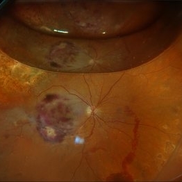
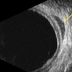 B-scan echography uveal lymphoma with orbital involvement
Jan 20 2023 by Elaine Michele Binkley, MD
B-scan echography (T10) shows a hypo-echoic mass posterior to the globe consistent with orbital involvement of uveal lymphoma.
Photographer: Laura Warner, University of Iowa
Condition/keywords: Uveal lymphoma
-
 Conjunctival involvement uveal lymphoma
Jan 20 2023 by Elaine Michele Binkley, MD
Slit lamp photograph shows characteristic "salmon-patch" conjunctival lesion in the setting of uveal marginal zone lymphoma with conjunctival involvement.
Photographer: Brice Critser, University of Iowa
Condition/keywords: Uveal Lymphoma
-
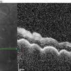 OCT Uveal Lymphoma
Jan 20 2023 by Elaine Michele Binkley, MD
OCT of a 71-year-old man with uveal marginal zone lymphoma shows characteristic "seasick" undulations with replacement of the choroid.
Photographer: Brice Critser
Condition/keywords: Uveal lymphoma
-
 Choroidal Lymphoma
Dec 10 2022 by Jordan D Deaner, MD
76-year-old man presented with progressive visual decline in his right eye found to have multifocal creamy yellow-white lesions in the choroid of the right eye. Indocyanine green angiography reveals numerous hypofluorescent lesions, many of which were subclinical on exam. He was eventually diagnosed with choroidal lymphoma.
Condition/keywords: Choroidal Lymphoma
-
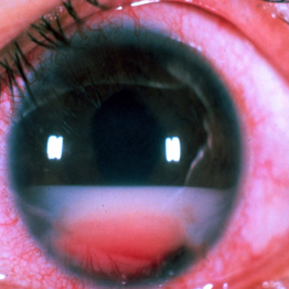
 Secondary intraocular lymphoma
Apr 11 2022 by Aniruddha K Agarwal, MD
A 65-year-old male underlying nasopharyngeal non-Hodgkin’s lymphoma presented with pseudo-hypopyon and infiltration of iris from tumor on his right eye. Aqueous tap showed atypical lymphocytes.
Photographer: Kessara Pathanapitoon MD, PhD Department of Ophthalmology, Faculty of Medicine Chiang Mai University, Chiang Mai, Thailand
Condition/keywords: lymphoma, masquerade syndrome, secondary iridocyclitis (noninfectious)
-
 Secondary intraocular lymphoma
Apr 11 2022 by Aniruddha K Agarwal, MD
A 65 year-old male underlying nasopharygeal non-Hodgkin’s lymhoma presented with pseudohypopyon and infiltration of iris from tumor on his right eye. Aqueous tap showed atypical lymphocytes.
Photographer: Kessara Pathanapitoon MD, PhD Department of Ophthalmology, Faculty of Medicine Chiang Mai University, Chiang Mai, Thailand
Condition/keywords: lymphoma, masquerade syndrome, secondary iridocyclitis (noninfectious)
-
 Subhyaloid Hemorrhage
Mar 1 2021 by Narciso F. Atienza, MD, MBA, FASRS, FPCS, FPAO.
Fundus photo of the left eye, 29-year-old male patient, with previous history of 6 cycles of chemotherapy from Hodgkins lymphoma. Photograph, however looks more like leukemic subhyaloid hemorrhage
Photographer: Narciso Atienza, Jr. MD, MBA. Legazpi Eye Center
Condition/keywords: Hodgkins lymphoma, subhyaloid hemorrhage
-
 Subhyaloid Hemorrhage
Mar 1 2021 by Narciso F. Atienza, MD, MBA, FASRS, FPCS, FPAO.
Fundus photo of the left eye, 29-year-old male patient, with previous history of 6 cycles of chemotherapy from Hodgkins lymphoma. Photograph, however looks more like leukemic subhyaloid hemorrhage.
Photographer: Narciso Atienza, Jr. MD, MBA. Legazpi Eye Center
Condition/keywords: Hodgkins lymphoma, subhyaloid hemorrhage
-
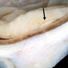 Primary Retinal and Vitreous Large B-Cell Lymphomas
May 18 2020 by McGill University Health Centre
These tumors are associated with intracranial nervous system lymphomas. The image shows the magnification of a nodule from the same specimen shows the thickening of the sensory retina. Note the infiltration of the subretinal pigment epithelium area (arrow).
Condition/keywords: large b cell lymphoma, large b cell lymphoma of the retina, lymphoma
-
 Primary Retinal and Vitreous Large B-Cell Lymphomas
May 18 2020 by McGill University Health Centre
These tumors are associated with intracranial nervous system lymphomas. The image shows an enucleation specimen with a multifocal, necrotic, and hemorrhagic whitish retinal tumor (arrow). Note the thickened, opaque cornea; the cataractous lens; the diffuse, flat retinal detachment; and the retinal hemorrhages overlying the tumor (arrowhead).
Condition/keywords: large b cell lymphoma of the retina, lymphoma
-
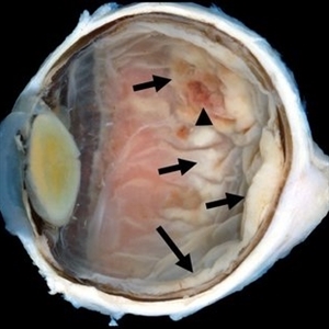
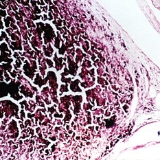
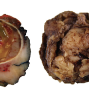 Uveal Tract Lymphoma
May 18 2020 by McGill University Health Centre
Uveal tract lymphoma most frequently occurs as a secondary location of systemic disease. The presence of a tumor in the choroid and ciliary body with an extensive orbital component may indicate a primary lymphoma or metastatic disease. In the image, the gross examination of a specimen shows a whitish, solid, and homogeneous tumor located at the posterior pole of the choroid and at the entire posterior orbit. Note that the tumor is also present in the ciliary body. In the second image, the tumor surrounds the optic nerve, mimicking a meningioma. In this patient, histopathological examination with immunohistochemistry confirmed the diagnosis of large B-cell lymphoma.
Condition/keywords: lymphoma
-
 Lymphoma Lesion at the Posterior Pole
Dec 27 2019 by Navneet Mehrotra, DNB
Fundus photograph of a 54-year-old female with posterior pole leopard skin lesion temporal to fovea. She underwent treatment for primary CNS lymphoma.
Photographer: Mitanshi
Imaging device: NIDEK MIRANTE
Condition/keywords: lymphoma, posterior pole lesion
-
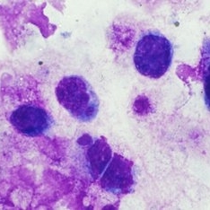
 Large B Cell Lymphoma of the Retina
Dec 13 2019 by McGill University Health Centre
65-year-old female with the clinical diagnosis of bilateral uveitis of unknown ethiology. Histopathology of a large B cell lymphoma in the anterior chamber, causing pseudohypopyon.
Photographer: Miguel N. Burnier, McGill University Health Center-McGill University Ocular Pathology & Translational Research Laboratory
Imaging device: Zeiss
Condition/keywords: large b cell lymphoma of the retina, masquerade syndrome, pseudohypopyon
-
 Large B Cell Lymphoma of the Retina
Dec 13 2019 by McGill University Health Centre
65-year-old female with the clinical diagnosis of bilateral uveitis of unknown etiology. Histopathology of the enucleated specimen showing large neoplastic cells in the anterior chamber, representing pseudohypopyon in a case of masquerade syndrome. The retina shows areas of necrosis with neoplastic large B cells.
Photographer: Miguel N. Burnier, McGill University Health Center-McGill University Ocular Pathology & Translational Research Laboratory
Imaging device: Zeiss
Condition/keywords: large b cell lymphoma, large b cell lymphoma of the retina, masquerade syndrome, pesudohypopyon
-
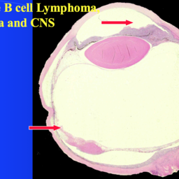
 Large B Cell Lymphoma of the Retina
Dec 13 2019 by McGill University Health Centre
65-year-old female with the clinical diagnosis of bilateral uveitis of unknown ethiology. The clinical picture shows a large pseudohypopyon, consistent with large B cell lymphoma of the retina, vitreous and CNS.
Photographer: Miguel N. Burnier, McGill University Health Center-McGill University Ocular Pathology & Translational Research Laboratory
Condition/keywords: large b cell lymphoma, pseudohypopyon, retina, uveitis, vitreous
-
 Primary intraocular Lymphoma
Nov 20 2019 by McGill University Health Centre
73-year-old man with retinal vasculitis and acute retinal lesions of the left eye. Vitrectomy specimen showing neoplastic cells with mitotic figures, consistent with large B cell lymphoma of the retina, vitreous and CNS.
Condition/keywords: large b cell lymphoma, primary intraocular lymphoma, vitrectomy
-
 Primary Intraocular Lymphoma
Nov 20 2019 by McGill University Health Centre
73-year-old man with retinal vasculitis and acute retinal lesions of the left eye. Optic nerve and retinal infiltrates consistent with acute retinal necrosis.
Condition/keywords: acute retinal necrosis, primary intraocular lymphoma
-
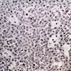 Slide 6-55
Mar 20 2019 by Lancaster Course in Ophthalmology
Burkitt's lymphoma. Undifferentiated, diffuse lymphosarcoma shows scattered macrophages engulfing nuclear debris, giving a "starry-sky" appearance to the tumor (H&E x252).
Condition/keywords: Burkitt's lymphoma, lymphosarcoma
-
 Slide 14-28
Mar 4 2019 by Lancaster Course in Ophthalmology
Other lesions mistaken for melanomas such as other tumors including hemangiomas, metastic tumors, melanocytoma of the disc, nevus of the choroid, hypertrophy of the pigment epithelium, adenoma and adenocarcinoma of the pigment epithelium, reactive proliferation of the pigment epithelium, and lymphoma and leukemia.
Condition/keywords: choroidal nevus, hemangioma, leukemia, lymphoma, melanoma, optic disc melanocytoma, retinal pigment epithelium (RPE) hypertrophy
-
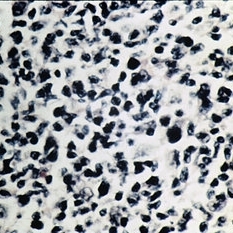 Slide 7-30
Feb 25 2019 by Lancaster Course in Ophthalmology
Pleomorphism, rather than polymorphism, characterizes a malignant lymphoma.
Condition/keywords: lymphoma, pleomorphism
 Loading…