Search results (136 results)
-
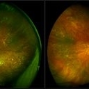
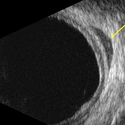 B-scan echography uveal lymphoma with orbital involvement
Jan 20 2023 by Elaine Michele Binkley, MD
B-scan echography (T10) shows a hypo-echoic mass posterior to the globe consistent with orbital involvement of uveal lymphoma.
Photographer: Laura Warner, University of Iowa
Condition/keywords: Uveal lymphoma
-
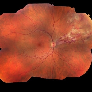
 Lymphoma Lesion at the Posterior Pole
Dec 27 2019 by Navneet Mehrotra, DNB
Fundus photograph of a 54-year-old female with posterior pole leopard skin lesion temporal to fovea. She underwent treatment for primary CNS lymphoma.
Photographer: Mitanshi
Imaging device: NIDEK MIRANTE
Condition/keywords: lymphoma, posterior pole lesion
-
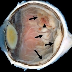 Primary Retinal and Vitreous Large B-Cell Lymphomas
May 18 2020 by McGill University Health Centre
These tumors are associated with intracranial nervous system lymphomas. The image shows an enucleation specimen with a multifocal, necrotic, and hemorrhagic whitish retinal tumor (arrow). Note the thickened, opaque cornea; the cataractous lens; the diffuse, flat retinal detachment; and the retinal hemorrhages overlying the tumor (arrowhead).
Condition/keywords: large b cell lymphoma of the retina, lymphoma
-
 Subhyaloid Hemorrhage
Mar 1 2021 by Narciso F. Atienza, MD, MBA, FASRS, FPCS, FPAO.
Fundus photo of the left eye, 29-year-old male patient, with previous history of 6 cycles of chemotherapy from Hodgkins lymphoma. Photograph, however looks more like leukemic subhyaloid hemorrhage.
Photographer: Narciso Atienza, Jr. MD, MBA. Legazpi Eye Center
Condition/keywords: Hodgkins lymphoma, subhyaloid hemorrhage
-
 Subhyaloid Hemorrhage
Mar 1 2021 by Narciso F. Atienza, MD, MBA, FASRS, FPCS, FPAO.
Fundus photo of the left eye, 29-year-old male patient, with previous history of 6 cycles of chemotherapy from Hodgkins lymphoma. Photograph, however looks more like leukemic subhyaloid hemorrhage
Photographer: Narciso Atienza, Jr. MD, MBA. Legazpi Eye Center
Condition/keywords: Hodgkins lymphoma, subhyaloid hemorrhage
-
 Primary Ocular Lymphoma
Jun 28 2012 by Suber S. Huang, MD, MBA, FASRS
Unilateral biopsy proven ocular lymphoma without systemic involvement. Father died of non-Hodgkins lymphoma. Older brother died of Hodgkin's lymphoma.
Photographer: Mark Harrod/Geoffrey Pankhurst, Case Western Reserve University/University Hospitals of Cleveland, Cleveland, OH
Imaging device: Topcon
-
 Cytomegalovirus Retinitis
Jan 16 2018 by Olivia Rainey
Color fundus montage of an 37-year-old, HIV positive male with CMV retinitis affecting his right eye. Patient's vision was sc20/20-1. He received an intravitreal Ganciclovir injection as well. The referring physcian suspects his condition is secondary to his chemotherapy for large B cell lymphoma or stomach cancer. The patient had not started taking oral Valgancyclovir.
Photographer: Olivia Rainey
Imaging device: Topcon 50dx
Condition/keywords: CMV retinitis, color fundus photograph, cytomegalovirus (CMV), HIV, montage
-
 Lymphoma
Sep 16 2012 by Ivan R. Batlle, MD
Color montage of 71-year-old female with non-Hodgkins lymphoma
Condition/keywords: non-Hodgkins lymphoma
-
 Ocular Lymphoma
Aug 22 2012 by Edwin H. Ryan, MD
Montage of 84-year-old man 12 years post vitrectomy-diagnosed intraocular lymphoma.
Photographer: Edwin Ryan Jr. MD, VitreoRetinal Surgery, PA
Condition/keywords: lymphoma
-
 choroidal lymphoma
Nov 25 2012 by Mallika Goyal, MD
Left eye of a 60-year-old lady shows multiple sub-retinal yellowish masses of choroidal lymphoma. Radiotherapy resulted in complete regression with recurrence after 10 months.
Photographer: Mallika Goyal, MD, Apollo Health City, Hyderabad, India
Condition/keywords: lymphoma
-
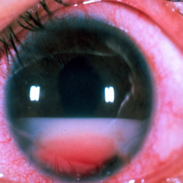 Large B Cell Lymphoma of the Retina
Dec 13 2019 by McGill University Health Centre
65-year-old female with the clinical diagnosis of bilateral uveitis of unknown ethiology. The clinical picture shows a large pseudohypopyon, consistent with large B cell lymphoma of the retina, vitreous and CNS.
Photographer: Miguel N. Burnier, McGill University Health Center-McGill University Ocular Pathology & Translational Research Laboratory
Condition/keywords: large b cell lymphoma, pseudohypopyon, retina, uveitis, vitreous
-
 Benign Uveal Lymphoid Hyperplasia
Jan 24 2024 by Michell Goyal
Fundus photograph of woman with benign uveal lymphoid hyperplasia. The patient had no symptoms and tested 20/20 vision.
Condition/keywords: benign uveal lymphoid hyperplasia, lymphoid hyperplasia, Uveal Lymphoma
-
 Bilateral Lymphoma Metastasis after Resolution with IVM
Sep 19 2018 by Olivia Rainey
Ultra-wide field, pseudocolor fundus images of an 86-year-old female treated with intravitreal methothrexate as a management of subretinal infiltrate in the macula of the right eye, as a manifestation of leukemia. Her last intravitreal methotrexate injection was 5/1/18.
Photographer: Olivia Rainey
Imaging device: Optos
Condition/keywords: bilateral, leukemia, methotrexate, Optos, pseudocolor, ultra-wide field imaging, uveitis
-
 Bilateral Lymphoma Metastasis after Resolution with IVM
Sep 19 2018 by Olivia Rainey
Ultra-wide field, autofluorescence images of an 86-year-old female treated with intravitreal methothrexate as a management of subretinal infiltrate in the macula of the right eye, as a manifestation of leukemia. Her last intravitreal methotrexate injection was 5/1/18.
Photographer: Olivia Rainey
Imaging device: Optos
Condition/keywords: bilateral, fundus autofluorescence (FAF), lymphoma, Optos, ultra-wide field imaging, uveitis
-
---thumb.JPG/image-square;max$300,300.ImageHandler) Choroidal Lymphoma
Choroidal Lymphoma
Nov 25 2012 by Mallika Goyal, MD
Left eye of a 60-year-old lady shows multiple sub-retinal yellowish masses. This is a recurrence 10 months following complete resolution of the lesions post-radiotherapy. Radiotherapy was repeated with regression of tumor.
Photographer: Mallika Goyal, MD, Apollo Health City, Hyderabad, India
-
---thumb.JPG/image-square;max$300,300.ImageHandler) choroidal lymphoma
choroidal lymphoma
Nov 25 2012 by Mallika Goyal, MD
Left eye of a 60-year-old lady shows areas of chorio-retinal atrophy corresponding to regression of choroidal lymphoma following external beam irradiation.
Photographer: Mallika Goyal, MD, Apollo Health City, Hyderabad, India
Condition/keywords: chorioretinal atrophy, lymphoma
-
---thumb.JPG/image-square;max$300,300.ImageHandler) choroidal lymphoma
choroidal lymphoma
Nov 25 2012 by Mallika Goyal, MD
Left eye of a 60-year-old lady shows multiple sub-retinal yellowish masses of choroidal lymphoma. Radiotherapy resulted in complete regression with recurrence after 10 months.
Photographer: Mallika Goyal, MD, Apollo Health City, Hyderabad, India
Condition/keywords: lymphoma
-
---thumb.JPG/image-square;max$300,300.ImageHandler) Choroidal Lymphoma
Choroidal Lymphoma
Nov 25 2012 by Mallika Goyal, MD
Right eye of a 60-year-old lady shows multiple sub-retinal yellowish masses. This is a recurrence 10 months following complete resolution of the lesions post-radiotherapy. Radiotherapy was repeated with regression of tumor.
Photographer: Mallika Goyal, MD, Apollo Health City, Hydeabad, India
-
 Choroidal Lymphoma
Dec 10 2022 by Jordan D Deaner, MD
76-year-old man presented with progressive visual decline in his right eye found to have multifocal creamy yellow-white lesions in the choroid of the right eye. Indocyanine green angiography reveals numerous hypofluorescent lesions, many of which were subclinical on exam. He was eventually diagnosed with choroidal lymphoma.
Condition/keywords: Choroidal Lymphoma
-
---thumb.JPG/image-square;max$300,300.ImageHandler) Choroidal lymphoma in remission following radiotherapy
Choroidal lymphoma in remission following radiotherapy
Nov 25 2012 by Mallika Goyal, MD
Left eye of a 60-year-old lady shows areas of chorio-retinal atrophy corresponding to regression of choroidal lymphoma following external beam irradiation.
Photographer: Mallika Goyal, MD, Apollo Health City, Hyderabad, India
Condition/keywords: lymphoma
-
 Conjunctival involvement uveal lymphoma
Jan 20 2023 by Elaine Michele Binkley, MD
Slit lamp photograph shows characteristic "salmon-patch" conjunctival lesion in the setting of uveal marginal zone lymphoma with conjunctival involvement.
Photographer: Brice Critser, University of Iowa
Condition/keywords: Uveal Lymphoma
-
 CSCR with PED Defect
Sep 3 2018 by John S. King, MD
Acute symptoms; HTN retinopathy and recent high BP readings; Hodgkins lymphoma (stable > 10 yrs), diabetic.
Photographer: Stacey Coleman
Imaging device: Cirrus
Condition/keywords: central serous chorioretinopathy (CSCR), pigment epithelial detachment (PED)
-
 CSCR with PED Defect
Sep 3 2018 by John S. King, MD
Acute symptoms; no steroids; HTN retinopathy and recent high BP readings; Hodgkins lymphoma (stable > 10 yrs), diabetic.
Photographer: Stacey Coleman
Imaging device: Cirrus
Condition/keywords: central serous chorioretinopathy (CSCR), pigment epithelial detachment (PED)
 Loading…