Search results (73 results)
-
 Angiographic Storm: Fluorescein Leakage in Retinal Vasculitis
Nov 17 2025 by SHRADDHA RAJ SHRIVASTAVA
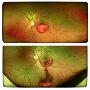
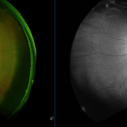
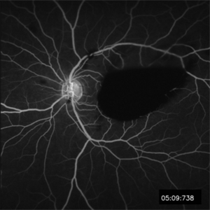
This left eye montage fundus fluorescein angiography (FFA) image of a 19 year old male with idiopathic retinal vasculitis, having skip vasculitic lesions predominantly involving retinal veins. There are areas of blocked fluorescence due to intraretinal hemorrhages, the involved veins have filling defects and occlusions, leading to formation of numerous collateral channels. The inflamed vessels also show perivascular fuzzy hyperfluorescent stain due to leakage of dye. We can also see multiple peripheral capillary non perfusion (CNP) areas, with a 'hot disc', suggestive of ongoing inflammation.
Photographer: Dr. Shraddha Raj Shrivastava
Imaging device: Nidek Mirante SLO/OCT (Confocal scanning/Spectral domain OCT)
Condition/keywords: FA late phase leakage, Fundus Fluorescein Angiography, idiopathic retinal vasculitis, optic disc leakage, VASCULITIS
-
 YAG Laser Hyaloidotomy
Aug 31 2025 by Giriraj Vibhute
A 24-year-old young man presented with sudden loss of vision in left eye following history of rigorous coughing. Visual acuity in RE was 6/6, LE was 6/60p. Fundoscopy showed bilateral multiple small intraretinal hemorrhages with LE large premacular subhyaloid hemorrhage just covering the fovea suggestive of bilateral valsalva retinopathy changes. Nd:YAG laser hyaloidotomy was performed to left eye the same day (A250; 2mJ;6 SHOTS). Visual acuity improved to 6/9 immediately following the procedure. After 1 week, the subhyaloid hemorrhage had completely cleared with dispersed intragel hemorrhage in the inferior vitreous cavity with visual acuity of 6/6 in left eye
Photographer: Dr Vani S. MM Joshi eye institute, Hubli
Condition/keywords: valsalva retinopathy, YAG HYALOIDOTOMY
-
 Commotio Retinae
Aug 7 2025 by Gabriel Costa Andrade, PhD
Color fundus photograph of a 13-year-old girl who was hit by accidental discharge of gel bullet in her right eye. She presented with retinal whitening with intraretinal hemorrhages in temporal inferior area of the peripheral retina.
Photographer: Gabriel Andrade
Condition/keywords: macula, Retina, Trauma
-
 Diabetic Retinopathy
Jun 4 2025 by Paulina Araujo
The 55-degree central fundus photograph of the right eye demonstrates numerous hard exudates, dot intraretinal hemorrhages, and microaneurysms.
Photographer: Paulina D.Araujo Martínez, Asociación para Evitar la Ceguera en México I.A.P., Hospital Dr Luis Sánchez Bulnes.
Condition/keywords: diabetic retinopathy
-
 Macular Edema
Jun 4 2025 by Paulina Araujo
The composite fundus photograph of the right eye demonstrates circinate hard exudates in the thickened macular area, along with flame-shaped intraretinal hemorrhages along the inferior temporal arcade.
Photographer: Paulina D.Araujo Martínez, Asociación para Evitar la Ceguera en México I.A.P., Hospital Dr Luis Sánchez Bulnes.
Condition/keywords: macular edema
-
 Retinal Macroaneurysm (Left Eye)
Apr 29 2025 by Daniela Bogenschutz
72 year-old female has visual complaints of central vision changes ongoing for 4 days. Patient was acutely symptomatic with an intraretinal hemorrhage due to the retinal macroaneurysm. We had a fun little laugh as this retinal macroaneurysm form a shape of a tick in her left eye. This photo is a side-by-side of the color photos and the autofluorescence done. She is being treated by her general doctor for elevated blood pressure.
Photographer: Daniela Bogenschutz, OSC; Retina Consultants of Carolina, P.A.
Imaging device: Optos
Condition/keywords: retinal macroaneurysm
-
 Coat's Disease with Exudative RD
Feb 12 2025 by Tejaswita Verma
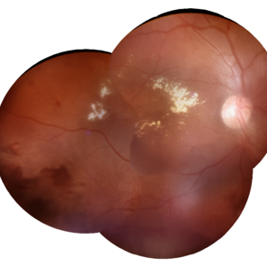
Fundus photo of a 7 year old boy with vision Counting fingers close to face in the right eye and intermittent outward deviation of the right eye observed by parents. Fundus examination shows subretinal exudates, telengiectatic vessels in superotemporal quadrant, intraretinal hemorrhages, macular scar, NVD.
Photographer: DR. TEJASWITA VERMA
Imaging device: MIRANTE
Condition/keywords: Coats' disease, exudative retinal detachment
-
 Leukemic Retinopathy
Nov 27 2024 by Ramses Rosales-Diaz
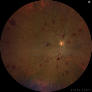
Fundus photograph of a 48-year-old woman showing venous dilatation and tortuosity, flame-shaped hemorrhages, intraretinal hemorrhages, sub-ILM hemorrhages, and Roth spots. Her complete blood count shows 425,540 lymphocytes/microliter, and the blood smear reveals Gumprecht shadows along with numerous lymphocytes with hypercondensed chromatin in their nuclei. She is diagnosed with chronic lymphocytic leukemia and receives appropriate treatment from the hematology team.
Photographer: Ramses Rosales-Diaz, Asociación Para Evitar la Ceguera en México
Imaging device: Zeiss Clarus 700
Condition/keywords: leukemia, sub ILM hemorrhage, white centered retinal hemorrhage (Roth Spot)
-
 Leukemic Retinopathy
Nov 27 2024 by Ramses Rosales-Diaz
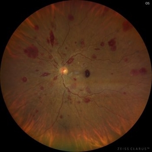
Fundus photograph of a 48-year-old woman showing venous dilatation and tortuosity, flame-shaped hemorrhages, intraretinal hemorrhages, sub-ILM hemorrhages, and Roth spots. Her complete blood count shows 425,540 lymphocytes/microliter, and the blood smear reveals Gumprecht shadows along with numerous lymphocytes with hypercondensed chromatin in their nuclei. She is diagnosed with chronic lymphocytic leukemia and receives appropriate treatment from the hematology team.
Photographer: Ramses Rosales-Diaz, Asociación Para Evitar la Ceguera en México
Imaging device: Zeiss Clarus 700
Condition/keywords: leukemia, sub ILM hemorrhage, white centered retinal hemorrhage (Roth Spot)
-
 Leukemic Retinopathy
Nov 27 2024 by Ramses Rosales-Diaz
Fundus photograph of a 48-year-old woman with venous dilatation and tortuosity, flame-shaped and intraretinal hemorrhages, Roth spots and sub-ILM hemorrhage. Her complete blood count reports 425,540 lymphocytes/microliter, and the blood smear reveals Gumprecht shadows and numerous lymphocytes with nuclei exhibiting hypercondensed chromatin. She is diagnosed with chronic lymphocytic leukemia and receives appropriate treatment from the hematology team
Photographer: Ramses Rosales-Diaz, Asociación Para Evitar la Ceguera en México
Imaging device: Clarus 700
Condition/keywords: leukemia, sub ILM hemorrhage, white centered retinal hemorrhage (Roth Spot)
-
 Valsalva Retinopathy
Oct 28 2024 by Andrew Jin, MD
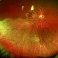
These photos depict the fundus photo and corresponding fluorescein angiogram for a 43 year old man with emesis after food poisoning. Note the blockage from the central preretinal hemorrhage and scattered peripheral intraretinal hemorrhages.
Condition/keywords: fluorescein angiogram (FA), fundus photograph, valsalva retinopathy
-
 Valsalva Retinopathy
Oct 28 2024 by Andrew Jin, MD
These photos depict the fundus photo and corresponding fluorescein angiogram for a 43 year old man with emesis after food poisoning. Note the blockage from the central preretinal hemorrhage and scattered peripheral intraretinal hemorrhages.
Condition/keywords: fluorescein angiogram (FA), fundus photograph, valsalva retinopathy
-
 Congenital Retinal Macrovessel
Oct 13 2023 by Jacob D. Grodsky, MD
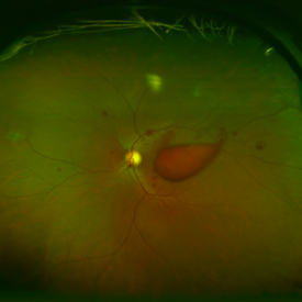
41 y/o male who presented with acute onset of blurred vision OD. Visual acuity was 20/200 OD; 20/25 OS. Examination was consistent with congenital retinal macrovessel through the macula with intraretinal hemorrhage as seen in the fundus photo. Intravitreal bevacizumab was injected, and visual acuity improved to 20/40 at 4-week follow-up. MRA head and neck was ordered to rule out other vascular anomalies.
Condition/keywords: congenital retinal macrovessel, RETINAL MACROVESSEL
-
 Idiophatic retinal vasculitis
Jul 9 2023 by Luiz A Zago, PhD
Idiophatic retinal vasculitis in a 45 year old woman
Photographer: Luiz Zago, PhD.
Imaging device: Topcon 50IX
Condition/keywords: diffuse vasculitis, intraretinal hemorrhage, optic disc leakage
-
 Idiophatic vasculitis OD
Jul 9 2023 by Luiz A Zago, PhD
Idiophatic vasculitis
Photographer: Luiz Zago, PhD.
Imaging device: Topcon 50IX
Condition/keywords: intraretinal hemorrhage, leaky parafoveal capillaries, optic disc leakage, vasculitis
-
 Broken macroaneurysm
Nov 27 2022 by Nassim Alejandro Abreu Arbaje, MD
Fundus video frame of a 58 year old male who had a PPV on his left eye because a retinal macroaneurysm that broke a bled on all 3 retinal planes.
Photographer: Nassim Abreu, Hospital Dr. Elías Santana
Imaging device: NGenuity 3D system
Condition/keywords: broken macroanerysm, intraretinal hemorrhage, macroaneurysm, subretinal hemorrhage, vitreous hemorrhage
-
 Diabetic Retinopahty
Nov 2 2022 by pedro fernandes souza neto
Fundus photograph of a 40-year-old man with diabetes and hypertension shows hard exudates, difuse intraretinal hemorrhages and splinter hemorrhages.
Photographer: Pedro Fernandes, Universidade Federal da Bahia, Brazil.
Condition/keywords: diabetic mellitus, hypertensive retinopathy, retinopathy
-
 Intraocular Foreign Body
Jul 18 2022 by Nelson Chamma Capelanes, MD
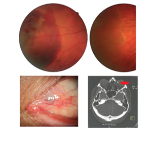
Intraocular foreign body after stone trauma. Foreign body is found in the choroid. - Fundus image on the upper left: one day after the trauma showing subretinal and intraretinal hemorrhage - Fundus image on the upper right: 40 days after laser photocoagulation. - Lower left image: 30 days after the trauma, showing part of the foreign body in the nasal region. - Lower right image showing CT scan and intraocular foreign body location.
Photographer: Nelson Chamma Capelanes, Promacula Indaiatuba, Brazil
Imaging device: Canon CX-2
Condition/keywords: intraocular foreign body
-
 Waldenstrom Macroglobulinemia
Mar 9 2022 by Austen N Knapp, MD
Ultra-widefield fundus photograph of a 67-year-old woman with peripheral intraretinal hemorrhages in the setting of Waldenstrom macroglobulinemia.
Condition/keywords: hyperviscosity retinopathy, intraretinal hemorrhages, Waldenstrom Macroglobulinemia
-
 Waldenstrom macroglobulinemia
Mar 9 2022 by Austen N Knapp, MD
Ultra-widefield fundus photograph of a 67-year-old woman with peripheral intraretinal hemorrhages in the setting of Waldenstrom macroglobulinemia.
Condition/keywords: hyperviscosity retinopathy, intraretinal hemorrhages, waldenstrom macroglubinemia
-
 Choroidal MRSA Abscess
Apr 15 2021 by Rui Zhang, BA
A 14-year-old boy receiving induction chemotherapy for acute lymphocytic leukemia (ALL) complained of floaters and central scotoma in his left eye. (A) Fundus photography showed sub-macular choroidal abscess with intraretinal hemorrhage and edema. (B) OCT confirmed that the abscess had not penetrated the retinal pigment epithelium (RPE). Due to systemic septic signs (fever, tachycardia, tachypnea, new-onset papules), blood cultures were drawn and they came back positive for methicillin-resistant staphylococcus aureus (MRSA). Patient was promptly treated with both IV and intravitreal antibiotics. This is a case of sub-macular choroidal MRSA abscess in the setting of systemic bacteremia in an immunocompromised host.
Photographer: Raymond Mok, CRA COMT (Dartmouth-Hitchcock Medical Center)
Imaging device: Optical coherence tomography
Condition/keywords: abscess, acute leukemia, MRSA sepsis
-
 Progressive Outer Retinal Necrosis
Nov 5 2019 by Nichole Lewis
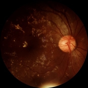
86-year-old male with progressive outer retinal necrosis, significant retinitis, retinal whitening, intraretinal hemorrhages and peripheral rpe changes. FA showed occlusive vasculitis with non-perfusion. Patient is immuno-suppressed with a history of renal transplant. VA 20/60.
Photographer: Nichole Lewis
Imaging device: Optos
Condition/keywords: intraretinal hemorrhage, occlusive vasculitis, progressive outer retinal necrosis (PORN), retinal pigment epithelium (RPE) changes, retinal whitening, retinitis
-
 Ruptured Macroaneurysm
May 22 2019 by Nichole Lewis
FA of a 91-year-old woman with a ruptured macroaneurysm, intraretinal hemorrhage and subretinal hemorrhage. VA 20/400.
Photographer: Nichole Lewis
Condition/keywords: intraretinal hemorrhage, ruptured macroaneurysm, subretinal hemorrhage
-
 Penetrating Trauma with Retinal Detachment
Apr 30 2019 by Olivia Rainey
Ultra-wide field pseudocolor image of a 39-year-old female with penetrating trauma resulting in a retinal detachment with an intraretinal hemorrhage affecting the left eye. Patient was struck with a champagne glass in October of 2018, which lacerated the eyelid and globe. Patient was "seeing red" when she first came to the office and after multiple surgeries she was seeing 20/20 at her last check in April 2019.
Photographer: Olivia Rainey
Imaging device: Optos
Condition/keywords: hemorrhage, left eye, Optos, penetrating trauma, ruptured globe, ultra-wide field imaging
-
 Multiple Myeloma
Apr 1 2019 by Gary R. Cook, MD, FACS
62-year-old white female with multiple cotton wool spots and intraretinal hemorrhages OD secondary to multiple myeloma; V.A. = 20/60+1.
Imaging device: Topcon VT-50
Condition/keywords: blot hemorrhages, cotton wool spots, myeloma, retinal hemorrhage, retinopathy
 Loading…