 Initializing download.
-
 By Nelson Chamma Capelanes, MD
Promedica Indaiatuba / UPO Oftalmologia
Co-author(s): Caio Regatieri - Uploaded on Jul 18, 2022.
- Last modified by Joshua Friedman on Jul 19, 2022.
- Rating
- Appears in
- Miscellaneous
- Condition/keywords
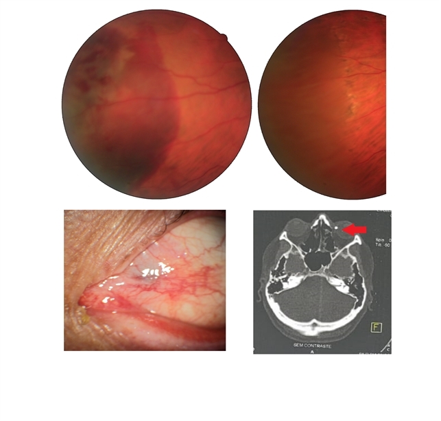
- intraocular foreign body
- Photographer
- Nelson Chamma Capelanes, Promacula Indaiatuba, Brazil
- Imaging device
-
Fundus camera
Canon CX-2 - Description
- Intraocular foreign body after stone trauma. Foreign body is found in the choroid. - Fundus image on the upper left: one day after the trauma showing subretinal and intraretinal hemorrhage - Fundus image on the upper right: 40 days after laser photocoagulation. - Lower left image: 30 days after the trauma, showing part of the foreign body in the nasal region. - Lower right image showing CT scan and intraocular foreign body location.


