 Initializing download.
-
 By John S. King, MD
Retina Associates, PA
Co-author(s): Kent Zocchi, MD - Uploaded on Jun 1, 2019.
- Last modified by Jennifer Carstens on Nov 23, 2021.
- Rating
- Appears in
- Presumptive FAME
- Condition/keywords
- macular edema, Gilenya, cystoid macular edema (CME)
- Photographer
- Kay Dalby
- Imaging device
-
Optical coherence tomography system
Cirrus - Description
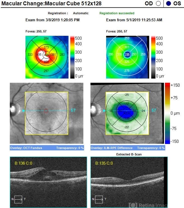
- 60 year old caucasian female with two week history of decreased vision in the left eye. Background history includes multiple sclerosis for which she uses Gileyna for the past five years, and no history of uveitis or recent MS relapse. Her vision in the left eye was 20/100 J5. The eye appeared overall "quiet" with the exception of rare cells in the anterior vitreous. The fundus appearance and FA can be seen in the images provided. OCT shows CME and SRF in OS only (left image). STK was administered and neurologist was able to discontinue the Gileyna. STK has been reported to be effective in FAME in patients who continue Gileyna (see below). 10 days later the CME had decreased significantly. 8 weeks later the edema had resolved as seen in the OCT images of the initial and latest appearance. Of note, this is a late presentation for FAME, and there was some rare debris in the anterior vitreous; it is possible, although no history of uveitis and the MS was inactive, that the CME may be related to other causes like uveitis (if there is recurrence of CME while patient is off Gileyna, then further work-up will be performed) Minuk A, Belliveau MJ, Almeida DR, Dorrepaal SJ, Gale JS. Fingolimod-associated macular edema: resolution by sub-tenon injection of triamcinolone with continued fingolimod use. JAMA ophthalmol 2013; 131(6): 802–804.

")
")
")
")
")
")


