Search results (171 results)
-
 ERMageddon - Wrinkle in the Space-time Fabric of Macula
Oct 29 2025 by SHRADDHA RAJ SHRIVASTAVA
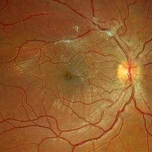
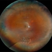
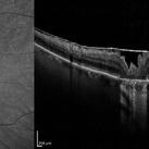
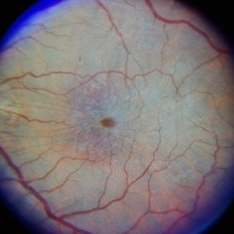
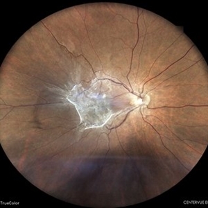
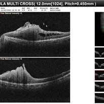
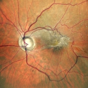
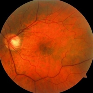
38 year old female with Epiretinal Membrane (ERM) over macula, post laser barrage for multiple symptomatic Horse-shoe Tears (HSTs) and Lattice Degenerations (seen on wide-field image). Posterior pole revealed tilted disc with peripapillary atrophy. There is thick opaque epiretinal membrane obscuring the underlying superior arcade vessels and causing foveal ectopia with distortion of perimacular vasculature. Patient was planned for Right Eye pars plana vitrectomy for ERM peeling.
Photographer: Dr. Shraddha Raj Shrivastava
Imaging device: Nidek Mirante SLO/OCT (Confocal scanning/Spectral domain OCT
Condition/keywords: BARRAGE LASER, ectopic fovea, epiretinal membrane (ERM), horseshoe tear, lattice degeneration, vitreomacular traction (VMT)
-
 ERMageddon - Wrinkle in the Space-time Fabric of Macula
Oct 29 2025 by SHRADDHA RAJ SHRIVASTAVA
38 year old female with Epiretinal Membrane (ERM) over macula, post laser barrage for multiple symptomatic Horse-shoe Tears (HSTs) and Lattice Degenerations. Posterior pole revealed tilted disc with peripapillary atrophy. There is thick opaque epiretinal membrane obscuring the underlying superior arcade vessels and causing foveal ectopia with distortion of perimacular vasculature. Patient was planned for Right Eye pars plana vitrectomy for ERM peeling.
Photographer: Dr. Shraddha Raj Shrivastava
Imaging device: Nidek Mirante SLO/OCT (Confocal scanning/Spectral domain OCT
Condition/keywords: ectopic fovea, epiretinal membrane (ERM), ERM, horseshoe tear, vitreomacular traction (VMT)
-
 Epiretinal Membrane
Aug 13 2025 by Ricardo Leitão Guerra
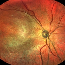
Idiopatic epiretinal membrane
Photographer: Ricardo Leitão Guerra
Imaging device: ZEISS CLARUS 700
Condition/keywords: epiretinal membrane (ERM)
-
 Lattice Degeneration With Atrophic Retinal Holes
Jan 30 2025 by Kimberly Wakester
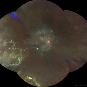
Ultra-wide field montage fundus photograph of a 56-year-old woman with lattice degeneration with atrophic holes statues post laser. Patient also has a small CHRPE temporal to macula and trace ERM that is not visually significant. Will continue follow up care to monitor and treat as needed.
Photographer: Kimberly Wakester, COA
Imaging device: Optos California
Condition/keywords: atrophic retinal hole, CHRPE, epiretinal membrane (ERM), lattice degeneration, montage photo
-
 Photic Retinopathy
Jan 30 2025 by Juan Alberto Olivera Cueva
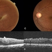
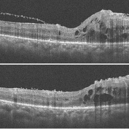
A 23-year-old male with a history of direct exposure to sunlight on several occasions, presenting blurred vision, comes for evaluation due to metamorphopsias of 3 months' evolution. The fundus photograph shows the presence of an epiretinal membrane. The OCT shows a hyperreflective line at the vitreomacular interface that causes traction to the layers of the inner retina, as well as distortion in the architecture of the macular region, with the presence of subfoveal detachment of the RPE.
Photographer: Dr. Juan Alberto Olivera Cueva, Escuela Militar de Medicina, Hospital Militar de Especialidades Oftalmológicas
Condition/keywords: epiretinal membrane (ERM)
-
 Branch Retinal Vein Occlusion with Multifactorial Macular Edema and Epiretinal Membrane
Oct 3 2024 by Logan ryzenga
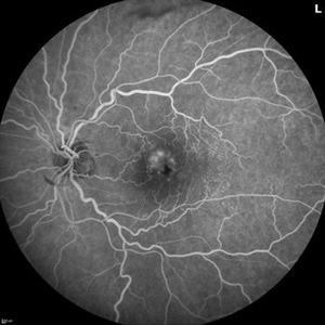
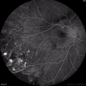
Fluorescein angiogram of a 62 year old woman with cystoid macular edema from concurrent Epiretinal Membrane and Branch Retinal Vein occlusion. She has an extensive history of anti-VEGF injections with stable but unresolved macular edema. Following angiography, it was determined that an epiretinal membrane peel would be indicated in an attempt to achieve resolution of macular edema.
Photographer: Logan Ryzenga
Imaging device: Heidelberg Spectralis
Condition/keywords: 55-degrees, branch retinal vein occlusion (BRVO), cystoid macular edema (CME), epiretinal membrane (ERM), Fluorescein angiography, heidelberg spectralis, hyperfluorescence, leakage, left eye, OS, wide angle imaging
-
 Epiretinal Membrane
Jan 30 2024 by Akansha Sharma
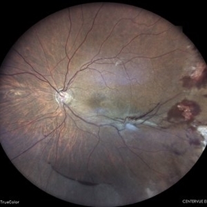
Color fundus photograph of a 65 year old hypertensive male with an epiretinal membrane in a case of old branch retinal vein occlusion with a subhyaloid hemorrhage seen inferiorly.
Photographer: Dr. Akansha Sharma, Bharati Eye Hospital
Condition/keywords: epiretinal membrane (ERM), ERM
-
 Bullseye Maculopathy
Jan 22 2024 by Kali Jend
Optical coherence tomography of a 73-year-old female with Bullseye Macular Changes affecting her left eye. Patient reports having a family history of this condition and denies prior Plaquenil or Elmiron use. Compared to previous imaging, the patient's condition progressed in the left eye from 2020 to 2023. Patient has a history of fluctuating Diabetic Macular Edema and a current Epiretinal Membrane as well. Patient's vision was Ncc20/60 at the time the image was taken.
Photographer: Kali Jend
Imaging device: Heidelberg Spectralis
Condition/keywords: bullseye maculopathy, epiretinal membrane (ERM), heidelberg spectralis, left eye, macular pucker, OCT, optical coherence tomography (OCT)
-
 Epiretinal membrane with pseudo-hole
Sep 12 2023 by Ben Serar
Fundus photograph of LE showing epiretinal membrane with pseudohole.
Condition/keywords: epiretinal membrane (ERM)
-
 EPIRETINAL MEMBRANE
Jun 6 2023 by Akansha Sharma
COLOUR FUNDUS PHOTOGRAPH OF A 40 YAER OLD MALE WITH EPIRETINAL MEMBRANE FORMATION
Photographer: Dr. Akansha Sharma, Dr. Denish Patel, Dr. Urmil Shah,
Condition/keywords: epiretinal membrane (ERM), ERM
-
 ERM with Retinal Striae
Apr 13 2023 by Virginia Gebhart
Right eye fundus photo of 65-year-old male with severe ERM with retinal striae s/p bilateral RD repair
Photographer: Virginia Gebhart, Retina Consultants of Carolina
Imaging device: Topcon TRC 50DX
Condition/keywords: epiretinal membrane (ERM), retinal strial
-
 Epiretinal Membrane causing Macular Pucker.
Dec 11 2022 by Anjana Mirajkar, MS Ophthalmology
OCT of LE in a 65 year old male case of Epiretinal Membrane causing Macular Pucker.
Photographer: Dr. Anjana Mirajkar -Retina Foundation, Ahmedabad.
Condition/keywords: epiretinal membrane (ERM), macular pucker
-
 Epiretinal Membrane causing Macular Pucker.
Dec 11 2022 by Anjana Mirajkar, MS Ophthalmology
Color Photo of LE in a 65 year old male case of Epiretinal Membrane causing Macular Pucker.
Photographer: Dr. Anjana Mirajkar -Retina Foundation, Ahmedabad.
Condition/keywords: epiretinal membrane (ERM), macular pucker
-
 Uveitis
Nov 25 2022 by Filip Kecer
Multicolor composite of an 15 year old boy with cyclitis posterior, first diagnosed with uveitis intermedia retinoschisis o.dx., since then he underwent several procedures, such as PPV, ILM peeling, capsulectomy, retinotomy, transscleral cryo
Photographer: Filip Kecer, National Institute of Childrens Diseases
Imaging device: Spectralis, Heidelberg Engineering
Condition/keywords: cyclitis, epiretinal membrane (ERM), uveitis
-
Epiretinal membrane and ILM peeling
Oct 24 2022 by Manish Nagpal, MD, FRCS (UK), FASRS
This video shows the technique of peeling a epiretinal membrane after triamcinilone staining followed by ILM removal.
Photographer: Manish Nagpal
Condition/keywords: epiretinal membrane (ERM), ILM, macular pucker, staining, video, vitrectomy
-
 Coats Disease Fluorescein Angiography
Sep 2 2022 by FLOR ANGELICA JACOME GUTIERREZ
Fluorescein angiography of a patient with Coats disease where we found telangiectatic vessels, aneurysms, peripheral capillary nonperfusion and perivascular leak.
Photographer: Dr.Guillermo Salcedo Villanueva
Imaging device: Zeiss CLARUS 700 (FA)
Condition/keywords: Coats' disease, epiretinal membrane (ERM)
-
 Coats disease
Sep 2 2022 by FLOR ANGELICA JACOME GUTIERREZ
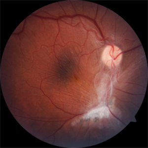
Fundus image of a 14 yo male with coats disease stage 2A and extensive epiretinal membrane. VA 20/80.
Photographer: Dr. Guillermo Salcedo Villanueva
Imaging device: Zeiss Clarus 700
Condition/keywords: Coats' disease, epiretinal membrane (ERM), exudates
-
 Epiretinal Membrane
May 14 2022 by Rinat Sutiushev
Female, born in 1961. Complains of decreased vision and distortion when reading text. The ocular fundus showed retinal surface wrinkling due to membrane contracture.
Photographer: Rinat Sutiushev
Condition/keywords: Cellophane Maculopathy, epiretinal membrane (ERM), macular pucker
-
 Spontaneous Regression of Idiopathic Epiretinal Membrane
Jan 13 2022 by Nasiq Hasan, MD
Fundus photograph and OCT of a 32-year-old man with idiopathic ERM which regressed after 2 years.
Photographer: Nasiq Hasan, Dr Agarwal's eye hospital, Tirunelveli
Imaging device: Zeiss FF4
Condition/keywords: epiretinal membrane (ERM)
-
Macular Hematoma Secondary Valsalva Maneuver
Oct 14 2021 by Islam bechakh
A 32-year-old man, who has presented for 02 months, a macular hematoma secondary to a Valsalva maneuver. He benefited from an attempt to open the hematoma with a Yag laser, but to no avail. We operated on and performed a 23G vitrectomy with posterior vitreous detachment, and discovered an epiretinal membrane which separated the hematoma from the posterior hyaloid. After removal of this membrane and aspiration of red blood cells and fibrin, the macula regained a normal appearance with good functional recovery.
Photographer: Islam Bechakh
Condition/keywords: epiretinal membrane (ERM), ERM, Macular hematoma, Valsalva maneuver
-
 Post Retinal Reattachment Surgery Epiretinal Membrane
Sep 14 2021 by Ogugua Ndubuisi Okonkwo, MD, FRCS (Edin), FASRS
Postoperative optical coherence tomography (OCT) of the right eye in a 65-year-old male who had retinal reattachment surgery for a macular hole retinal detachment. This OCT scan shows epiretinal membrane and intraretinal cystic fluid spaces.
Photographer: Oreoluwa Olabode , Eye Foundation Hospital, Lagos.
Imaging device: Optovue Avanti RTVue.
Condition/keywords: epiretinal membrane (ERM), macular hole retinal detachment, Retinal Reattachment surgery
-
 Epiretinal Membrane
Sep 6 2021 by Ricardo Leitão Guerra
65-year-old woman with an asymptomatic ERM (BCVA=20/20).
Imaging device: Zeiss Clarus 700
Condition/keywords: epiretinal membrane (ERM)
-
 Combined Hamartoma of Retina and Retinal Pigment Epithelium
Apr 30 2021 by ARVIND JAIN M
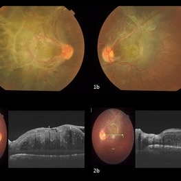
A 26-year-old gentlemen came with complains of defective vision in both eyes since childhood. His BCVA was right eye 5/60 and left eye 6/60. His anterior segment examination showed no abnormality with posterior segment examination showed both eyes (1a and 1b) greyish white elevated lesion involving the macula with thick fibrotic epiretinal membrane causing the macular drag temporally in right eye and supero-temporally in left eye. (2a and 2b) showing the thick ERM with the hamartoma of the retina and RPE.
Photographer: DR ARVIND JAIN, ARAVIND EYE HOSPITAL, COIMBATORE,INDIA
Condition/keywords: combined hamartoma, congenital hypertrophy of the retinal pigment epithelium (CHRPE), epiretinal membrane (ERM), retinal pigment epithelium (RPE) hamartoma
-
 Macular Traction Related to Toxoplasma Chorioretinitis
Jan 7 2021 by Lucas Zago Ribeiro, MD
Fundus image of a 50-year-old woman with macular traction and epiretinal membrane after toxoplasma chorioretinitis.
Photographer: Lucas Zago Ribeiro, UNIFESP / EPM, Brazil
Condition/keywords: epiretinal membrane (ERM), toxoplasmosis
 Loading…