Search results (94 results)
-
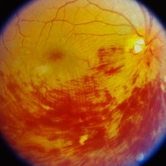
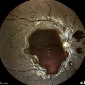
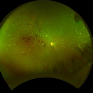
 Lower Temporal Branch Retinal Vein Occlusion
Sep 16 2025 by Seif Allah Anwar
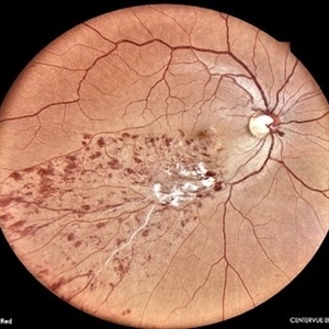
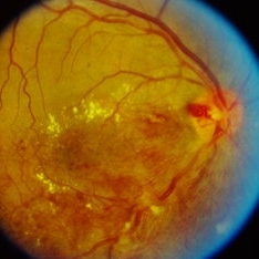
Fundus photograph of a 46-year old hypertensive male patient showing sheathed lower temporal retinal vein with whitish cotton wool spots and hemorrhages ( dots, blots and flame shaped) along the area drained by the obstructed vein with vein.
Photographer: Dr Seif Anwar. FRCSEd
Imaging device: CENTERVUE EIDON
Condition/keywords: Lower temporal branch retinal vein occlusion
-
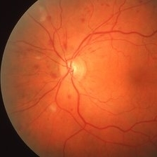
 Myelinated Nerve Fibers
Apr 18 2025 by DR Rohit Gupta
The **myelinated nerve fibers of the optic disc** (also known as **medullated nerve fibers**) are retinal nerve fibers that retain their myelin sheath as they pass through the optic nerve head. Normally, retinal nerve fibers are unmyelinated to allow for light transparency, but in some cases, myelination extends anteriorly into the retina, appearing as a striking white, feathery patch on the optic disc or peripapillary retina. ### **Key Features:** 1. **Appearance:** - Dense, white, striated patches with feathery edges. - Typically located at the superior or inferior pole of the optic disc. - May obscure retinal vessels underneath. 2. **Clinical Significance:** - Usually **benign** and asymptomatic. - **Congenital** (present at birth or early childhood). - Rarely associated with **visual field defects** (e.g., scotomas corresponding to the area of myelination). - Occasionally linked with **high myopia** or **amblyopia** if extensive. 3. **Pathophysiology:** - Failure of oligodendrocytes or Schwann cells to stop myelination at the lamina cribrosa. - Normally, myelination stops at the optic nerve head, but in this condition, it extends into the retina. 4. **Diagnosis:** - **Fundoscopy:** Classic white, feathery appearance. - **Optical Coherence Tomography (OCT):** Shows thickened retinal nerve fiber layer (RNFL). - **Visual Field Testing:** May detect defects if large. 5. **Differential Diagnosis:** - Optic disc edema - Cotton wool spots - Retinoblastoma (rarely, but must be ruled out in children) 6. **Management:** - No treatment required if asymptomatic. - Monitor for amblyopia in children. - Rare cases with significant visual impairment may need further evaluation. ### **Fun Fact:** Myelinated nerve fibers are seen in **~0.5-1%** of the population and are usually an incidental finding.
Photographer: Dr Rohit gupta
Imaging device: Samsung S21
Condition/keywords: Medulated Nerve fibre, Medullated Nerve fibres, myelinated nerve fibers, Myelinated Nerve Fibres, optic disc drusen
-
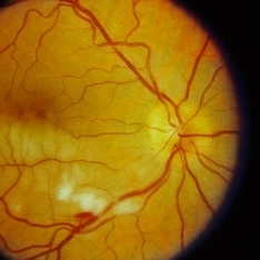
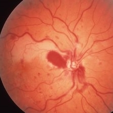
 PDR with NVD
Dec 5 2024 by Tejaswita Verma
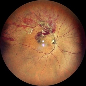
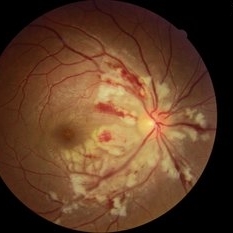
Fundus image of a middle aged male with NVD, multiple dot blot and flame shaped hemorrhages, cotton wool spots, hard exudates at the posterior pole in a case of PDR . Vision was 6/9.
Photographer: DR. TEJASWITA VERMA
Imaging device: MIRANTE
Condition/keywords: NEOVASCULARISATION OF DISC, proliferative diabetic retinopathy (PDR)
-
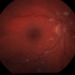
 Proliferative Diabetic Retinopathy
May 24 2024 by Anjana Mirajkar, MS Ophthalmology
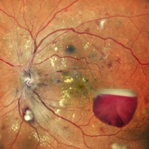
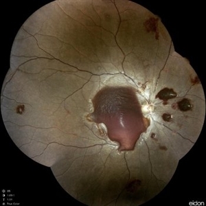
A central photo of a 50 year old male case of PDR showing a sub-hyaloid hemorrhage with cotton wool spots , hard exudates at the fovea with dot and blot hemorrhages.
Photographer: Dr. Anjana Mirajkar -Retina Foundation, Ahmedabad
Imaging device: Mirante-Nidek
Condition/keywords: proliferative diabetic retinopathy (PDR)
-
 Branch Retinal Vein Occlusion
Apr 28 2024 by Anjana Mirajkar, MS Ophthalmology
A widefield color photo of a 55 year old male case of supero-temporal BRVO showing venous tortuosity, cotton wool spots, flame shaped hemorrhages and macular edema.
Photographer: Dr. Anjana Mirajkar -Retina Foundation, Ahmedabad
Imaging device: Mirante-Nidek
Condition/keywords: branch retinal vein occlusion (BRVO), ST BRVO
-
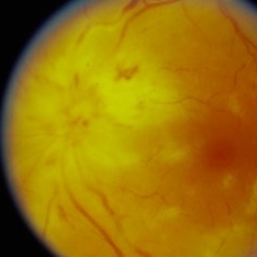
 Florid Disc Edema Hemorrhage
Apr 1 2024 by Dr Venkatasubramaniam G
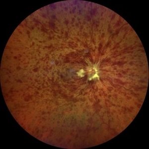
Fundus photograph of a 55 yr old male with Florid disc edema Hemorrhage and dense cotton wool spots with ischemia.
Photographer: Venkatsubramaniam
Imaging device: Remedio fundus on phone
Condition/keywords: Central vein oclussion, Diabetic Retinopathy
-
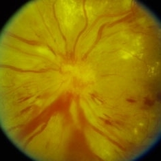
 Florid Disc Edema Hemorrhage
Apr 1 2024 by Dr Venkatasubramaniam G
Fundus photograph of a 55 yr old male with Florid disc edema Hemorrhage and dense cotton wool spots with ischemia.
Photographer: Venkatasubramaniam
Imaging device: Remedio fundus on phone
Condition/keywords: Central vein oclussion, diabetic retinopathy
-
 Central Retinal Vein Occlusion associated with disc edema
Oct 19 2023 by Gabriel Costa Andrade, PhD
53-year-old woman with an acute CRVO. The patient has a history of breast cancer undergoing treatment with systemic chemotherapy. Notice the peripapillary cotton wool spots, superficial flame shaped hemorrhages and deeper dot and blot hemorrhages in all 4 quadrants.
Photographer: Gabriel Andrade
Condition/keywords: central retinal vein occlusion (CRVO), macular edema, Retina
-
 Branch Retinal Vein Occlusion (BRVO)
Sep 26 2023 by Ben Serar
Fundus Photograph of RE showing superficial retinal haemorrhages ,with cotton wool spots, along the inferotemporal vessel arcade, in a case of inferotemporal branch retinal vein occlusion.
Condition/keywords: branch retinal vein occlusion (BRVO)
-
 Branch Retinal Artery Occlusion (BRAO)
Sep 21 2023 by Ben Serar
Fundus photograph of RE showing retinal edema and opacification along the inferotemporal vessel arcade, with cotton wool spots and flame shaped haemorrhage, in a case of Branch Retinal Artery Occlusion (BRAO).
Condition/keywords: branch retinal artery occlusion (BRAO)
-
 Disc edema
Sep 12 2023 by Ben Serar
Fundus photograph showing Disc edema, with splinter haemorrhages and cotton wool spots.
Condition/keywords: disc edema, splinter haemorrhages
-
 Disc edema
Sep 12 2023 by Ben Serar
Fundus photograph showing Disc edema, with splinter haemorrhages and cotton wool spots.
Condition/keywords: disc edema, splinter haemorrhages
-
 Branch Retinal Vein Occlusion (BRVO)
Sep 12 2023 by Ben Serar
Fundus Photograph of RE showing superficial retinal haemorrhages, with exudates at the macula, with cotton wool spots, with venous engorgement along the inferotemporal vessel arcade, in a case of inferotemporal branch retinal vein occlusion.
Condition/keywords: branch retinal vein occlusion (BRVO)
-
 Branch Retinal Artery Occlusion (BRAO)
Sep 12 2023 by Ben Serar
Fundus photograph of the LE showing arterial occlusion along the inferotemporal vessel arcade with surrounding retinal edema and cotton-wool spots, in a case of Branch Retinal Artery Occlusion (BRAO).
Condition/keywords: branch retinal artery occlusion (BRAO), cotton wool spots, retinal edema
-
 Retinal Lupus Vasculitis
Sep 25 2021 by Denis Jusufbegovic, MD
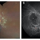
27-year-old woman with a history of systemic lupus erythematosus (SLE) presented with decreased vision to counting fingers at 2’ in the right eye. Funduscopic examination of the right eye (A) demonstrated retinal thickening and whitening of the macula, numerous cotton-wool spots, intra-retinal hemorrhages, sclerotic vessels, and vascular sheathing. Fluorescein angiography (B) demonstrated extensive vessel wall leakage (red asterisk) and large areas of capillary non-perfusion (white asterisk). These findings were consistent with severe vaso-occlusive retinopathy, a serious ophthalmologic manifestation of SLE. She was also diagnosed with concomitant cerebral lupus vasculitis. She was treated with intravenous methylprednisolone followed by oral prednisone taper and aspirin therapy. Mycophenolate mofetil was titrated to 1500 mg twice daily. Upon follow-up vision improved to 20/200.
Imaging device: Zeiss Clarus 500
Condition/keywords: cerebral lupus vasculitis, cotton wool spots, systemic lupus erythematosus (SLE) vasculitis, vaso-occlusive disease
-
 Right Eye-Post Seg
Aug 10 2020 by RITESH VERMA
Fundus photograph of a 27-year-old male with multiple cotton wool spots and hemorrhages in NF layer 6 days post roadside accident.
Photographer: Dr. Ritesh Verma, Regional institute of Ophthalmology, Rohtak, Haryana, India
Imaging device: CR-2AF CANON
Condition/keywords: cotton wool spots, hemorrhage
-
 Anemic Retinopathy Related Retinal Hemorrhages
Nov 5 2019 by Chinmayi Vyas
Anemic retinopathy related retinal hemorrhages in a 24 years old male with Hb of 4.2gm/ dl. The manifestations of anemic retinopathy are nonspecific and may closely simulate hypertensive or diabetic retina. Retinal changes in anemia are cotton wool spots, venous tortuosity, and hemorrhages which may be present at all levels of the retina and choroid. All retinal hemorrhages can occur when Hb falls below 8 g/100 ml or if the platelet count falls below 50,000/cumm. The combination of severe anemia and thrombocytopenia is likely to produce retinal hemorrhages. The Roth’s spots or white centre hemorrhages are typically associated with bacterial endocarditis , anemia and other systemic conditions. The white center is suspected to represents focal ischemia, inflammatory or infectious infiltrate, fibrin or accumulation of neoplasticism cells.
Photographer: Dr Chinmayi Vyas, Nethradhama superspeciality eye hospital , banglore, india
Imaging device: Eidon fundus imaging
Condition/keywords: anaemic retinopathy
-
 Anemic Retinopathy Related Retinal Hemorrhages
Nov 5 2019 by Chinmayi Vyas
Anemic retinopathy related retinal hemorrhages in a 24 years old male with Hb of 4.2gm/ dl. The manifestations of anemic retinopathy are nonspecific and may closely simulate hypertensive or diabetic retina. Retinal changes in anemia are cotton wool spots, venous tortuosity, and hemorrhages which may be present at all levels of the retina and choroid. All retinal hemorrhages can occur when Hb falls below 8 g/100 ml or if the platelet count falls below 50,000/cumm. The combination of severe anemia and thrombocytopenia is likely to produce retinal hemorrhages. The Roth’s spots or white centre hemorrhages are typically associated with bacterial endocarditis , anemia and other systemic conditions. The white center is suspected to represents focal ischemia, inflammatory or infectious infiltrate, fibrin or accumulation of neoplasticism cells.
Photographer: Dr Chinmayi Vyas
Condition/keywords: retinal hemorrhage
-
 Multiple Myeloma
Apr 1 2019 by Gary R. Cook, MD, FACS
62-year-old white female with multiple cotton wool spots and intraretinal hemorrhages OD secondary to multiple myeloma; V.A. = 20/60+1.
Imaging device: Topcon VT-50
Condition/keywords: blot hemorrhages, cotton wool spots, myeloma, retinal hemorrhage, retinopathy
-
 Combined CRVO and BRAO
Mar 27 2019 by Gary R. Cook, MD, FACS
Right eye of a 56-year-old white male with a combined perfused CRVO (venous dilation and dot & blot hemorrhages in all 4 quadrants) and a superotemporal BRAO with peripapillary hemorrhages and cotton wool spots, and an area of retinal whitening inside of the ST arcade. V.A.= 20/70.
Imaging device: Topcon VT-50
Condition/keywords: branch retinal artery occlusion (BRAO), central retinal vein occlusion (CRVO)
-
 Multiple Myeloma
Mar 26 2019 by Gary R. Cook, MD, FACS
62-year-old white female with multiple CWS and intraretinal hemorrhages OS secondary to multiple myeloma; VA= 20/20-2.
Imaging device: Topcon VT-50
Condition/keywords: blot hemorrhages, cotton wool spots, myeloma, retinal hemorrhage
-
 Cotton Wool Spots
Aug 23 2018 by Nichole Lewis
Cotton Wool Spots
Photographer: Nichole Lewis
Condition/keywords: cotton wool spots
-
 SLE Retinopathy
Jul 10 2018 by Deepak Bhojwani, MS
Colour fundus montage image of a 33-year-old young lady with history of Systemic Lupus Erythematosus of 6 years showing classic SLE retinopathy with multiple cotton wool spots , few haemorrhages and multiple small vessel sheathing s/o SLE vasculitis.
Photographer: Deepak Bhojwani
Condition/keywords: systemic lupus erythematosus (SLE) retinopathy, systemic lupus erythematosus (SLE) vasculitis
-
 Lupus Hemorrhagic Occlusive Vasculitis
Apr 23 2018 by Frank Chin
Fundus photograph of the right eye of a 24-year-old woman with history of systemic lupus erythematosus who presented with decreased visual acuity for 2-3 days found to have lupus hemorrhagic occlusive vasculitis with mild disc elevation, diffuse punctate cotton wool spots and dot blot hemorrhages, and a hemorrhage occlusive vasculitis along the superior branch of the superotemporal arcade involving the artery and vein.
Photographer: Frank Chin, MD, George Washington University
Imaging device: Optos 200Tx
Condition/keywords: blot hemorrhages, cotton wool spots, occlusive vasculitis, systemic lupus erythematosus (SLE) vasculitis
-
 Hypertensive Retinopathy
Oct 19 2017 by Nichole Lewis
Fundus photography of a 41-year-old male with hypertensive retinopathy, disc edema, cotton wool spots, subretinal hemorrhage and lipid. BP 136/79
Photographer: Nichole Lewis
Condition/keywords: cotton wool spots, disc edema, hypertensive retinopathy, lipid, subretinal hemorrhage
 Loading…