Search results (50 results)
-
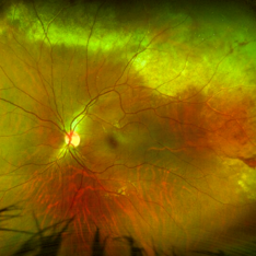
 Intraocular Foreign Body Scleral Lac
Nov 19 2025 by Nikhil Das, M.D.
A 34-year-old man presented with a right intraocular foreign body after hammering a carbon-steel chisel 12 hours after injury. CT orbits showed a 3-mm hyperattenuating foreign body within the right globe, centered in the vitreous cavity. BCVA was 20/40. Anterior segment examination revealed a 2.8-mm scleral laceration. DFE demonstrated a metallic IOFB, a superior air bubble, superior commotio retinae, and Berlin’s edema involving the macula.
Photographer: Nikhil Das, Saint Louis University School of Medicine
Imaging device: iPhone
Condition/keywords: intraocular foreign body, iofb, metallic foreign body, scleral laceration
-
 Intraocular Foreign Body CT Coronal
Nov 19 2025 by Nikhil Das, M.D.
A 34-year-old man presented with a right intraocular foreign body after hammering a carbon-steel chisel 12 hours after injury. CT orbits showed a 3-mm hyperattenuating foreign body within the right globe, centered in the vitreous cavity. BCVA was 20/40. Anterior segment examination revealed a 2.8-mm scleral laceration. DFE demonstrated a metallic IOFB, a superior air bubble, superior commotio retinae, and Berlin’s edema involving the macula.
Photographer: Nikhil Das, Saint Louis University School of Medicine
Imaging device: CT Scan
Condition/keywords: intraocular foreign body, iofb, metallic foreign body, scleral laceration
-
 Intraocular Foreign Body CT Axial
Nov 19 2025 by Nikhil Das, M.D.
A 34-year-old man presented with a right intraocular foreign body after hammering a carbon-steel chisel 12 hours after injury. CT orbits showed a 3-mm hyperattenuating foreign body within the right globe, centered in the vitreous cavity. BCVA was 20/40. Anterior segment examination revealed a 2.8-mm scleral laceration. DFE demonstrated a metallic IOFB, a superior air bubble, superior commotio retinae, and Berlin’s edema involving the macula.
Photographer: Nikhil Das, Saint Louis University School of Medicine
Imaging device: CT Scan
Condition/keywords: intraocular foreign body, iofb, metallic foreign body, scleral laceration
-
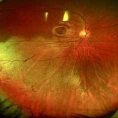
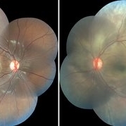
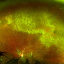
 Intraocular Foreign Body
Nov 19 2025 by Nikhil Das, M.D.
A 34-year-old man presented with a right intraocular foreign body after hammering a carbon-steel chisel 12 hours after injury. CT orbits showed a 3-mm hyperattenuating foreign body within the right globe, centered in the vitreous cavity. BCVA was 20/40. Anterior segment examination revealed a 2.8-mm scleral laceration. DFE demonstrated a metallic IOFB, a superior air bubble, superior commotio retinae, and Berlin’s edema involving the macula.
Photographer: Nikhil Das, Saint Louis University School of Medicine
Condition/keywords: intraocular foreign body, metallic foreign body, scleral laceration
-
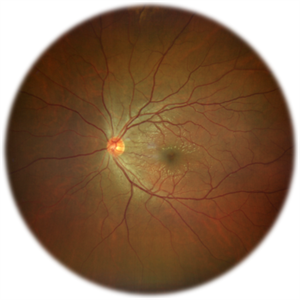
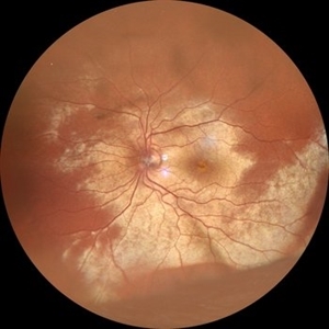
 Commotio Retinae
Aug 7 2025 by Gabriel Costa Andrade, PhD
Color fundus photograph of a 13-year-old girl who was hit by accidental discharge of gel bullet in her right eye. She presented with retinal whitening with intraretinal hemorrhages in temporal inferior area of the peripheral retina.
Photographer: Gabriel Andrade
Condition/keywords: macula, Retina, Trauma
-
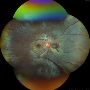
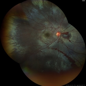
 Commotio Retinae
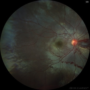
Jun 10 2025 by CUI YUELING
The patient presented 2 hours after sustaining a left eye injury caused by a stick. Visual acuity in the left eye was 0.2 without improvement upon correction, and intraocular pressure measured 15 mmHg. Examination of the anterior segment revealed ciliary conjunctival injection accompanied by patchy subconjunctival hemorrhage. The corneal surface remained smooth, and the anterior chamber was deep with hyphema characterized by blood-tinged aqueous humor predominantly settled inferiorly. The pupil was slightly irregular, approximately 3 mm in diameter, with a superotemporal notch; pupillary light reflex was intact. The lens appeared clear. Fundus examination showed well-defined optic disc margins with normal coloration and a cup-to-disc ratio of 0.2. Retinal arteries and veins were normally distributed with an artery-to-vein ratio of 2:3. At the posterior pole, the foveal reflex exhibited concentric ripple-like changes centered on the fovea, accompanied by localized pigment attenuation and reduced reflex intensity. Irregular reflectivity was noted in the superotemporal and inferotemporal nerve fiber layers.
Photographer: Yueling Cui
Imaging device: Zeiss Clarus 500
Condition/keywords: commotio retinae
-
 Firework Injury
Feb 13 2025 by Virginia Gebhart
44 year old male presented New Year's Day for trauma after fireworks injury. Choroidal rupture temporal macula, inferior vitreous hemorrhage, and extensive RPE changes in the macula. Significant improvement since initial presentation. Limited central vision, guarded prognosis due to extensive blunt trauma.
Photographer: Virginia Gebhart, Retina Consultants of Carolina
Imaging device: Optos California
Condition/keywords: blunt trauma, choroidal rupture, commotio retinae, firework injury, secondary glaucoma, subretinal hemorrhage, VH, vitreous hemorrhage
-
 Choroidal-rupture
Jan 2 2024 by Tahsin Khundkar, MD
37-year-old male with blunt ocular trauma presented with a choroidal rupture, pre -retinal and sub-retinal heme, and a heart shaped patch of commotio retinae.
Photographer: Jeffrey Zeigler, Concord Eye Center
Imaging device: Topcon
Condition/keywords: Choroidal Rupture, commotio retinae, Trauma
-
 Choroidal Rupture
Sep 30 2023 by Jacob D. Grodsky, MD
24 year old female who presented after being hit in the head with a metal softball bat after an altercation. The patient reported blurred vision as well as a zig-zag line described as a “lightning strike” across her vision. Examination was significant for a choroidal rupture OD as well as commotio retinae OU.
Condition/keywords: choroidal rupture, commotio retinae, trauma
-
 Berlin's edema
Sep 21 2023 by Vaidehi Sathaye
Widefield photograph of LE of a 28 year male with Berlin's edema, status post blunt trauma with tennis ball
Photographer: Dr. Vaidehi Sathaye
Imaging device: Mirante
Condition/keywords: Berlin's edema, commotio retinae
-
 Berlin's edema
Sep 21 2023 by Vaidehi Sathaye
Fundus photograph of LE of a 28 year male with Berlin's edema, status post blunt trauma with tennis ball
Photographer: Dr. Vaidehi Sathaye
Imaging device: Mirante
Condition/keywords: Berlin's edema, commotio retinae
-
 Blunt Trauma with Berlin's Edema
Aug 21 2023 by Harsh Vardhan Singh, MS
A 13-year-old male with history of trauma with cricket ball presented with inferior dialysis with commotio retinae & retinal hemorrhage
Photographer: Harsh Vardhan Singh, Iva R Kalita
Condition/keywords: Berlin's edema, blunt trauma, commotio retinae
-
 Blunt Trauma with Berlin's Edema
Aug 21 2023 by Harsh Vardhan Singh, MS
A 13-year-old male with history of trauma with cricket ball presented with inferior dialysis with commotio retinae & retinal hemorrhage
Photographer: Harsh Vardhan Singh
Condition/keywords: Berlin's edema, blunt trauma, commotio retinae
-
 BERLIN'S EDEMA
Nov 21 2022 by Akansha Sharma
COLOUR FUNDUS PHOTOGRAPH OF A 35 YEAR OLD MALE WITH BERLIN'S EDEMA STATUS POST FIRE-CRACKER INJURY
Photographer: Dr. Akansha Sharma-Retina Foundation, Ahmedabad
Condition/keywords: Berlin's edema, commotio retinae, firework injury
-
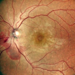
 Commotio retinae
Apr 29 2022 by Otakar Dušek, M.D. Ph.D.
Color fundus photograph of a 24-year-old woman who was hit by a volleyball in her right eye. This caused whitening of the lower peripheral retina (Berlin's edema) i.e. commotio retinae.
Photographer: Otakar Dušek, Charles University, Prague
Imaging device: Zeiss Clarus
Condition/keywords: Berlin's edema, blunt trauma, commotio retinae
-
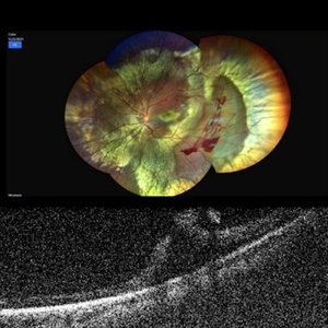 Traumatic Retinal Tear
Jan 20 2022 by Aditya S Kelkar, MS, FRCS, FASRS,FRCOphth
Color fundus photograph of a 34-year old man's left eye, 2 hours after a tennis ball injury, showing commotio retinae with Berlin's edema and cherry red spot in the fovea along with linear retinal tears in the temporal equatorial zone. Adjoining OCT slice taken through the break shows full thickness retinal tear without any underlying choroidal rupture.
Photographer: Dr Sukanya Mondal, National Institute of Ophthalmology, Pune. India
Imaging device: Zeiss Clarus 500
Condition/keywords: Berlin's edema, cherry red spot, commotio retinae, retinal tear
-
 Traumatic Retinal Tear
Dec 5 2021 by Aditya S Kelkar, MS, FRCS, FASRS,FRCOphth
Color fundus photograph of a 34-year old man's left eye, 2 hours after a tennis ball injury, showing commotio retinae with Berlin's edema and cherry red spot in the fovea along with linear retinal tears in the temporal equatorial zone.
Photographer: Dr Sukanya Mondal. National Institute of Ophthalmology, Pune. India.
Imaging device: Zeiss Clarus 500
Condition/keywords: Berlin's edema, cherry red spot, commotio retinae, retinal tear
-
 Commotio-Retinae
Sep 22 2021 by Luiz Guilherme Freitas, MD, MsC, PhD
Fundus photograph of a 30-year-old male patient with blunt injury to the globe. Commotio retinae is retinal whitening/opacification that results from a blunt injury. The ocular findings will often resolve in a matter of days to weeks. Vision loss can result from commotio involving the posterior pole (historically referred to as Berlin’s edema). Clinical findings of commotio include the characteristic retinal whitening. Commotio may result in significant vision loss that can be transient. Healing can result in pigmentary changes and retinal thinning which may be associated with poor visual recovery if the area of involvement is macular.
Photographer: Diogo Melo, Santa Luzia Eye Hospital Recife - PE – Brazil
Condition/keywords: Berlin's edema, blunt trauma, commotio retinae, retinal whitening
-
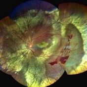
 Commotio Retinae
Apr 27 2021 by Priya Rasipuram Chandrasekaran, MBBS, DO, DNB, FRCS
This is the fundus photo montage of a 11-year-old boy showing extensive commotio retinae evidenced by the absence of greyish opacification of the retina with the absence of the usual red reflex following blunt trauma to the left eye. There is no pseudo cherry red spot. The right eye has been added for comparison.
Condition/keywords: commotio retinae
-
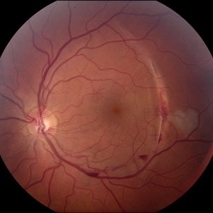
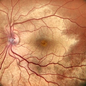
 Massive Commotio Retinae
Oct 20 2020 by Veronika Yehezkeli
Fundus photograph of a 24-year-old male, made after blunt trauma with a plastic bottle. Note massive commotio retinae and preretinal hemorrhages in the contralateral to trauma area.
Photographer: Veronika Yehezkeli, Meir medical center, Israel
Condition/keywords: blunt trauma, commotio retinae, preretinal hemorrhage, trauma
-
 Massive Commotio Retinae
Oct 20 2020 by Veronika Yehezkeli
Fundus photograph of a 24-year-old male, made after blunt trauma with a plastic bottle. Note massive commotio retinae and preretinal hemorrhages in the contralateral to trauma area.
Photographer: Veronika Yehezkeli, Meir medical center, Israel
Condition/keywords: blunt trauma, commotio retinae, preretinal hemorrhage
-
 Massive Commotio Retinae
Oct 20 2020 by Veronika Yehezkeli
24-year-old man was injured from an explosion of a plastic bottle towards the nasal conjunctiva of his left eye. A massive commotio retinae was diagnosed superotemporally.
Photographer: Veronika Yehezkeli, Meir medical center, Israel
Condition/keywords: blunt trauma, commotio retinae, preretinal hemorrhage
-
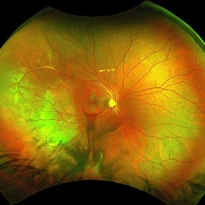
 Blunt Ocular Trauma Due to Firework Injury
Jun 9 2020 by Brittany Rota
Ultra- widefield pseudocolor image of an 18-year-old male with blunt ocular trauma in the right eye due to a firework injury. The patient presented with commotio retinae (sclopteria), an acute vitreous hemorrhage, choroidal rupture, and a subretinal hemorrhage. The referring physician performed surgery on the lateral rectus muscle which was macerated but not severed, and several orbital fibrous foreign bodies were removed from the posterior orbit. The globe was intact. There is no evidence of retinal tear in the region of sclopetaria; however, there is complete necrosis of the temporal peripheral choroid and retina. The vitreous hemorrhage was slowly clearing on his exam 6-9-2020. The patient is developing subretinal fibrosis. The physician is concerned about the choroidal rupture that is visible through the submacular hemorrhage. There is one rupture that appears to course directly under the fovea. The physician states that if this is the case, his vision most likely will be 20/200 or worse. His vision was hand motion in all fields except nasally, which he was unable to see hand motion at his visit on 6-9-2020.
Photographer: Brittany Rota
Imaging device: Optos California
Condition/keywords: blunt trauma, choroidal rupture, commotio retinae, fibrosis, firework injury, fundus photograph, hand motion, necrotizing retina, Optos, pseudocolor, subretinal hemorrhage, vitreous hemorrhage
 Loading…