Search results (87 results)
-
 The Great Disc-guise
Nov 12 2025 by SHRADDHA RAJ SHRIVASTAVA
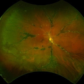
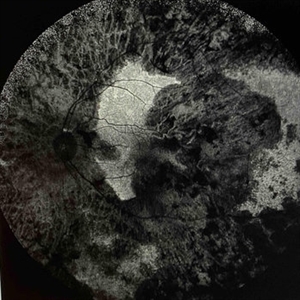
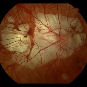
Right eye pseudocolor fundus photo of a 20 year old with Both eyes Pathological Myopia (spherical refractive error of - 18.00 DS in BE), showing a tilted myopic disc with peripapillary atrophy, and extensive posterior staphyloma baring the underlying choroidal vessels and scleral tissue. We can also see a well-defined round chorioretinal atrophic (CRA) patch superonasal to the disc, giving the illusion of double disc on cursory fundus examination.
Photographer: Dr. Shraddha Raj Shrivastava
Imaging device: Nidek Mirante SLO/OCT (Confocal scanning/Spectral domain OCT)
Condition/keywords: chorioretinal atrophy, High Myopia, pathologic myopia, peripapillary atrophy, posterior staphyloma
-
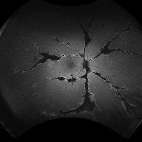
 Pigmented Paravenous Retinochoroidal Atrophy (PPRCA)
Jun 30 2025 by Maria Letícia Costa Holanda
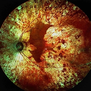
Fundoscopy of a 42-year-old asymptomatic man with pigmented paravenous chorioretinal atrophy. Pigmented paravenous retinochoroidal atrophy (PPRCA) is a rare disorder of unknown etiology. The disease is characterized by pigment accumulation along the distribution of retinal veins. The findings are usually incidental with minimal effect on vision.
Photographer: Guilherme da Cruz Reis, CLINOS Eye Hospital - Feira de Santana (BA),Brazil
Condition/keywords: pigmented paravenous chorioretinal atrophy (PPCRA)
-
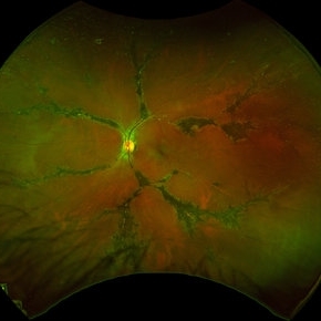
 Pigmented Paravenous Retinochoroidal Atrophy (PPRCA)
Jun 27 2025 by Maria Letícia Costa Holanda
Fundoscopy of a 42-year-old asymptomatic man with pigmented paravenous chorioretinal atrophy. Pigmented paravenous retinochoroidal atrophy (PPRCA) is a rare disorder of unknown etiology. The disease is characterized by pigment accumulation along the distribution of retinal veins. The findings are usually incidental with minimal effect on vision.
Photographer: Guilherme da Cruz Reis, CLINOS Eye Hospital - Feira de Santana (BA),Brazil
Condition/keywords: pigmented paravenous chorioretinal atrophy (PPCRA)
-
 Pigmented Paravenous Retinochoroidal Atrophy (PPRCA)
Jun 27 2025 by Maria Letícia Costa Holanda
Fundoscopy of a 42-year-old asymptomatic man with pigmented paravenous chorioretinal atrophy. Pigmented paravenous retinochoroidal atrophy (PPRCA) is a rare disorder of unknown etiology. The disease is characterized by pigment accumulation along the distribution of retinal veins. The findings are usually incidental with minimal effect on vision.
Photographer: Guilherme da Cruz Reis, CLINOS Eye Hospital - Feira de Santana (BA),Brazil
Condition/keywords: Pigmented Paravenous Retinochoroidal Atrophy
-
 Pigmented Paravenous Retinochoroidal Atrophy (PPRCA)
Jun 27 2025 by Maria Letícia Costa Holanda
Fundoscopy of a 42-year-old asymptomatic man with pigmented paravenous chorioretinal atrophy. Pigmented paravenous retinochoroidal atrophy (PPRCA) is a rare disorder of unknown etiology. The disease is characterized by pigment accumulation along the distribution of retinal veins. The findings are usually incidental with minimal effect on vision.
Photographer: Guilherme da Cruz Reis, CLINOS Eye Hospital - Feira de Santana (BA),Brazil
Condition/keywords: pigmented paravenous chorioretinal atrophy (PPCRA)
-
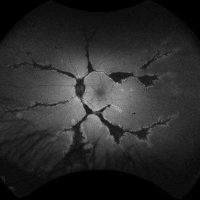
 Pigmented Paravenous Chorioretinal Atrophy (PPCRA)
Jun 27 2025 by Maria Letícia Costa Holanda
Fundoscopy of a 42-year-old asymptomatic man with pigmented paravenous chorioretinal atrophy. Pigmented paravenous retinochoroidal atrophy (PPRCA) is a rare disorder of unknown etiology. The disease is characterized by pigment accumulation along the distribution of retinal veins. The findings are usually incidental with minimal effect on vision.
Photographer: Guilherme da Cruz Reis, CLINOS Eye Hospital - Feira de Santana (BA),Brazil
Condition/keywords: pigmented paravenous chorioretinal atrophy (PPCRA)
-
 Pigmented Paravenous Retinochoroidal Atrophy
Jun 18 2025 by César Adrián Gómez Valdivia, MD
Fundus Autofluorescence Image of a 42YO female patient diagnosed with Pigmented Paravenous Retinochoroidal Atrophy. Findings were bilateral. Image shows hypoautofluorescence in the affected areas due to overall loss of RPE cells and thus lower lipofuscin levels.
Photographer: @eyemissu2
Imaging device: California ICG OPTOS
Condition/keywords: pigmented paravenous chorioretinal atrophy (PPCRA)
-
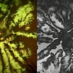 Fractal Pattern of Chronic Serpiginous Choroiditis
Jun 17 2025 by Guilherme Sturzeneker, MD, MSc
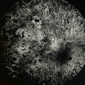
Ultra-widefield fundus photograph and autofluorescence of a 33-year-old woman with longstanding serpiginous choroiditis in the right eye. The image reveals centrifugal chorioretinal atrophy forming a dramatic fractal-like pattern, sparing the fovea. The patient is several years post-onset, with repeated negative workups, including for tuberculosis. Despite extensive lesions, the patient retains 20/20 vision in both eyes. Management included azathioprine monotherapy, as systemic steroids were contraindicated due to bipolar disorder.
Photographer: Andrea Almeida, IPEPO - Instituto da Visão
Imaging device: Optos Silverstone
Condition/keywords: autoimmune uveitis, azathioprine, chorioretinal atrophy, serpiginous choroiditis, ultra-wide field imaging
-
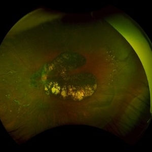
 Extensive Chorioretinal Scarring With Partial Macular Sparing
Apr 22 2025 by Maxwell J Wingelaar, MD
Fundus autofluorescence of extensive chorioretinal scarring in the left eye.
Photographer: Killian Roberts
Imaging device: Heidelberg Spectralis AF
Condition/keywords: chorioretinal atrophy, chorioretinal inflammations
-
 Extensive Chorioretinal Scarring with Partial Macular Sparring
Apr 22 2025 by Maxwell J Wingelaar, MD
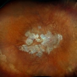
A multicolor photo showing chorioretinal scarring with partial macular sparing in the left eye.
Photographer: Killian Roberts
Imaging device: Heidelberg Spectralis Multicolor Photo
Condition/keywords: chorioretinal atrophy, chorioretinal inflammations
-
 Extensive Chorioretinal Scarring in the Right Eye
Apr 22 2025 by Maxwell J Wingelaar, MD
Fundus autofluorescence of Extensive chorioretinal scarring in the right eye.
Photographer: Killian Roberts
Imaging device: Heidelberg Spectralis AF
Condition/keywords: chorioretinal atrophy, chorioretinal inflammations
-
 Extensive Chorioretinal Scarring in the Right Eye
Apr 22 2025 by Maxwell J Wingelaar, MD
A multicolor photo showing chorioretinal scarring with macular involvement in the right eye
Photographer: Killian Roberts
Imaging device: Heidelberg Spectralis Multicolor Photo
Condition/keywords: chorioretinal atrophy, chorioretinal inflammations
-
 Myopic Degeneration
Dec 9 2024 by Virginia Gebhart
67 year old female with myopic degeneration. Posterior staphylomas are stable. VA limited by extensive chorioretinal atrophy. BCVA 20/160 (ecc)
Photographer: Virginia Gebhart, Retina Consultants of Carolina
Imaging device: Optos California
Condition/keywords: chorioretinal atrophy, myopic degeneration, staphyloma
-
 Peripheral Retinal Degeneration (L-ORD)
Apr 17 2024 by Virginia Gebhart
92 year old female with bilateral patchy, sharply demarcated circular areas of chorioretinal atrophy with hyperpigmented margins in the mid to far periphery. Labs showed normal plasma ornithine levels ruling out generalized gyrate atrophy. Also intermediate uveitis and CMD/CME. FTA-ABS, Quant gold, and HLA-A29 labs all negative.
Photographer: Virginia Gebhart
Imaging device: Optos California
Condition/keywords: cystoid macular degeneration, cystoid macular edema (CME), FA, Fluorescein angiography, peripheral retinal degeneration
-
 Diffuse Chorioretinal Atrophy
Feb 21 2024 by Virginia Gebhart
61 year male with myopic degeneration and diffuse chorioretinal atrophy. BCVA 20/200.
Photographer: Virginia Gebhart
Imaging device: Topcon TRC 50DX
Condition/keywords: chorioretinal atrophy, myopic degeneration
-
 PPCRA
PPCRA
Jan 31 2024 by Pallavi Goel
A 14-year-old male, presented to our clinic for a regular ophthalmic examination. Both Eyes Best Corrected Visual Acuity was 6/6, N6. The Indirect Ophthalmoscopic examination revealed an incidental finding in both eyes with patches of chorioretinal atrophy and pigment clumps along the veins consistent with pigmented paravenous chorioretinal atrophy (PPCRA) with early attenuation of retinal vessels, normal discs, and macula. ERG was normal. The patient was counseled and explained the nature of his condition. He was asked to be in yearly follow-up.
Photographer: Pallavi Goel, Dr. Shroff's Charity eye hospital,Delhi
Condition/keywords: ERG
-
 Macular Dystrophy vs Myopic Degeneration
Dec 22 2023 by Virginia Gebhart
35 year old female with myopic degeneration (-18.00 OU). BCVA 20/100 OU. RPE atrophy present in both eyes, but no significant chorioretinal atrophy. OCT not consistent with degenerative myopia due to dome shape appearance rather than posterior bowing. Possible macular dystrophy over degeneration. Will observe
Photographer: Virginia Gebhart
Imaging device: Topcon
Condition/keywords: Macular Dystrophy, myopic degeneration
-
 Pigmented Paravenous Retinochoroidal Atrophy
Nov 25 2023 by Jane-Ming Lin
A 27-year-old male patient complained with gradual progressive loss of vision in both eyes for 4 years. He had no family history of inherited ocular diseases and was diagnosed with Pigmented Paravenous Retinochoroidal Atrophy.
Condition/keywords: pigmented paravenous chorioretinal atrophy (PPCRA)
-
 Sympathetic Ophthalmia
May 24 2023 by Niloofar Piri, MD
Montage fundus photograph of the left eye with end stage Sympathetic Ophthalmia, demonstrating optic nerve pallor, severe arterial attenuation, extensive chorioretinal atrophy (sclera is exposed in most areas), and peripheral RPE hyperplasia. Patient is a 50 yo Asian female with history of multiple vitrectomies due to retinal detachment and loss of vision who developed Sympathetic Ophthalmia in the other eye. This picture is 20 years after the disease process started, with end stage picture and HM vision.
Photographer: Sean Kelso, Saint louis university
Condition/keywords: sympathetic ophthalmia
-
 Gyrate Atrophy
Apr 12 2023 by Ahmed Abbas Hashmi, OD
Left eye fundus of a 53-year-old male patient with advanced gyrate atrophy of the choroid and retina with macular sparing. Optic nerve head is healthy.
Photographer: Ahmed Abbas Hashmi
Imaging device: Topcon TRC-NW8F
Condition/keywords: chorioretinal atrophy
-
 Degenerative Myopia
Apr 12 2023 by Ahmed Abbas Hashmi, OD
Right eye Fundus photograph of a 61-year-old female with pathological myopia.
Condition/keywords: chorioretinal atrophy, high myopia, pathologic myopia
-
 Choroideremia
Apr 4 2023 by Ian C Han, MD
Widefield color fundus photograph of a teenage male with molecularly-confirmed choroideremia shows lobular areas of chorioretinal atrophy with sparing of the macula.
Photographer: Nicole Radunzel, University of Iowa, Department of Ophthalmology and Visual Sciences
Condition/keywords: choroid, choroideremia, dystrophy, retina
-
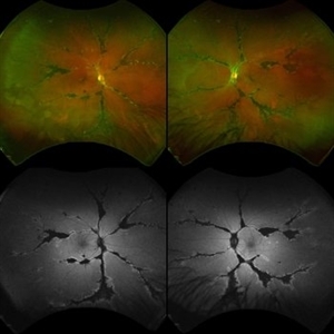
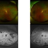 Ultra-wide images of Paravenous chorioretinal atrophy (PPCRA)
Dec 11 2022 by Suhwan Lee, MD
Ultra-wide fundus and autofluorescence images of a 41-year-old woman with PPCRA.
Imaging device: Optos california
Condition/keywords: pigmented paravenous chorioretinal atrophy (PPCRA)
-
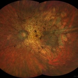 Rod Cone dystrophy
Nov 29 2022 by Niloofar Piri, MD
Fundus photograph of the left eye in a 58 yo male with rod cone dystrophy. He presented with night blindness and peripheral vision loss since youth and recent decrease in central vision for the past 10 years. Notice waxy pallor of the nerve, severe arterial narrowing and chorioretinal atrophy mainly around the arcades as well as posterior pole along with RPE hyperplastic changes and atrophy. RPE atrophy in midperiphery has coin shaped appearance. FAF has characteristic appearance (uploaded separately) He has one pathogenic variants of both CEP290 and PRPH2 genes.
Photographer: Sean Kelso, Saint Louis University
Condition/keywords: hereditary retinal deg, hereditary retinal dystrophy, Rod cone dystrophy
 Loading…