 Initializing download.
-
 By Pallavi Goel
Shroff charity eye hospital
Co-author(s): Tanya Jain, Consultant, Dr. Shroff's Charity Eye Hospital, Delhi - Uploaded on Jan 31, 2024.
- Last modified by Joshua Friedman on Feb 1, 2024.
- Rating
- Appears in
- 31-Jan-2024
- Condition/keywords
- ERG
- Photographer
- Pallavi Goel, Dr. Shroff's Charity eye hospital,Delhi
- Imaging device
- Fundus camera
- Description
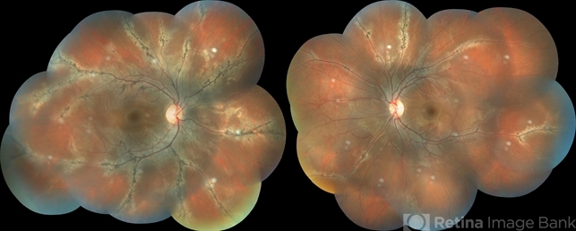
- A 14-year-old male, presented to our clinic for a regular ophthalmic examination. Both Eyes Best Corrected Visual Acuity was 6/6, N6. The Indirect Ophthalmoscopic examination revealed an incidental finding in both eyes with patches of chorioretinal atrophy and pigment clumps along the veins consistent with pigmented paravenous chorioretinal atrophy (PPCRA) with early attenuation of retinal vessels, normal discs, and macula. ERG was normal. The patient was counseled and explained the nature of his condition. He was asked to be in yearly follow-up.

