Search results (29 results)
-
 T-Cell Lymphoma
Jul 3 2025 by Virginia Gebhart
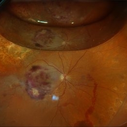
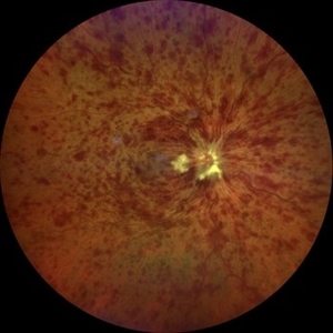
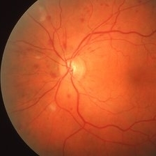
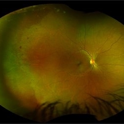
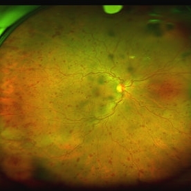
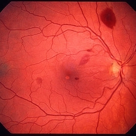
78 year old male s/p vitreous biopsy for T-Cell lymphoma. Pt presented with peripheral blot hemorrhages and numerous white subretinal infiltrates. Retinal pallor and thickening temporally. History of cutaneous T-cell lymphoma. PPV/vitreous biopsy performed to find differential diagnosis. Silicone oil was placed for 6 weeks, then removed and exchanged with a gas bubble. Hematology pathologist and Emory reviewed path report and agrees it is consistent with T-cell lymphoma. Pt received intravitreal Methotrexate and will be scheduled for weekly treatments. BCVA CF
Photographer: Virginia Gebhart, Retina Consultants of Carolina
Imaging device: Optos California
Condition/keywords: biopsy, gas bubble, lymphoma
-
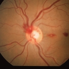
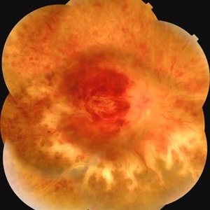 A Vein in Vain: Ischemic CRVO
Dec 6 2024 by Jasmeet Kaur Chandi
Fundus photo of a 55 year-old female with extensive flame-shaped and dot-blot hemorrhages in all four quadrants. Tortuous and dilated veins with cotton-wool spots. Optic disc swelling with hyperemia and macular edema.
Photographer: Dr. Jasmeet Kaur Chandi
Condition/keywords: Ischemic Central Retinal Vein Occlusion
-
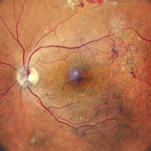
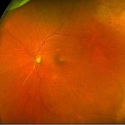
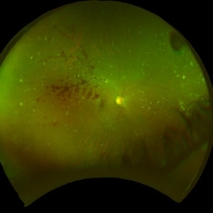
 Torpedo Retinopathy
Oct 31 2024 by AVIK DEY SARKAR, MS, FVRS, FAICO(VR)
This is a 42 year old male with known history of diabetes mellitus for past 10 years. Patient presented with complains regarding presbyopia. On dilated fundoscopy, along with dot and blot hemorrhages, in the infero-temporal near-periphery outside the vascular arcade a hypopigmented torpedo-shaped lesion was noted. On OCT, outer retinal attenuation with sublesional choriocappilaris layer thinning was noted. The lesion is diagnosed as torpedo retinopathy. Torpedo maculopathy is rare in clinical practice and usually is found at the margin of temporal arcade over "Temporal Bulge". But this lesion is seen well away from the posterior pole. This case indicates the necessity of substituting the terminology "Torpedo Maculopathy" with "Torpedo Retinopathy" as mentioned earlier in ophthalmic literature.
Photographer: Dr. Avik Dey Sarkar, MBBS, MS, FVRS, FAICO, Consultant, Department of Vitreoretinal Services, Aravind Eye Hospital, Madurai, India
Imaging device: Wide angled Fundus imaging with Clarus 300
Condition/keywords: torpedo maculopathy, torpedo Retinopathy
-
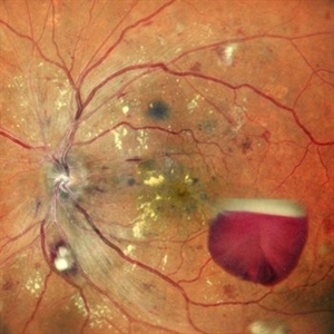
 Proliferative Diabetic Retinopathy
May 24 2024 by Anjana Mirajkar, MS Ophthalmology
A central photo of a 60 year old male of left eye case of neovascularization elsewhere with dot and blot hemorrhages in a case of proliferative diabetic retinopathy.
Photographer: Dr. Anjana Mirajkar -Retina Foundation, Ahmedabad
Imaging device: Mirante-Nidek
Condition/keywords: NVE, pre-proliferative diabetic retinopathy
-
 Proliferative Diabetic Retinopathy
May 24 2024 by Anjana Mirajkar, MS Ophthalmology
A central photo of a 50 year old male case of PDR showing a sub-hyaloid hemorrhage with cotton wool spots , hard exudates at the fovea with dot and blot hemorrhages.
Photographer: Dr. Anjana Mirajkar -Retina Foundation, Ahmedabad
Imaging device: Mirante-Nidek
Condition/keywords: proliferative diabetic retinopathy (PDR), Sub hyaloid haemorrhage
-
 Proliferative Diabetic Retinopathy
Apr 20 2024 by Tejaswita Verma
Widefield fundus photograph of the left eye of a 62 year old female with left eye lasered proliferative diabetic retinopathy showing neovascularisation elsewhere with few dot-blot hemorrhages.
Photographer: DR. TEJASWITA VERMA
Condition/keywords: Neovascularisation elsewhere (NVE), pan-retinal photocoagulation (PRP), proliferative diabetic retinopathy (PDR)
-
 Central Retinal Vein Occlusion associated with disc edema
Oct 19 2023 by Gabriel Costa Andrade, PhD
53-year-old woman with an acute CRVO. The patient has a history of breast cancer undergoing treatment with systemic chemotherapy. Notice the peripapillary cotton wool spots, superficial flame shaped hemorrhages and deeper dot and blot hemorrhages in all 4 quadrants.
Photographer: Gabriel Andrade
Condition/keywords: central retinal vein occlusion (CRVO), macular edema, Retina
-
 Branch retinal artery occlusion
Jan 24 2023 by Rayna Marshall
Widefield fundus image of a 54-year-old female with an asymptomatic chronic branch retinal artery occlusion in the left eye. Peripheral schisis-like changes with pigmentation and temporal dot-blot hemorrhages. Vision was 20/20.
Photographer: Drew H. Scoles, MD, PhD, University of Pennsylvania
Condition/keywords: branch retinal artery occlusion (BRAO), BRAO, embolus
-
 Macular Pucker
Jan 7 2020 by RAFAEL REIS PEREIRA, MD
A clinical grading system was proposed by Gass in 1987 describe the different stages of the epiretinal membrane. Grade 2 Macular pucker consists of a thick fibroglial membrane that contracts and produces obscuration of underlying vessels and marked full-thickness retinal distortion. Sometimes associated with cotton-wool spots, exudates, blot hemorrhages, microaneurysms, and cystoid macular edema.
Photographer: Rafael Reis, Retina Clinic - Brazil
Condition/keywords: macular pucker
-
 Multiple Myeloma
Apr 1 2019 by Gary R. Cook, MD, FACS
62-year-old white female with multiple cotton wool spots and intraretinal hemorrhages OD secondary to multiple myeloma; V.A. = 20/60+1.
Imaging device: Topcon VT-50
Condition/keywords: blot hemorrhages, cotton wool spots, myeloma, retinal hemorrhage, retinopathy
-
 Combined CRVO and BRAO
Mar 27 2019 by Gary R. Cook, MD, FACS
Right eye of a 56-year-old white male with a combined perfused CRVO (venous dilation and dot & blot hemorrhages in all 4 quadrants) and a superotemporal BRAO with peripapillary hemorrhages and cotton wool spots, and an area of retinal whitening inside of the ST arcade. V.A.= 20/70.
Imaging device: Topcon VT-50
Condition/keywords: branch retinal artery occlusion (BRAO), central retinal vein occlusion (CRVO)
-
 Multiple Myeloma
Mar 26 2019 by Gary R. Cook, MD, FACS
62-year-old white female with multiple CWS and intraretinal hemorrhages OS secondary to multiple myeloma; VA= 20/20-2.
Imaging device: Topcon VT-50
Condition/keywords: blot hemorrhages, cotton wool spots, myeloma, retinal hemorrhage
-
 Anemia
Mar 26 2019 by Gary R. Cook, MD, FACS
Retinal hemorrhages OS in a 45 year old female secondary to iron deficiency anemia; VA= 20/20.
Imaging device: Topcon VT-50
Condition/keywords: anemia, blot hemorrhages, hemorrhage, white centered retinal hemorrhage (Roth Spot)
-
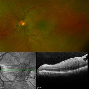
 Ocular Ischemic Syndrome
Jun 20 2018 by Andreas Ebneter, MD, PhD, FASRS
Ocular ischemic syndrome can present with a wide variety of ocular findings in both the anterior and posterior segments. The color fundus image of this 77-year-old male shows scattered blot hemorrhages in the deep retinal layers of the posterior pole that are only occasionally confluent. Commonly, these typical hemorrhages are predominantly found in the mid-periphery. Fluorescein angiography helps in confirming the diagnosis. Choroidal filling time is frequently somewhat delayed and patchy. Arteriovenous transit time is clearly prolonged. Staining of both veins and arteries in late images (top right) reflects diffuse endothelial cell damage with compromise of the blood-retina barrier. The peripheral retina is affected by extensive non-perfusion.
Photographer: Eva Steffen, Bern University Hospital, Switzerland
Imaging device: Optos 200Tx and Heidelberg Spectralis OCT
Condition/keywords: ocular ischemic syndrome
-
 Lupus Hemorrhagic Occlusive Vasculitis
Apr 23 2018 by Frank Chin
Fundus photograph of the right eye of a 24-year-old woman with history of systemic lupus erythematosus who presented with decreased visual acuity for 2-3 days found to have lupus hemorrhagic occlusive vasculitis with mild disc elevation, diffuse punctate cotton wool spots and dot blot hemorrhages, and a hemorrhage occlusive vasculitis along the superior branch of the superotemporal arcade involving the artery and vein.
Photographer: Frank Chin, MD, George Washington University
Imaging device: Optos 200Tx
Condition/keywords: blot hemorrhages, cotton wool spots, occlusive vasculitis, systemic lupus erythematosus (SLE) vasculitis
-
 White Without Pressure
Jan 31 2018 by Olivia Rainey
Ultra-wide field pseudocolor photograph of a 57-year-old female with white without pressure affecting her left eye. Patient will be having bloodwork done to rule out possible sarcoidosis or sickle cell.
Photographer: Olivia Rainey
Imaging device: Optos
Condition/keywords: blot hemorrhages, color fundus photograph, left eye, Optos, ultra-wide field imaging, white without pressure
-
 White Without Pressure/Dot Blot Hemorrhages
Jan 31 2018 by Olivia Rainey
Ultra-wide field pseudocolor photograph of a 57-year-old female with white without pressure affecting her right eye. Patient will be having bloodwork done to rule out possible sarcoidosis or sickle cell.
Photographer: Olivia Rainey
Imaging device: Optos
Condition/keywords: blot hemorrhages, Optos, ultra-wide field imaging
-
 Multiple Blot Hemorrhages and Roth Spots
Jan 24 2018 by Gabriel Costa Andrade, PhD
Multiple blot hemorrhages and Roth spots in a patient with acute leukemia.
Photographer: Gabriel Andrade, MD
Condition/keywords: leukemia, Roth spots
-
 Proliferative Diabetic Retinopathy
Mar 3 2017 by Nichole Lewis
61-year-old female with proliferative diabetic retinopathy, neovascularization, microaneurysms, dot blot hemorrhages and capillary nonperfusion.
Photographer: Nichole Lewis
Condition/keywords: microaneurysms, neovascularization elsewhere (NVE), proliferative diabetic retinopathy (PDR)
-
 CRVO
CRVO
Sep 7 2015 by Andrea Arriola-Lopez, MD MSc
Color fundus photography of right eye with central retinal vein occlusion; BCVA 20/25; IOP 16mmHg. No macular edema or neovascularization.
Photographer: Andrea Elizabeth Arriola López, MD, MSc
Imaging device: OPTOS Dakota
Condition/keywords: blot hemorrhages, central retinal vein occlusion (CRVO), venous tortuosity
-
 Terson's Syndrome
Jan 7 2015 by H. Michael Lambert, MD
Color photograph- flame and blot hemorrhages
Condition/keywords: Terson's Syndrome
-
 Terson's Syndrome
Jan 7 2015 by H. Michael Lambert, MD
Color photograph- premacular and blot hemorrhages
Condition/keywords: Terson's Syndrome
-
 Leukemia
Mar 29 2013 by Henry J. Kaplan, MD
Multiple blot hemorrhages in a patient with leukemia #1.
Condition/keywords: leukemia
-
---thumb.jpg/image-square;max$300,300.ImageHandler) SLE Retinopathy
SLE Retinopathy
Feb 26 2013 by Henry J. Kaplan, MD
SLE retinopathy,l eft eye: multiple cotton wool spots and blot hemorrhages. #2
Condition/keywords: blot hemorrhages, cotton wool spots, systemic lupus erythematosus (SLE) retinopathy
-
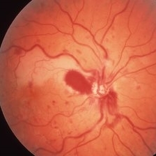
---thumb.jpg/image-square;max$300,300.ImageHandler) Central Retinal Vein Occlusion
Central Retinal Vein Occlusion
Oct 30 2012 by Lihteh Wu, MD
35-year-old hypertensive man with an acute CRVO. Notice the peripapillary cotton wool spots, superficial flame shaped hemorrhages and deeper dot and blot hemorrhages in all 4 quadrants. This is the typical blood and thunder appearance of a CRVO.
Condition/keywords: central retinal vein occlusion (CRVO), cotton wool spots
 Loading…