Search results (181 results)
-
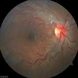
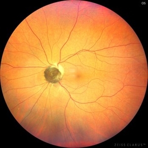
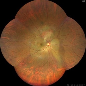
 Leber’s Miliary Aneurysm
Dec 12 2025 by KANWALJEET HARJOT MADAN, M.S. (Ophthalmology); FAICO (Vitreous - Retina)
A 34 year-old male presented with decrease vision in right eye for 3 months. Anterior segment exam was normal. Fundus exam in RE revealed presence of macular edema which was evident on OCT. Multiple retinal vascular aneurysmal dilatations with telangiectasia of the retina blood vessels noted superiorly which was evident on FFA. These aneurysms were multiple, tiny and leaky on FFA. He was diagnosed to have Leber’s miliary aneurysms. It is a rare, typically unilateral eye condition, often seen in young males, characterized by multiple tiny, leaky aneurysms in the retinal blood vessels, leading to deposits of hard exudates and potential vision loss, especially if it affects the macula. It is considered a milder form of Coats' disease.
Photographer: Dr. Kanwaljeet Harjot Madan, Thind Eye Hospital, Jalandhar City (Punjab) INDIA.
Imaging device: Zeiss Fundus Camera
Condition/keywords: FFA, Leber's miliary aneurysm
-
 Congenital Cataract
Dec 8 2025 by Rinat Sutiushev
M. 29 years, the variant of the lamellar cataract affects only the outer layer of the embryonic nucleus. The clinical case demonstrates photographic data, anterior segment OCT, and IOL-master 700 data. Visual acuity Vis OD 20/80 Vis OS 20/80. Surgical treatment of congenital cataracts is recommended. p.s. At first glance, I thought of space.
Condition/keywords: congenital cataract
-
 Congenital Cataract
Dec 8 2025 by Rinat Sutiushev
M. 29 years, the variant of the lamellar cataract affects only the outer layer of the embryonic nucleus. The clinical case demonstrates photographic data, anterior segment OCT, and IOL-master 700 data. Visual acuity Vis OD 20/80 Vis OS 20/80. Surgical treatment of congenital cataracts is recommended. p.s. At first glance, I thought of space.
Photographer: Rinat Sutiushev, Ophthalmological center “Vision”, Saint Petersburg
Condition/keywords: congenital cataract
-
 Ozurdex
Nov 22 2025 by Gabriel Costa Andrade, PhD
Anterior segment photograph of a Ozurdex implant in a 53-year-old man with macular edema due to intermediate uveitis.
Photographer: Gabriel Andrade
Condition/keywords: Ozurdex implant
-
 Intraocular Foreign Body Scleral Lac
Nov 19 2025 by Nikhil Das, M.D.
A 34-year-old man presented with a right intraocular foreign body after hammering a carbon-steel chisel 12 hours after injury. CT orbits showed a 3-mm hyperattenuating foreign body within the right globe, centered in the vitreous cavity. BCVA was 20/40. Anterior segment examination revealed a 2.8-mm scleral laceration. DFE demonstrated a metallic IOFB, a superior air bubble, superior commotio retinae, and Berlin’s edema involving the macula.
Photographer: Nikhil Das, Saint Louis University School of Medicine
Imaging device: iPhone
Condition/keywords: intraocular foreign body, iofb, metallic foreign body, scleral laceration
-
 Intraocular Foreign Body CT Coronal
Nov 19 2025 by Nikhil Das, M.D.
A 34-year-old man presented with a right intraocular foreign body after hammering a carbon-steel chisel 12 hours after injury. CT orbits showed a 3-mm hyperattenuating foreign body within the right globe, centered in the vitreous cavity. BCVA was 20/40. Anterior segment examination revealed a 2.8-mm scleral laceration. DFE demonstrated a metallic IOFB, a superior air bubble, superior commotio retinae, and Berlin’s edema involving the macula.
Photographer: Nikhil Das, Saint Louis University School of Medicine
Imaging device: CT Scan
Condition/keywords: intraocular foreign body, iofb, metallic foreign body, scleral laceration
-
 Intraocular Foreign Body CT Axial
Nov 19 2025 by Nikhil Das, M.D.
A 34-year-old man presented with a right intraocular foreign body after hammering a carbon-steel chisel 12 hours after injury. CT orbits showed a 3-mm hyperattenuating foreign body within the right globe, centered in the vitreous cavity. BCVA was 20/40. Anterior segment examination revealed a 2.8-mm scleral laceration. DFE demonstrated a metallic IOFB, a superior air bubble, superior commotio retinae, and Berlin’s edema involving the macula.
Photographer: Nikhil Das, Saint Louis University School of Medicine
Imaging device: CT Scan
Condition/keywords: intraocular foreign body, iofb, metallic foreign body, scleral laceration
-
 Intraocular Foreign Body
Nov 19 2025 by Nikhil Das, M.D.
A 34-year-old man presented with a right intraocular foreign body after hammering a carbon-steel chisel 12 hours after injury. CT orbits showed a 3-mm hyperattenuating foreign body within the right globe, centered in the vitreous cavity. BCVA was 20/40. Anterior segment examination revealed a 2.8-mm scleral laceration. DFE demonstrated a metallic IOFB, a superior air bubble, superior commotio retinae, and Berlin’s edema involving the macula.
Photographer: Nikhil Das, Saint Louis University School of Medicine
Condition/keywords: intraocular foreign body, metallic foreign body, scleral laceration
-
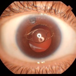
 Gravity = 1, Zonules = 0 : Cionni ring-IOL-Bag complex subluxation
Nov 7 2025 by SHRADDHA RAJ SHRIVASTAVA
Right eye anterior segment slit-lamp image of a 30 year old male, who was operated for spontaneous bilateral inferior subluxation of crystalline lens. Primary surgery was performed almost 20 years ago in which lens extraction was done followed by IOL placement in bag after stabilising it with Cionni ring. The patient presented to us recently with right eye diminution of vision and was noted to have inferiorly subluxated IOL-capsular bag complex, with vitreous in AC coming from the superior aphakic area. Interestingly, we have also captured in this image - the capsular tension ring (Cionni ring) with its central fixation eyelet.
Photographer: Dr. Shraddha Raj Shrivastava
Condition/keywords: dislocated IOL, dropped capsular IOL bag complex, IOL drop, Subluxated IOL, zonular dehiscence
-
 Papillophlebitis Salauno
Sep 3 2025 by Pablo Angel Garcia Uribe
Fundus photograph of a 24-year-old woman, previously healthy, with a history of recreational inhaled cannabis use, presented with a 24-hour history of photopsias and mild decrease in visual acuity, associated with a subtle relative central scotoma in the right eye. On ophthalmic examination, the anterior segment of both eyes was unremarkable. Best-corrected visual acuity was slightly reduced in the right eye and normal in the left. Fundus biomicroscopy of the right eye revealed moderate disc edema with hyperemia and well-defined margins, accompanied by venous engorgement and tortuosity, predominantly affecting the venules. No retinal hemorrhages were observed. Additionally, retinal thickening was noted along the temporal arcades, with apparent foveal sparing. The left eye showed no pathological findings. Based on the patient’s age, the acute onset of symptoms, the fundoscopic features, and the absence of systemic risk factors, the clinical presentation was consistent with papillophlebitis.
Photographer: Clínica Oftalmológica Salauno
Imaging device: Visucam 524, Carl Zeiss Meditec AG, Jena, Germany
Condition/keywords: papillophlebitis
-
Ocular Toxocariasis
Jun 30 2025 by ASRS Staff
Ocular Toxocariasis found in a 46 year-old female patient with decreased vision. Findings were unilateral. Fundus photographs, echography and anterior segment ultrabiomicroscopy can be found at @eyemissu2
Condition/keywords: Ocular Toxocariasis
-
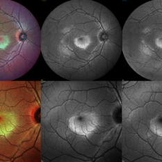 Berlin's Edema
Jun 12 2025 by Shivankar Sen, MS, FVRS
A 22 year old male came with history of sports injury to the right eye with the nose of shuttlecock while playing badminton. On examination, right eye anterior segment shows conjunctival congestion with brisk pupillary reaction and quiet anterior chamber. His best corrected visual acuity was 6/12; N6 in the right eye and 6/6; N6 in the left eye. Retinal examination revealed OD Berlin's Edema, OS within normal limits. Image Description (From Left to Right) Multicolor Reflectance (Blue-Green Enhanced) shows well defined yellowish discoloration Green reflectance and blue reflectance show corresponding whitish discoloration at the area of edema
Photographer: Dr. Shivankar Sen
Imaging device: Heidelberg Spectralis HRA+OCT
Condition/keywords: Shuttlecock Injury
-
 Commotio Retinae
Jun 10 2025 by CUI YUELING
The patient presented 2 hours after sustaining a left eye injury caused by a stick. Visual acuity in the left eye was 0.2 without improvement upon correction, and intraocular pressure measured 15 mmHg. Examination of the anterior segment revealed ciliary conjunctival injection accompanied by patchy subconjunctival hemorrhage. The corneal surface remained smooth, and the anterior chamber was deep with hyphema characterized by blood-tinged aqueous humor predominantly settled inferiorly. The pupil was slightly irregular, approximately 3 mm in diameter, with a superotemporal notch; pupillary light reflex was intact. The lens appeared clear. Fundus examination showed well-defined optic disc margins with normal coloration and a cup-to-disc ratio of 0.2. Retinal arteries and veins were normally distributed with an artery-to-vein ratio of 2:3. At the posterior pole, the foveal reflex exhibited concentric ripple-like changes centered on the fovea, accompanied by localized pigment attenuation and reduced reflex intensity. Irregular reflectivity was noted in the superotemporal and inferotemporal nerve fiber layers.
Photographer: Yueling Cui
Imaging device: Zeiss Clarus 500
Condition/keywords: commotio retinae
-
 Anterior Iris Claw Artisan Lens
May 14 2025 by Moazzam Parvez
Anterior segment image of a 40 year old gentleman with a anteriorly placed iris claw lens post retinal detachment surgery.
Photographer: Dr Moazzam Parvez, Netralayam , Kolkata
Imaging device: Topcon DC-4
Condition/keywords: Anteriorly placed iris claw lens
-
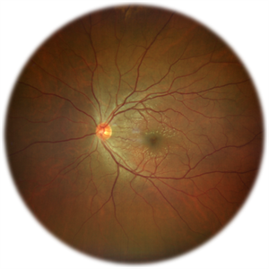
 Optic Nerve Melanocytoma
May 4 2025 by KANWALJEET HARJOT MADAN, M.S. (Ophthalmology); FAICO (Vitreous - Retina)
This is a fundus picture of a young 42-year male who visited for a routine eye exam. His BCVA was 20/20 in both eyes. Anterior segment examination was normal. His left eye showed grey-black pigmentation at the infero-nasal margin of the optic disc. Fundus of the right eye was normal. The patient was diagnosed to have optic disc melanocytoma on multimodal imaging and was advised regular follow-up. Optic nerve melanocytoma is typically a benign tumor made up of melanocytes and melanin. It can grow, but rarely transforms into a malignancy. Patients with Optic Nerve Melanocytoma should be periodically examined for evidence of growth, loss of visual field and optic nerve compression.
Photographer: Dr. Kanwaljeet Harjot Madan, Thind Eye Hospital, Jalandhar City (Punjab) INDIA.
Imaging device: Zeiss Fundus Camera
Condition/keywords: melanocytoma, melanoma, optic nerve
-
 Hourglass in an Eye
Apr 22 2025 by KRISHNENDU NANDI, MS
A twenty-five-year-young male presented with a decrease in vision in the right eye following a blunt trauma with a football. On examination the BCVA in the right eye was CFCF and the left eye was 6/6, N6. The anterior segment was within normal limits. AT was 12 and 10 mm of Hg in the right and left eyes, respectively. Fundus examination reveals subhyaloid haemorrhage in the right eye with an attached retina. The fundus of the left eye was within normal limits. YAG laser hyaloidotomy was done with an energy of 2 mJ in the right eye. After 3 weeks the BCVA in the right eye improved to 6/9, N6.
Photographer: Dr. Krishnendu Nandi
Imaging device: Topcon
Condition/keywords: Trauma, YAG HYALOIDOTOMY, Young Male
-
 Ozurdex
Mar 20 2025 by T. P . VIGNESH, MBBS,MS
Photo of the anterior segment of left eye of a 50 year old woman with intravitreal Ozurdex implant seen attached to the posterior capsule.
Photographer: T.P. VIGNESH
Condition/keywords: ozurdex, Ozurdex implant
-
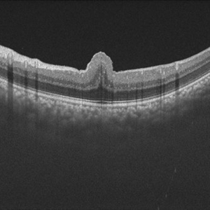
 Retinal Fold in Posterior Microphthalmos
Mar 1 2025 by Hemanth Murthy, MBBS, MD, FASRS
Swept source OCT image of left eye of 34 year male patient with high hypermetropia(+14). BCVA 20/20 in right eye and 20/60 in left eye. Anterior segment was normal. There is loss of foveal pit with omega shaped elevation of inner retinal layers.
Photographer: Mr Veda Vyas
Condition/keywords: posterior microphthalmos
-
 Retinal Fold in Posterior Microphthalmos
Mar 1 2025 by Hemanth Murthy, MBBS, MD, FASRS
Fundus photo of left eye of 34 year male patient with high hypermetropia(+14). BCVA 20/20 in right eye and 20/60 in left eye. Anterior segment was normal. There is loss of foveal pit with omega shaped elevation of inner retinal layers.
Photographer: Mr Veda Vyas
Condition/keywords: posterior microphthalmos
-
 Retinal Fold in Posterior Microphthalmos
Mar 1 2025 by Hemanth Murthy, MBBS, MD, FASRS
Fundus photo of Right eye of 34 year male patient with high hypermetropia(+14). BCVA 20/20 in right eye and 20/60 in left eye. Anterior segment was normal. There is loss of foveal pit with omega shaped elevation of inner retinal layers.
Photographer: Mr Veda Vyas
Condition/keywords: posterior microphthalmos
-
 Retinal Fold in Posterior Microphthalmos
Mar 1 2025 by Hemanth Murthy, MBBS, MD, FASRS
Swept source OCT image of Right eye of 34 year male patient with high hypermetropia(+14). BCVA 20/20 in right eye and 20/60 in left eye. Anterior segment was normal. There is loss of foveal pit with omega shaped elevation of inner retinal layers.
Photographer: Mr Veda Vyas
Condition/keywords: posterior microphthalmos
-
 Vortex-pattern Exudative Retinal Detachment
Feb 22 2025 by CUI YUELING
Patient: Male, 40 years old. Chief Complaint: Blurred vision and metamorphopsia in the left eye for more than 10 days. Past Medical History Hypertension for 4 years, with a highest recorded blood pressure of 160/80 mmHg. Currently controlled with oral "Nifedipine Sustained-Release Tablets, 2 tablets daily." Long-term history of heavy alcohol consumption and smoking. Ophthalmic Examination: Visual Acuity: Right eye (OD): 0.4 (uncorrected, no improvement with correction). Left eye (OS): 0.5 (-1.5DS = 1.0). Intraocular Pressure (IOP): OD: 15 mmHg. OS: 17 mmHg. Anterior Segment:Unremarkable. Fundus Examination: Right eye: Optic disc margins are clear, with a slightly reddish hue. Cup-to-disc ratio (C/D) = 0.2. A scalloped, orange-red elevated lesion is observed superior to the optic disc, with anterior displacement of the focal point. This is accompanied by a secondary, turbine-like exudative retinal detachment centered around the optic disc, involving the macula. The macular region shows scattered punctate yellow-white exudates. Diagnosis: Choroidal hemangioma with secondary exudative retinal detachment(OD).
Photographer: Cui yueling The First People's Hospital of Zunyi, Guizhou, Zunyi, China
Imaging device: Zeiss Clarus 500
Condition/keywords: choroidal hemangioma, exudative retinal detachment
-
 A Classic Case of Retinal Ora Serrata Imaging
Jan 16 2025 by yuan duo
A 5-year-old girl, born full-term with no history of systemic disease, presented with poor vision since early childhood and underwent fundus examination. Anterior segments of both eyes showed no significant abnormalities. Fundus examination revealed retinal folds extending from the optic disc to the temporal peripheral retina, with blood vessels coursing through the folds (A, B). Avascular zones were observed in the peripheral retina, and the ora serrata’s boundaries were clearly visible, displaying dentate processes and bays (C, D). Retinal pigmentation was evident. Genetic testing confirmed the final diagnosis of bilateral Familial Exudative Vitreoretinopathy (FEVR).
Condition/keywords: Retinal Ora Serrata
-
 Familial Exudative Vitreoretinopathy
Jan 16 2025 by yuan duo
A 5-year-old girl, born full-term with no history of systemic disease, presented with poor vision since early childhood and underwent fundus examination. Anterior segments of both eyes showed no significant abnormalities. Fundus examination revealed retinal folds extending from the optic disc to the temporal peripheral retina, with blood vessels coursing through the folds (A, B). Avascular zones were observed in the peripheral retina, and the ora serrata’s boundaries were clearly visible, displaying dentate processes and bays (C, D). Retinal pigmentation was evident. Genetic testing confirmed the final diagnosis of bilateral Familial Exudative Vitreoretinopathy (FEVR).
Condition/keywords: Retinal Ora Serrata
 Loading…