Search results (30 results)
-
 Sea Fan Neovascular Frond in Rare Case of Fanconi Anemia
May 18 2024 by Amol yuvraj ganvir
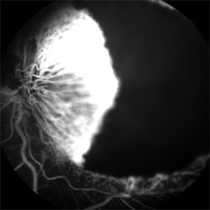
The left eye fundus shows a typical sea fan-shaped hyperfluorescent area, confirming neovascularization along the major temporal arcade and blocked fluorescence due to intraretinal and subretinal hemorrhage.
Photographer: Dr. Amol Ganvir
Condition/keywords: Fanconi Anemia, sea fan
-
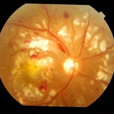
 Sea Fan Neovascular Frond in Rare Case of Fanconi Anemia
May 18 2024 by Amol yuvraj ganvir
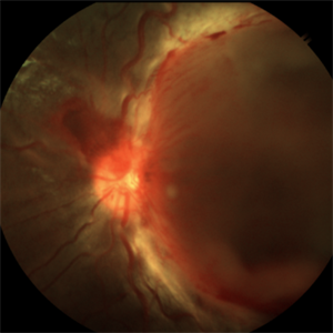
A 15-year-old female patient brought by her parents with defective vision in left eye for 15 days. The patient had a known case of Fanconi Anemia. Left eye tortuous vessels and new retinal vessels along the major temporal arcade, with intraretinal and subretinal hemorrhage covering the macula.
Photographer: Dr. Amol Ganvir
Condition/keywords: Fanconi Anemia, sea fan
-
 Sub ILM Hemorrhage
May 19 2023 by Rahul Bhatia, MS, DNB
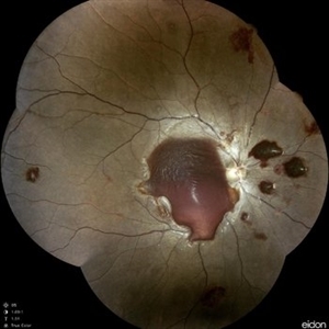
A 10-year-old male with Aplastic Anemia presented to Retina Clinic. Fundus Photograph and OCT line scan suggestive of Sub ILM Hemorrhage
Photographer: Dr Rahul Bhatia, LHMC, Delhi, India
Imaging device: Iphone
Condition/keywords: sub internal limiting membrane haemorrhage
-
 Combined Central Retinal Artery Occlusion with Central Retinal Venous Occlusion
Mar 22 2023 by VIRAL SHAH
26 YEARS OLD MALE PATIENTS HAS COMPLAIN OF DIMNESS OF VISION SINCE 3 DAYS IN RIGHT EYE. HE IS SUFFERING FROM ANEMIA
Photographer: VIRAL SHAH
Condition/keywords: VASCULAR SHEATHING WITH HOLLENHORST PLAQUE
-
 ROP 4B late Retinal Findings
Mar 31 2022 by Franco Benvenuto, MD
A 9-year-old male, that was born at 30 weeks of gestation with birth weight of 1500 g and history of hospitalization for 20 days with respiratory distress and packed red blood cell transfusion for anemia. At the first exam, both eyes were with stage 4B ROP. Vitrectomy with 25 G was done in both eyes. The flat fibrosis dragged the macula nasally in both the eyes.
Photographer: Franco Benvenuto, Universidad de Buenos Aires, Argentina; Universidad de Guadalajara, México.
Condition/keywords: cicatricial retinopathy of prematurity, retinopathy of prematurity (ROP)
-
 Anemic Retinopathy Related Retinal Hemorrhages
Nov 5 2019 by Chinmayi Vyas
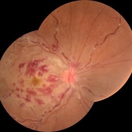
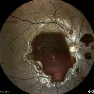
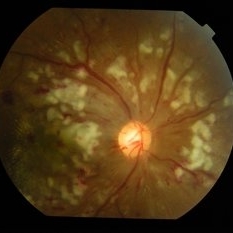
Anemic retinopathy related retinal hemorrhages in a 24 years old male with Hb of 4.2gm/ dl. The manifestations of anemic retinopathy are nonspecific and may closely simulate hypertensive or diabetic retina. Retinal changes in anemia are cotton wool spots, venous tortuosity, and hemorrhages which may be present at all levels of the retina and choroid. All retinal hemorrhages can occur when Hb falls below 8 g/100 ml or if the platelet count falls below 50,000/cumm. The combination of severe anemia and thrombocytopenia is likely to produce retinal hemorrhages. The Roth’s spots or white centre hemorrhages are typically associated with bacterial endocarditis , anemia and other systemic conditions. The white center is suspected to represents focal ischemia, inflammatory or infectious infiltrate, fibrin or accumulation of neoplasticism cells.
Photographer: Dr Chinmayi Vyas, Nethradhama superspeciality eye hospital , banglore, india
Imaging device: Eidon fundus imaging
Condition/keywords: anaemic retinopathy
-
 Anemic Retinopathy Related Retinal Hemorrhages
Nov 5 2019 by Chinmayi Vyas
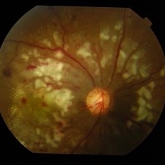
Anemic retinopathy related retinal hemorrhages in a 24 years old male with Hb of 4.2gm/ dl. The manifestations of anemic retinopathy are nonspecific and may closely simulate hypertensive or diabetic retina. Retinal changes in anemia are cotton wool spots, venous tortuosity, and hemorrhages which may be present at all levels of the retina and choroid. All retinal hemorrhages can occur when Hb falls below 8 g/100 ml or if the platelet count falls below 50,000/cumm. The combination of severe anemia and thrombocytopenia is likely to produce retinal hemorrhages. The Roth’s spots or white centre hemorrhages are typically associated with bacterial endocarditis , anemia and other systemic conditions. The white center is suspected to represents focal ischemia, inflammatory or infectious infiltrate, fibrin or accumulation of neoplasticism cells.
Photographer: Dr Chinmayi Vyas
Condition/keywords: retinal hemorrhage
-
 Anemia
Mar 26 2019 by Gary R. Cook, MD, FACS
Retinal hemorrhages OS in a 45 year old female secondary to iron deficiency anemia; VA= 20/20.
Imaging device: Topcon VT-50
Condition/keywords: anemia, blot hemorrhages, hemorrhage, white centered retinal hemorrhage (Roth Spot)
-
 Slide 9-13
Feb 26 2019 by Lancaster Course in Ophthalmology
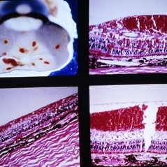
Superficial retinal hemorrhages. These hemorrhages have a flame-shaped appearance and are located just beneath the internal limiting membrane of the retina in a 61-year-old man with severe anemia and thrombocytopenia.
Condition/keywords: retinal hemorrhage, thrombocytopenia
-
 Proliferative Sickle Cell Retinopathy, Late FA OS
May 23 2018 by Hosam Attia, MD
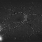
Fluorescein angiogram photograph of a 45-year-old African American, male with sickle cell anemia (SC disease), depicting peripheral capillary non-perfusion, with multiple, small area of mild late leakage consistent with active NVEs/ early Seafans OS.
Imaging device: Optos California Ultra-Wide Field Fundus Camera
Condition/keywords: neovascularization elsewhere (NVE), proliferative retinopathy, sea fan, sickle cell, sickle cell retinopathy
-
 Proliferative Sickle Cell Retinopathy, Late phase FA OD
May 23 2018 by Hosam Attia, MD
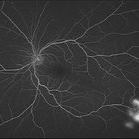
Fluorescein angiogram photograph of a 45-year-old African American, male with sickle cell anemia (SC disease), depicting extensive peripheral capillary non-perfusion, with late staining and diffuse leakage consistent with partially auto-infarcted, but active NVE/sea fan OD.
Imaging device: Optos California Ultra-Wide Field Fundus Camera
Condition/keywords: neovascularization elsewhere (NVE), proliferative retinopathy, sea fan, sickle cell, sickle cell retinopathy
-
 Proliferative Sickle Cell Retinopathy, Mid phase FA OS
May 23 2018 by Hosam Attia, MD
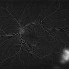
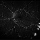
Fluorescein angiogram photograph of a 45-year-old African American, male with sickle cell anemia (SC disease), depicting peripheral capillary non-perfusion, with multiple, small area of early to mid phase hyperfluorescence over the ischemic retina temporally, with mild late leakage consistent with active NVEs/ early sea fans OS.
Imaging device: Optos California Ultra-Wide Field Fundus Camera
Condition/keywords: neovascularization elsewhere (NVE), proliferative retinopathy, sea fan, sickle cell, sickle cell retinopathy
-
 Proliferative Sickle Cell Retinopathy, Early phase FA OS
May 23 2018 by Hosam Attia, MD
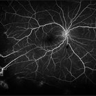
Fluorescein angiogram photograph of a 45-year-old African American, male with cell anemia (SC disease ), depicting peripheral capillary non-perfusion, with multiple, small area of early to mid phase hyperfluorescence over the ischemic retina temporally, with mild late leakage consistent with active NVEs/ early sea fans OS.
Imaging device: Optos California Ultra-Wide Field Fundus Camera
Condition/keywords: neovascularization elsewhere (NVE), proliferative retinopathy, sea fan, sickle cell, sickle cell retinopathy
-
 Proliferative Sickle Cell Retinopathy, Early phase FA OD
May 23 2018 by Hosam Attia, MD
Fluorescein angiogram photograph of a 45-year-old African American, male with sickle cell anemia (SC disease), depicting extensive peripheral capillary non-perfusion, with early hyperfluorescence over the ischemic retina temporally, with late staining and diffuse leakage consistent with partially auto-infarcted, but active NVE/sea fan OD.
Imaging device: Optos California Ultra-Wide Field Fundus Camera
Condition/keywords: neovascularization elsewhere (NVE), proliferative retinopathy, sea fan, sickle cell, sickle cell retinopathy
-
 Proliferative Sickle Cell Retinopathy, Red Free OS
May 23 2018 by Hosam Attia, MD
Red free fundus photograph of a 45-year-old African American, male with sickle cell anemia (SC disease) with arteriolar attenuation, mild venous tortuosity, peripheral arterio-venous anastomoses (Inferotemporally), multiple small NVEs/ early sea fans OS.
Photographer: Aaron Appiah, M.D.
Imaging device: Optos California Ultra-Wide Field Fundus Camera
Condition/keywords: neovascularization elsewhere (NVE), proliferative retinopathy, sea fan, sickle cell, sickle cell retinopathy
-
 Proliferative Sickle Cell Retinopathy, Red Free OD
May 23 2018 by Hosam Attia, MD
Red free fundus photo of a 45-year-old African American, male with sickle cell anemia (SC Disease ) with arteriolar attenuation, mild venous tortuosity, Sunburst (S) and large, partially auto-infarcted sea fan, OD.
Imaging device: Optos California Ultra-Wide Field Fundus Camera
Condition/keywords: neovascularization elsewhere (NVE), proliferative retinopathy, sea fan, sickle cell, sickle cell retinopathy
-
 Proliferative Sickle Cell Retinopathy, Color OS
May 23 2018 by Hosam Attia, MD
45-year-old African American, male with sickle cell anemia (SC disease ) with arteriolar attenuation, mild venous tortuosity, peripheral arterio-venous anastomoses (shown better on red free), multiple small NVEs/ early sea fans (one w/ early auto-infarction) and sunburst (S) - (Not showing very well in photos) OS.
Imaging device: Optos California Ultra-Wide Field Fundus Camera
Condition/keywords: neovascularization elsewhere (NVE), proliferative retinopathy, sea fan, sickle cell, sickle cell retinopathy
-
 Proliferative Sickle Cell Retinopathy, Color OD
May 23 2018 by Hosam Attia, MD
45-year-old African American, male with sickle cell anemia (SC disease) with arteriolar attenuation, mild venous tortuosity, Sunburst (S) and large, partially auto-infarcted Seafan with fresh heme, OD.
Imaging device: Optos California Ultra-Wide Field Fundus Camera
Condition/keywords: neovascularization elsewhere (NVE), proliferative retinopathy, sea fan, sickle cell, sickle cell retinopathy
-
 Proliferative Sickle Cell Retinopathy, Color OD
May 23 2018 by Hosam Attia, MD
45-year-old African American, male with sickle cell anemia (SC disease) with arteriolar attenuation, mild venous tortuosity, Sunburst (S) and large, partially auto-infarcted sea fan with fresh heme, OD.
Imaging device: Optos California Ultra-Wide Field Fundus Camera
Condition/keywords: neovascularization elsewhere (NVE), proliferative retinopathy, sea fan, sickle cell, sickle cell retinopathy
-
 Sickle Cell Anemia
Feb 22 2018 by Nichole Lewis
Sickle Cell Anemia
Photographer: Nichole Lewis
Condition/keywords: sickle cell
-
 Hypertensive Retinopathy
Feb 10 2016 by Mallika Goyal, MD
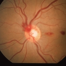
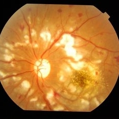
Bilateral hypertensive retinopathy in a 19-year-old girl with renal disease, hypertension and anemia.
Photographer: Mallika Goyal, MD, Apollo Health City, Hyderabad, India
Condition/keywords: hypertensive retinopathy
-
 Hypertensive Retinopathy
Feb 10 2016 by Mallika Goyal, MD
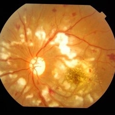
Bilateral hypertensive retinopathy in a 19-year-old girl with renal disease, hypertension and anemia.
Photographer: Mallika Goyal, MD, Apollo Health City, Hyderabad, India
Condition/keywords: hypertensive retinopathy
-
 Hypertensive Retinopathy
Feb 10 2016 by Mallika Goyal, MD
Bilateral hypertensive retinopathy in a 19-year-old girl with renal disease, hypertension and anemia.
Photographer: Mallika Goyal, MD, Apollo Health City, Hyderabad, India
Condition/keywords: hypertensive retinopathy
-
 Hypertensive Retinopathy
Feb 10 2016 by Mallika Goyal, MD
Bilateral hypertensive retinopathy in a 19-year-old girl with renal disease, hypertension and anemia.
Photographer: Mallika Goyal, MD, Apollo Health City, Hyderabad, India
Condition/keywords: hypertensive retinopathy
-
 Hypertensive Retinopathy
Feb 10 2016 by Mallika Goyal, MD
Bilateral hypertensive retinopathy in a 19-year-old girl with renal disease, hypertension and anemia.
Photographer: Mallika Goyal, MD, Apollo Health City, Hyderabad, India
Condition/keywords: hypertensive retinopathy
 Loading…