Search results (30 results)
-
 Sickle Cell Sea Fan Retinopathy
Jun 4 2014 by Henry J. Kaplan, MD
Sea fan peripheral retinal neovascularization in sickle cell anemia.
Condition/keywords: sea fan, sickle cell retinopathy
-
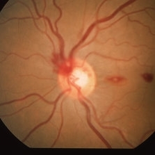
 Hypertensive Retinopathy
Jun 28 2013 by Jason S. Calhoun
Patient came in complaining of spots in vision in both eyes. VA was 20/25 - right eye and 20/20- left eye. Fundus exam reveals little hemorrhages with cotton wool spots due to hypertension and anemia.
Photographer: Jason S. Calhoun, Mayo Clinic Jacksonville, Florida
Imaging device: TOPCON TRC 50-EX
Condition/keywords: hypertensive retinopathy
-
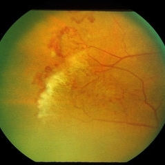
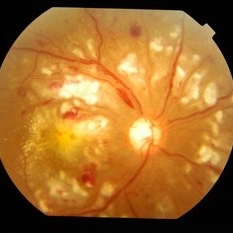
 Proliferative Sickle Cell Retinopathy, Color OD
May 23 2018 by Hosam Attia, MD
45-year-old African American, male with sickle cell anemia (SC disease) with arteriolar attenuation, mild venous tortuosity, Sunburst (S) and large, partially auto-infarcted sea fan with fresh heme, OD.
Imaging device: Optos California Ultra-Wide Field Fundus Camera
Condition/keywords: neovascularization elsewhere (NVE), proliferative retinopathy, sea fan, sickle cell, sickle cell retinopathy
-
 Hypertensive Retinopathy
Feb 10 2016 by Mallika Goyal, MD
Bilateral hypertensive retinopathy in a 19-year-old girl with renal disease, hypertension and anemia.
Photographer: Mallika Goyal, MD, Apollo Health City, Hyderabad, India
Condition/keywords: hypertensive retinopathy
-
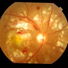
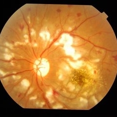
 Anemic Retinopathy Related Retinal Hemorrhages
Nov 5 2019 by Chinmayi Vyas
Anemic retinopathy related retinal hemorrhages in a 24 years old male with Hb of 4.2gm/ dl. The manifestations of anemic retinopathy are nonspecific and may closely simulate hypertensive or diabetic retina. Retinal changes in anemia are cotton wool spots, venous tortuosity, and hemorrhages which may be present at all levels of the retina and choroid. All retinal hemorrhages can occur when Hb falls below 8 g/100 ml or if the platelet count falls below 50,000/cumm. The combination of severe anemia and thrombocytopenia is likely to produce retinal hemorrhages. The Roth’s spots or white centre hemorrhages are typically associated with bacterial endocarditis , anemia and other systemic conditions. The white center is suspected to represents focal ischemia, inflammatory or infectious infiltrate, fibrin or accumulation of neoplasticism cells.
Photographer: Dr Chinmayi Vyas
Condition/keywords: retinal hemorrhage
-
 Hypertensive Retinopathy
Feb 10 2016 by Mallika Goyal, MD
Bilateral hypertensive retinopathy in a 19-year-old girl with renal disease, hypertension and anemia.
Photographer: Mallika Goyal, MD, Apollo Health City, Hyderabad, India
Condition/keywords: hypertensive retinopathy
-
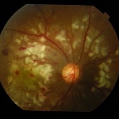
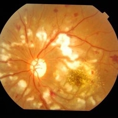
 Proliferative Sickle Cell Retinopathy, Color OS
May 23 2018 by Hosam Attia, MD
45-year-old African American, male with sickle cell anemia (SC disease ) with arteriolar attenuation, mild venous tortuosity, peripheral arterio-venous anastomoses (shown better on red free), multiple small NVEs/ early sea fans (one w/ early auto-infarction) and sunburst (S) - (Not showing very well in photos) OS.
Imaging device: Optos California Ultra-Wide Field Fundus Camera
Condition/keywords: neovascularization elsewhere (NVE), proliferative retinopathy, sea fan, sickle cell, sickle cell retinopathy
-
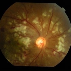
 Proliferative Sickle Cell Retinopathy, Color OD
May 23 2018 by Hosam Attia, MD
45-year-old African American, male with sickle cell anemia (SC disease) with arteriolar attenuation, mild venous tortuosity, Sunburst (S) and large, partially auto-infarcted Seafan with fresh heme, OD.
Imaging device: Optos California Ultra-Wide Field Fundus Camera
Condition/keywords: neovascularization elsewhere (NVE), proliferative retinopathy, sea fan, sickle cell, sickle cell retinopathy
-
 Anemic Retinopathy Related Retinal Hemorrhages
Nov 5 2019 by Chinmayi Vyas
Anemic retinopathy related retinal hemorrhages in a 24 years old male with Hb of 4.2gm/ dl. The manifestations of anemic retinopathy are nonspecific and may closely simulate hypertensive or diabetic retina. Retinal changes in anemia are cotton wool spots, venous tortuosity, and hemorrhages which may be present at all levels of the retina and choroid. All retinal hemorrhages can occur when Hb falls below 8 g/100 ml or if the platelet count falls below 50,000/cumm. The combination of severe anemia and thrombocytopenia is likely to produce retinal hemorrhages. The Roth’s spots or white centre hemorrhages are typically associated with bacterial endocarditis , anemia and other systemic conditions. The white center is suspected to represents focal ischemia, inflammatory or infectious infiltrate, fibrin or accumulation of neoplasticism cells.
Photographer: Dr Chinmayi Vyas, Nethradhama superspeciality eye hospital , banglore, india
Imaging device: Eidon fundus imaging
Condition/keywords: anaemic retinopathy
-
 Hypertensive Retinopathy
Feb 10 2016 by Mallika Goyal, MD
Bilateral hypertensive retinopathy in a 19-year-old girl with renal disease, hypertension and anemia.
Photographer: Mallika Goyal, MD, Apollo Health City, Hyderabad, India
Condition/keywords: hypertensive retinopathy
-
 Anemia
Mar 26 2019 by Gary R. Cook, MD, FACS
Retinal hemorrhages OS in a 45 year old female secondary to iron deficiency anemia; VA= 20/20.
Imaging device: Topcon VT-50
Condition/keywords: anemia, blot hemorrhages, hemorrhage, white centered retinal hemorrhage (Roth Spot)
-
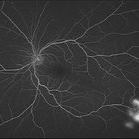
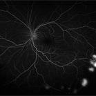
 Proliferative Sickle Cell Retinopathy, Early phase FA OD
May 23 2018 by Hosam Attia, MD
Fluorescein angiogram photograph of a 45-year-old African American, male with sickle cell anemia (SC disease), depicting extensive peripheral capillary non-perfusion, with early hyperfluorescence over the ischemic retina temporally, with late staining and diffuse leakage consistent with partially auto-infarcted, but active NVE/sea fan OD.
Imaging device: Optos California Ultra-Wide Field Fundus Camera
Condition/keywords: neovascularization elsewhere (NVE), proliferative retinopathy, sea fan, sickle cell, sickle cell retinopathy
-
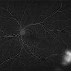
 Proliferative Sickle Cell Retinopathy, Red Free OD
May 23 2018 by Hosam Attia, MD
Red free fundus photo of a 45-year-old African American, male with sickle cell anemia (SC Disease ) with arteriolar attenuation, mild venous tortuosity, Sunburst (S) and large, partially auto-infarcted sea fan, OD.
Imaging device: Optos California Ultra-Wide Field Fundus Camera
Condition/keywords: neovascularization elsewhere (NVE), proliferative retinopathy, sea fan, sickle cell, sickle cell retinopathy
-
 Proliferative Sickle Cell Retinopathy, Red Free OS
May 23 2018 by Hosam Attia, MD
Red free fundus photograph of a 45-year-old African American, male with sickle cell anemia (SC disease) with arteriolar attenuation, mild venous tortuosity, peripheral arterio-venous anastomoses (Inferotemporally), multiple small NVEs/ early sea fans OS.
Photographer: Aaron Appiah, M.D.
Imaging device: Optos California Ultra-Wide Field Fundus Camera
Condition/keywords: neovascularization elsewhere (NVE), proliferative retinopathy, sea fan, sickle cell, sickle cell retinopathy
-
 Proliferative Sickle Cell Retinopathy, Mid phase FA OS
May 23 2018 by Hosam Attia, MD
Fluorescein angiogram photograph of a 45-year-old African American, male with sickle cell anemia (SC disease), depicting peripheral capillary non-perfusion, with multiple, small area of early to mid phase hyperfluorescence over the ischemic retina temporally, with mild late leakage consistent with active NVEs/ early sea fans OS.
Imaging device: Optos California Ultra-Wide Field Fundus Camera
Condition/keywords: neovascularization elsewhere (NVE), proliferative retinopathy, sea fan, sickle cell, sickle cell retinopathy
-
 Hypertensive Retinopathy
Feb 10 2016 by Mallika Goyal, MD
Bilateral hypertensive retinopathy in a 19-year-old girl with renal disease, hypertension and anemia.
Photographer: Mallika Goyal, MD, Apollo Health City, Hyderabad, India
Condition/keywords: hypertensive retinopathy
-
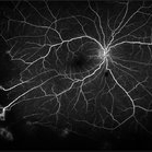
 Slide 9-13
Feb 26 2019 by Lancaster Course in Ophthalmology
Superficial retinal hemorrhages. These hemorrhages have a flame-shaped appearance and are located just beneath the internal limiting membrane of the retina in a 61-year-old man with severe anemia and thrombocytopenia.
Condition/keywords: retinal hemorrhage, thrombocytopenia
-
 Sickle Cell Anemia
Feb 22 2018 by Nichole Lewis
Sickle Cell Anemia
Photographer: Nichole Lewis
Condition/keywords: sickle cell
-
 Proliferative Sickle Cell Retinopathy, Late FA OS
May 23 2018 by Hosam Attia, MD
Fluorescein angiogram photograph of a 45-year-old African American, male with sickle cell anemia (SC disease), depicting peripheral capillary non-perfusion, with multiple, small area of mild late leakage consistent with active NVEs/ early Seafans OS.
Imaging device: Optos California Ultra-Wide Field Fundus Camera
Condition/keywords: neovascularization elsewhere (NVE), proliferative retinopathy, sea fan, sickle cell, sickle cell retinopathy
-
 Hypertensive Retinopathy
Feb 10 2016 by Mallika Goyal, MD
Bilateral hypertensive retinopathy in a 19-year-old girl with renal disease, hypertension and anemia.
Photographer: Mallika Goyal, MD, Apollo Health City, Hyderabad, India
Condition/keywords: hypertensive retinopathy
-
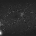 Proliferative Sickle Cell Retinopathy, Late phase FA OD
May 23 2018 by Hosam Attia, MD
Fluorescein angiogram photograph of a 45-year-old African American, male with sickle cell anemia (SC disease), depicting extensive peripheral capillary non-perfusion, with late staining and diffuse leakage consistent with partially auto-infarcted, but active NVE/sea fan OD.
Imaging device: Optos California Ultra-Wide Field Fundus Camera
Condition/keywords: neovascularization elsewhere (NVE), proliferative retinopathy, sea fan, sickle cell, sickle cell retinopathy
-
 Proliferative Sickle Cell Retinopathy, Early phase FA OS
May 23 2018 by Hosam Attia, MD
Fluorescein angiogram photograph of a 45-year-old African American, male with cell anemia (SC disease ), depicting peripheral capillary non-perfusion, with multiple, small area of early to mid phase hyperfluorescence over the ischemic retina temporally, with mild late leakage consistent with active NVEs/ early sea fans OS.
Imaging device: Optos California Ultra-Wide Field Fundus Camera
Condition/keywords: neovascularization elsewhere (NVE), proliferative retinopathy, sea fan, sickle cell, sickle cell retinopathy
-
 Hypertensive Retinopathy
Feb 10 2016 by Mallika Goyal, MD
Bilateral hypertensive retinopathy in a 19-year-old girl with renal disease, hypertension and anemia.
Photographer: Mallika Goyal, MD, Apollo Health City, Hyderabad, India
Condition/keywords: hypertensive retinopathy
-
 Anemia
Aug 21 2013 by Howard Schatz, MD
Twenty seven year old black female, 20/25.
Condition/keywords: anemia
-
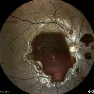
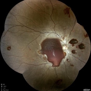
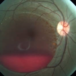 Subhyaloid Hemorrhage
Sep 10 2014 by Mehul A Shah
20-year-female patient presented with sudden loss of vision following blood transfusion for severe anemia.
Photographer: Drashti Netralaya,Dahod
Imaging device: Zeiss ff450
Condition/keywords: subhyaloid hemorrhage
 Loading…