Search results (25 results)
-
 Branch Retinal Vein Occlusion with Multifactorial Macular Edema and Epiretinal Membrane
Oct 3 2024 by Logan ryzenga
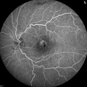
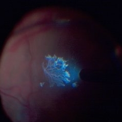
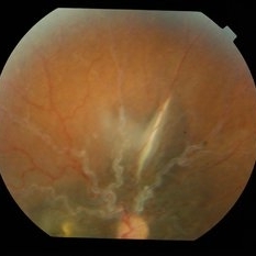
Fluorescein angiogram of a 62 year old woman with cystoid macular edema from concurrent Epiretinal Membrane and Branch Retinal Vein occlusion. She has an extensive history of anti-VEGF injections with stable but unresolved macular edema. Following angiography, it was determined that an epiretinal membrane peel would be indicated in an attempt to achieve resolution of macular edema.
Photographer: Logan Ryzenga
Imaging device: Heidelberg Spectralis
Condition/keywords: 55-degrees, branch retinal vein occlusion (BRVO), cystoid macular edema (CME), epiretinal membrane (ERM), Fluorescein angiography, heidelberg spectralis, hyperfluorescence, leakage, left eye, OS, wide angle imaging
-
Tractional RD-Making the Decision When and Where to Stop
May 23 2024 by ARVIND JAIN M
This is a young gentlemen with defective vision for 3 months in his right eye. He gave the history of recurrent redness of the right past few months. he was diagnosed to have right eye vasculitis with tractional detachment. He underwent uveitic workup and under steroid cover right eye paraplana vitrectomy with membrane peeling with endolaser with c3f8 gas was planned. patient improved significantly. this surgical video demonstrates when and where to stop during membrane peeling and get good results.
Condition/keywords: Eales disease, retinal vasculitis, tractional retinal detachment
-
 PDR- Post Membrane Peeling
Apr 30 2024 by Eesh Nigam, MS
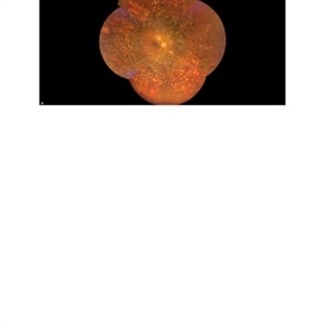
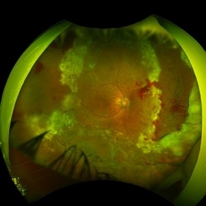
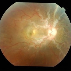
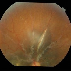
Fundus image of right eye of 47 years old female known Diabetic, underwent Vitrectomy with Membrane peeling with endolaser for vitreous hemorrhage and Tractional retinal detachment. This the post operative image of the eye.
Photographer: Ms Sushmita Ghosh
Imaging device: Clarus 700
Condition/keywords: Attached Retina after Vitrectomy with Membrane peeling with endolaser for PDR with Tractional detachment of Retina
-
 PDR with Vitreous Hemorrhage
Apr 30 2024 by Eesh Nigam, MS
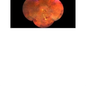
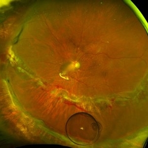
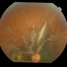
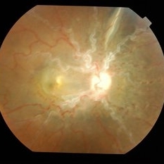
Fundus image of left eye of an 56 years old female known Diabetic underwent vitrectomy and membrane peeling with endolaser for PDR with vitreous hemorrhage.
Photographer: Susmita Saha
Imaging device: Clarus 700
Condition/keywords: Attached retina with Endolaser marks
-
 Before and After Vitrectomy
Nov 17 2023 by Bradley T. Smith, MD, FASRS
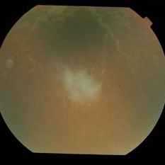
Middle age male diabetic retinopathy and resolving exudate following repair of tractional detachment with membrane peeling.
Condition/keywords: coats-like response, Diabetes, fibrotic neovascularization, fibrovascular proliferation, pars plana vitrectomy (PPV), proliferative diabetic retinopathy (PDR), tractional retinal detachment
-
 Crystallized silicone oil particles in the anterior chamber
Oct 26 2023 by Anmol Naik, MS, DNB, FMRF, FICO, MNAMS
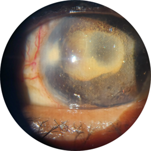
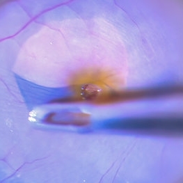
Anterior segment image of a 67-year-old Indian woman who had proliferative diabetic retinopathy with traction retinal detachment with neovascular glaucoma. Patient underwent vitrectomy with membrane peeling with endolaser followed by silicone oil injection 1 year back. Patient was lost to follow up and presented a year later with this picture. She had crystallized silicone oil particles in the anterior chamber rendering a polychromatic lustre like appearance; a unique and rare finding.
Photographer: Anmol Naik
Condition/keywords: Polychromatic lustre in Anterior Chamber
-
 Chronic Full Thickness Macular Hole
Oct 25 2023 by Jessica Hampton, BS
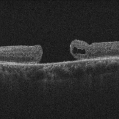
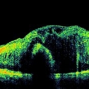
Optical-coherence tomography image of a 65-year old woman with a chronic full-thickness macular hole in the left eye, recurred following three attempts at repair with pars plana vitrectomy, membrane peel, and gas tamponade.
Photographer: Dr. Diana Do, Stanford Medicine, Byers Eye Institute
Condition/keywords: full thickness macular hole, optical coherence tomography (OCT)
-
 Radial Scan Macular Hole
May 25 2023 by Annaka Gooding
Optical Coherence Tomography of a 69 year old male with a Macular Hole affecting his right eye. Patient presented at the clinic following visual distortion that had been ongoing for two weeks. Patients vision was Dcc20/200 PHNI. The Physician recommended Pars Plana Vitrectomy/ Membrane Peel Surgery.
Photographer: Annaka Gooding
Imaging device: Heidelberg Spectralis
Condition/keywords: macular hole, OCT
-
 Epiretinal Membrane Peeling during Vitrectomy Surgery | Intra-Operative Still
Apr 28 2023 by Veer Singh, MS, FVRS, FMRF, FICO (Retina)
Epiretinal Membrane Peeling during Vitrectomy Surgery | Intra-Operative Still
Photographer: Dr. Veer Singh
Condition/keywords: ERM, peeling, vitreomacular surgery
-
 Internal Limiting Membrane Peeling
Feb 2 2022 by Manish Nagpal, MD, FRCS (UK), FASRS
Intraoperative photo of an ILM peeling being done after brilliant blue staining with 25 gauge forceps.
Photographer: Manish Nagpal, Retina Foundation, Ahmedabad, India
Imaging device: Sony PMW -10 MD surgical camera
Condition/keywords: ILM flap, ILM staining, internal limiting membrane (ILM) peeling
-
 Retinal Detachment Following Scleral Buckling, Retinectomy, Laser, and Oil
Jan 31 2022 by Ahmad B. Tarabishy, MD
Ultra wide-field fundus photograph of a 55-year-old gentleman who is 4 days after surgery with scleral buckling, pars plana vitrectomy, perfluoron tamponade, membrane peeling, direct fluid-PFO-oil exchange, nasal and temporal retinectomies, and endolaser photocoagulation. Visual acuity was 20/150 under oil.
Photographer: Megan McLandsborough, Lakeland Eye Clinic
Imaging device: Optos California UWF Camera
Condition/keywords: endolaser, Membrane Peel, PPV, proliferative retinopathy, proliferative vitreoretinopathy (PVR), Retinal Detachment, retinal detachment with retinal defect, scleral buckle, submacular perfluorocarbon liquid (PFO)
-
 Methotrexate Bubble in Silicone Oil Filled Eye: Proliferative Vitreoretinopathy Prevention
Jan 22 2022 by Yoshihiro Yonekawa, MD, FASRS
A middle-aged man underwent complex retinal detachment repair with vitrectomy, membrane peeling, relaxing retinectomy, and silicone oil tamponade. This is a wide-field image immediately after methotrexate injection during a postoperative clinic visit, for the prevention of proliferative vitreoretinopathy. The methotrexate bubble is seen inferiorly.
Photographer: Christina Rowland
Imaging device: Optos California
Condition/keywords: methotrexate, proliferative vitreoretinopathy (PVR), retinectomy, vitrectomy
-
 Internal Limiting Membrane Peeling
Jan 10 2022 by Manish Nagpal, MD, FRCS (UK), FASRS
Intraoperative image of internal limiting membrane being peeled using a 25 gauge ILM forceps. Brilliant blue dye has been used to stain the ILM.
Photographer: Manish Nagpal, Director, Retina Foundation, Ahmedabad
Imaging device: Sony PMW -10 MD surgical camera
Condition/keywords: internal limiting membrane (ILM) peeling
-
 Scleral Buckle with 360 Degree Laser
Sep 22 2021 by Ahmad B. Tarabishy, MD
56 year-old male with a history of retinal detachment OD, complicated by recurrent RD with PVR and cataract. He underwent multiple surgical procedures that included scleral buckling using a 3mm silicone sponge, membrane peel, inferior retinectomy, endolaser, lensectomy, and secondary IOL placement. Current VA is 20/60 OD.
Photographer: Michelle Howarth, Lakeland Eye Clinic
Imaging device: Optos P200TDx
Condition/keywords: encircling scleral buckle, proliferative vitreoretinopathy (PVR)
-
 Detached NVE During PVD induction
Apr 27 2018 by Michael J. Koss, MD, PhD, MBA
A 73-year-old woman with macular pucker underwent a pars plana vitrectomy with membrane peeling. Additionally the patient suffers from diabetic retinopathy after being diagnosed with type 2 diabetes mellitus sixteen years ago. Prior to the procedure she was treated with a series of intravitreal Bevacizumab-injections due to diabetic macular edema. There was no history of a proliferative DRP. During the vitrectomy a branch of an obliterated NVE spontaneously detached and floated freely in the vitreous. The 3D shot was captured via Alcon’s NGENUITY® 3D Visualization System in form of photograph and video providing an outstandingly detailed image of the branched NVE.
Photographer: Michael Koss, Augenzentrum Nymphenburger Hoefe
Imaging device: Alcon’s NGENUITY® 3D Visualization System
Condition/keywords: diabetes, diabetic retinopathy, neovascularization elsewhere (NVE), pars plana vitrectomy (PPV), PVD induction
-
 Biclonal Gammopathy
May 4 2014 by Mallika Goyal, MD
Right eye fundus of a 17-year-old female patient with biclonal gammopathy 2 months following vitrectomy and epimacular membrane peeling. OCT shows no change in macular thickness compared to pre-operative OCT. There was also no visual improvement following surgery.
Photographer: Mallika Goyal, MD, Apollo Health City, Jubilee Hills, Hyderabad, India
Condition/keywords: biclonal gammopathy
-
 Biclonal Gammopathy
May 4 2014 by Mallika Goyal, MD
Right eye superior fundus of a 17-year-old female patient with biclonal gammopathy 2 months following vitrectomy and epimacular membrane peeling.
Photographer: Mallika Goyal, MD, Apollo Health City, Jubilee Hills, Hyderabad, India
Condition/keywords: biclonal gammopathy
-
 Biclonal Gammopathy
May 4 2014 by Mallika Goyal, MD
Right eye inferior fundus of a 17-year-old female patient with biclonal gammopathy 2 months following vitrectomy and epimacular membrane peeling.
Condition/keywords: biclonal gammopathy
-
 Biclonal Gammopathy
May 4 2014 by Mallika Goyal, MD
Right eye superior fundus of a 17-year-old female patient with biclonal gammopathy 2 months following vitrectomy and epimacular membrane peeling.
Photographer: Mallika Goyal, MD, Apollo Health City, Jubilee Hills, Hyderabad, India
Condition/keywords: biclonal gammopathy
-
 Biclonal Gammopathy
May 4 2014 by Mallika Goyal, MD
Right eye fundus of a 17-year-old female patient with biclonal gammopathy 2 months following vitrectomy and epimacular membrane peeling. OCT shows no change in macular thickness compared to pre-operative OCT. There was also no visual improvement following surgery.
Photographer: Mallika Goyal, MD, Apollo Health City, Jubilee Hills, Hyderabad, India
Condition/keywords: biclonal gammopathy
-
 Biclonal Gammopathy
May 4 2014 by Mallika Goyal, MD
Right eye OCT of a 17-year-old female patient with biclonal gammopathy 2 months following vitrectomy and epimacular membrane peeling shows no change in macular thickness compared to pre-operative OCT. There was also no visual improvement following surgery.
Photographer: Mallika Goyal, MD, Apollo Health City, Jubilee Hills, Hyderabad, India
Condition/keywords: biclonal gammopathy
-
---thumb.jpg/image-square;max$300,300.ImageHandler) ERM-Post-Op-20100
ERM-Post-Op-20100
Feb 24 2014 by Susanna S. Park, MD, PhD
Cirrus OCT of the macula of a 60-year-old woman taken 1 year after vitrectomy and ICG dye stained membrane peeling showing persistent foveal puckering and loss of vision despite successful removal of the epiretinal membrane.
Condition/keywords: macular pucker, optical coherence tomography (OCT), post-op, vision loss
-
---thumb.jpg/image-square;max$300,300.ImageHandler) Macular Hole Post-op
Macular Hole Post-op
Jan 10 2014 by Susanna S. Park, MD, PhD
Cirrus OCT of a 65-year-old woman 6 months after vitrectomy and internal limiting membrane peeling with ICG staining and gas tamponade for full-thickness macular hole. The macular hole is closed with excellent foveal morphology and intact photoreceptor layer but mild macular surface irregularity is noted.
Condition/keywords: optical coherence tomography (OCT)
-
 Epiretinal Membrane
Oct 11 2012 by Michael P. Kelly, FOPS
This is a patient with idiopathic panuveitis who developed a visually significant epiretinal membrane. Pars plana vitrectomy with membrane peeling was performed to remove the epiretinal proliferation. I recommend magnifying the image to see the exquisite detail centrally.
Photographer: Michael P. Kelly, FOPS Director, Duke Eye Center Labs, Duke Universtiy Hospital
Imaging device: Zeiss 450Plus
Condition/keywords: epiretinal membrane (ERM), panuveitis
 Loading…