Search results (436 results)
-
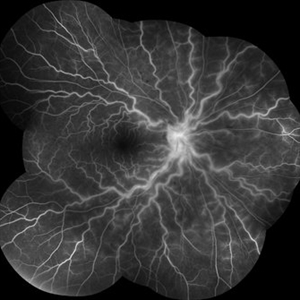
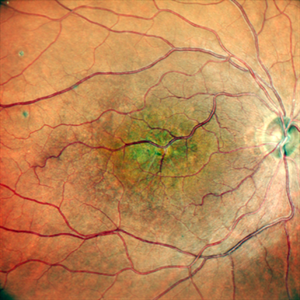
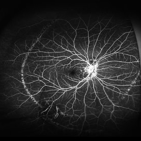
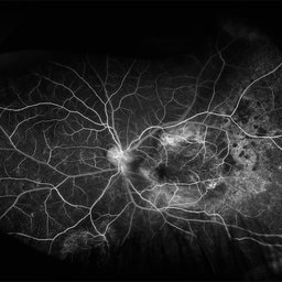
 Lower Nasal Venous Branch Obstruction
Nov 20 2025 by Vicente Nicanor Mancilla Guerrero
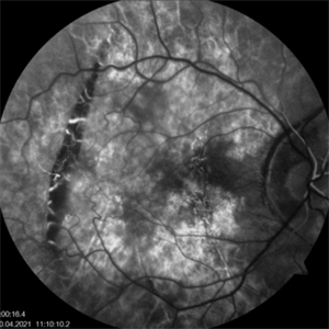
Early fluorescein angiography of the left eye of a 57-year-old patient who presents with a long-standing inferior nasal venous branch occlusion. Dilation and tortuosity can be seen in the lower arch, along with the classic areas of retinal ischemia.
Photographer: Vicente Mancilla Guerrero, Ophthalmic Medical Technologist
Imaging device: Canon cx-1
Condition/keywords: branch retinal vein occlusion (BRVO)
-
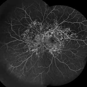
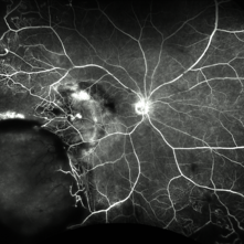
 Angiographic Storm: Fluorescein Leakage in Retinal Vasculitis
Nov 17 2025 by SHRADDHA RAJ SHRIVASTAVA
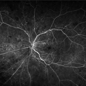
This left eye montage fundus fluorescein angiography (FFA) image of a 19 year old male with idiopathic retinal vasculitis, having skip vasculitic lesions predominantly involving retinal veins. There are areas of blocked fluorescence due to intraretinal hemorrhages, the involved veins have filling defects and occlusions, leading to formation of numerous collateral channels. The inflamed vessels also show perivascular fuzzy hyperfluorescent stain due to leakage of dye. We can also see multiple peripheral capillary non perfusion (CNP) areas, with a 'hot disc', suggestive of ongoing inflammation.
Photographer: Dr. Shraddha Raj Shrivastava
Imaging device: Nidek Mirante SLO/OCT (Confocal scanning/Spectral domain OCT)
Condition/keywords: FA late phase leakage, Fundus Fluorescein Angiography, idiopathic retinal vasculitis, optic disc leakage, VASCULITIS
-
 Twin Maps of Ischemia: A Fluorescein–OCT Angiography Mirror in Post-Transplant Vasculopathy
Nov 13 2025 by Guilherme Sturzeneker, MD, MSc
Fluorescein angiography (top) and optical coherence tomography angiography (bottom) from both eyes of a 29-year-old woman, obtained 49 days after haploidentical hematopoietic stem cell transplantation, demonstrating marked bilateral macular hypoperfusion with perivascular leakage. OCT-A confirms extensive capillary dropout in both eyes. These findings are consistent with atypical bilateral ischemic retinal vasculopathy unrelated to graft-versus-host disease. Management included oral corticosteroids and intravitreal anti-VEGF injections, with partial anatomical and functional response.
Photographer: Patrick Oikawa, IPEPO - Instituto da Visão
Imaging device: Intalight Dream OCT
Condition/keywords: Fluorescein angiography, Hematopoietic stem cell transplantation, Ischemic retinal vasculopathy, Non-graft-versus-host disease, OCT Angiography, oncology
-
 Retinal Hypotonia
Oct 16 2025 by Oftalmontt Clínica Láser
Fluorescein angiography of a 57-year-old male patient. The image is presented in arterial time and shows numerous hyperfluorescent folds in the papillomacular sector together with a marked hypofluorescence in the nasal sector.
Photographer: Ophthalmic Medical Technologist
Imaging device: Canon cx-1
Condition/keywords: angiography with fluorescein, retinal hypotonia
-
 Large Leaking Optic Disc Neovascularization
Sep 9 2025 by Seif Allah Anwar
Large leaking optic disc neovascularization.
Photographer: Dr Seif Anwar
Imaging device: Topcon
Condition/keywords: Fundus Fluorescein Angiography
-
 Fluorescein Angiography Papillophlebitis Salauno
Sep 3 2025 by Pablo Angel Garcia Uribe
In the arteriovenous phase, fluorescein angiography demonstrated venous engorgement and tortuosity, with relative incompetence of the venous walls leading to mild leakage. Optic disc staining with late leakage was also observed. There was no evidence of significant capillary non-perfusion, and only subtle perivenous leakage was noted. The foveal region remained spared.
Photographer: Optom. Marilyn Alvarez Monroy, Clínica Oftalmológica Salauno
Imaging device: Visucam 524, Carl Zeiss Meditec AG, Jena, Germany
Condition/keywords: FA late phase leakage, retina
-
 Snaking Away
Sep 1 2025 by Malvika Singh
Fluorescein angiography montage of a 45 year old man showing areas of staining in a case of healed choroiditis.
Photographer: Dr Malvika Singh, Retina Foundation, Ahmedabad, India
Imaging device: Mirante SLO/OCT
Condition/keywords: fluorescein angiogram (FA), healed choroiditis, serpiginous choroiditis
-
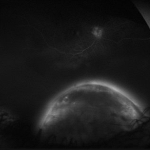 Full Moon
Aug 20 2025 by Gustavo Uriel Fonseca Aguirre
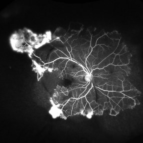
This ultra-widefield fluorescein angiography reveals a hyperfluorescent peripheral inferior choroidal melanoma. The lesion demonstrates early heterogeneous hyperfluorescence with progressive late staining and diffuse leakage.
Photographer: Gustavo U. Fonseca Aguirre, Hospital Conde de Valenciana, Ciudad de México
Condition/keywords: choroidal melanoma, FLUORESCEIN ANGIOGRAPHY
-
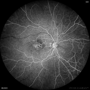 Congenital Retinal Macrovessel
Aug 7 2025 by Cesar Valdez, MD
Fundus photograph and fluorescein angiography of a 43-year-old man with typical findings of congenital retinal macrovessel and RPE atrophy due to chronic macular edema.
Photographer: César Valdez, Instituto Mexicano de Oftalmología, IAP. Querétaro, México.
Imaging device: Zeiss Clarus 700
Condition/keywords: congenital retinal macrovessel
-
 Congenital Retinal Macrovessel
Aug 7 2025 by Cesar Valdez, MD
Fundus photograph and fluorescein angiography of a 43-year-old man with typical findings of congenital retinal macrovessel and RPE atrophy due to chronic macular edema.
Photographer: César Valdez, Instituto Mexicano de Oftalmología, IAP. Querétaro, México.
Imaging device: Nidek Mirante
Condition/keywords: Retina
-
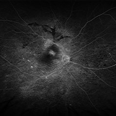
 Black Swan - Optic Disc Melanocytoma
Aug 5 2025 by SHRADDHA ASHOK CHANDORKAR, DNB DO FVRS
Just like the Black Swan which signifies an event that comes as a surprise, can have a major effect, and is often inappropriately rationalized after the fact with the benefit of hindsight, a 50 yr old presbyopic lady came to OPD with complains of diminution of vision - BCVA being 6/6 N6 in both eyes. Fundus examination revealed a pigmented nodule covering the optic disc .In most cases, fluorescein angiography of a melanocytoma of the optic disk demonstrates hypofluorescence throughout the angiogram. OCT disc showed elevated lesion, OCT macula normal and USG B scan with measurements were done to corroborate the posterior extension and to note increase in size if any on follow ups, perimetry was done to check for any field defects. All tests seemingly within normal limits - Patient was counselled and asked for 6 monthly follow up. Optic Disc Melanocytoma usually unilateral known to be a benign lesion that carries an excellent prognosis, the malignancy of this specific condition is rare 1-2%. The mean age at diagnosis of optic disk melanocytoma is 50 years with a median of 52 and range of 1–91 years. It is possible that melanocytoma is a congenital lesion but may not become clinically apparent until later in life, perhaps due to acquisition of pigment in a previously amelanotic lesion.
Photographer: Dr.Shraddha A. Chandorkar
Imaging device: topcon
Condition/keywords: optic disc melanocytoma
-
 Branch Retinal Vein Occlusion
Jul 23 2025 by Malvika Singh
Fluorescein angiogram of a 52 year old man showing capillary non perfusion areas and leakages along the superotemporal arcade and at the macula.
Photographer: Dr Malvika Singh, Retina Foundation, Ahmedabad, India
Imaging device: Mirante SLO/OCT
Condition/keywords: branch retinal vein occlusion (BRVO), CNP areas, FLUORESCEIN ANGIOGRAPHY, fluorescein leakage, macular edema
-
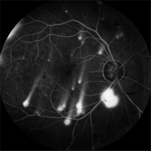 Shooting Stars
Jul 9 2025 by Majda Hadziahmetovic, MD
Fluorescein angiography image demonstrating multiple areas of neovascularization in a middle-aged male patient with long-standing diabetes.
Condition/keywords: proliferative diabetic retinopathy (PDR)
-
 Schlaegel Line
Jul 7 2025 by César Adrián Gómez Valdivia, MD
Fluorescein Angiography showing a Schlaegel Line in a patient with suspected PHOS
Photographer: @eyemissu2
Imaging device: California ICG OPTOS
Condition/keywords: Schlaegel Line
-
 Fluorescein Angiography (FA) of a Primary Retinal Vasoproliferative Tumor
Jun 29 2025 by Marcelo Zas, MD PhD
We present a case of a 33-year-old male patient, who presented with decreased visual acuity in his right eye with 20/80, presenting a primary retinal vasoproliferative tumor in the lower temporal quadrant. The fluorescein angiography findings are: 1. Early hyperfluorescence due to its rich intrinsic vascularity and often has dilated feeding arterioles and draining venules. 2. Marked progressive leakage from the tumor vessels. 3. The late leakage often obscures fine vascular details in the late phase and corresponds to exudation and macular edema seen clinically. 4. Staining of surrounding exudates, RPE disturbances and gliosis. 5. In our case also a marked peripheral capillary closure in the areas adjacent to the tumor and in other quadrants as well.
Photographer: Marcelo Zas MD PhD
Condition/keywords: RETINAL VASOPROLIFERATIVE TUMOR
-
 BRVO-MCR-FFA
Jun 27 2025 by Gayathri M S
Case of impending Macular BRVO. 52 year old female on medication for Hypertension and Diabetes Mellitus since 2 years. BCVA 6/18,N6. IOP 16 mmHg. Multicolor Reflectance and Fundus Fluorescein Angiography picture shows mild dilated tortuous inferior vessels, small areas of capillary non perfusion and few microanurysms.
Photographer: Gayathri MS
Imaging device: Heidelberg spectralis
Condition/keywords: fluorescein angiogram (FA), macular branch retinal vein occlusion (BRVO), multicolor
-
 Fluorescein Angiography in Choroidal Rupture
Jun 26 2025 by Hector Gabriel Moreno Solano, MD, MHA
Fluorescein angiography of the right eye reveals three linear hypofluorescent lesions with progressive staining at the edges, consistent with choroidal ruptures. These lesions are temporally located in the posterior pole, with one of them situated near the fovea but without direct foveal involvement. The pattern is suggestive of previous blunt ocular trauma.
Photographer: Héctor Gabriel Moreno Solano, Instituto Mexicano de Oftalmología “IMO I.A.P”
Imaging device: CLARUS
Condition/keywords: Choroidal Rupture, fluorescein angiogram (FA)
-
 Post-traumatic Choroida Rupture-Fluorescein Angiography
Jun 20 2025 by Alexander Babaev
Fluorescein angiography of an 46-year-man with a choroidal rupture after blunt trauma, complicated CNV. 07.15, Dye leakage is visible along the edges of the rupture
Photographer: Babaev Alexander, Saint-Petersburg, medical clinic "Vision"
Imaging device: Carl Zeiss Visucam 500
Condition/keywords: blunt trauma
-
 Post-traumatic Choroidal Rupture-Fluorescein Angiography
Jun 20 2025 by Alexander Babaev
Fluorescein angiography of an 46-year-man with a choroidal rupture after blunt trauma, complicated CNV. 00.31s, Dye leakage is visible along the edges of the rupture
Photographer: Babaev Alexander, Saint-Petersburg, medical clinic "Vision"
Imaging device: Carl Zeiss Visucam 500
Condition/keywords: fluorescein angiogram (FA)
-
 Post-traumatic Choroidal Rupture-Fluorescein Angiography
Jun 20 2025 by Alexander Babaev
Fluorescein angiography of an 46-year-man with a choroidal rupture after blunt trauma, complicated CNV. 00.16s
Photographer: Babaev Alexander, Saint-Petersburg, medical clinic "Vision"
Imaging device: Carl Zeiss Visucam 500
Condition/keywords: blunt trauma
-
 Central Retinal Vein Occlusion With Waldenstroms macroglobulinemia
Jun 18 2025 by Korey Starkey
64-year-old patient presents with CRVO with secondary macular edema in both eyes. Venous beading present in 2/4 quadrants OU. Patient diagnosed with Waldenstroms macroglobulinemia, found on SPEP and bone marrow biopsy. Treatment recommended of anti-vegF intravitreal injections OU.
Photographer: Korey Starkey
Imaging device: Optos
Condition/keywords: attenuated vessels, central retinal vein occlusion (CRVO), CRVO, FA early phase, FLUORESCEIN ANGIOGRAPHY, macular edema, Optomap, OPTOS CALIFORNIA, severe NPDR, venous beading, Waldenstroms macroglobulinemia
-
 Proliferative Sickle Retinopathy
Jun 13 2025 by Brandon I Fram, MD
30 year-old with HbSC sickle retinopathy found to have profound retinal ischemia and florid peripheral neovascularization.
Imaging device: Fluorescein Angiography
Condition/keywords: proliferative sickle retinopathy, retinal ischemia, sea fan, sickle cell retinopathy
-
 Uveal Effusion Syndrome
Jun 13 2025 by Brandon I Fram, MD
75 year-old with bilateral inferior serous detachments, right more than left. Scleral window with biopsy showed scleral thickening with stromal deposits of amorphous glycosaminoglycan-like material.
Imaging device: Fluorescein Angiography
Condition/keywords: exudative retinal detachment, idiopathic uveal effusion syndrome, leopard spots, uveal effusion, uveal effusion syndrome
-
 Coats Disease
May 27 2025 by César Adrián Gómez Valdivia, MD
Fluorescein Angiography on an 8 year-old male patient with Coats disease. Vascular leakage causes hard exudates which may be peripheral (near the vascular abnormalities) or midperipheral and central (at the macula. Findings were bilateral.
Photographer: @eyemissu2
Imaging device: California ICG OPTOS
Condition/keywords: Coats disease
-
 VKH Pseudotumor – Fluorescein Angiography
May 11 2025 by Felipe Murati
Fluorescein angiography image from a 36-year-old woman with chronic Vogt-Koyanagi-Harada (VKH) syndrome showing a pseudotumor-like lesion with late-phase staining and no active leakage. The image highlights subretinal fibrosis in the right eye, stable under long-term immunosuppressive therapy with mycophenolate mofetil and adalimumab. No signs of active choroiditis are present, confirming a quiescent phase.
Photographer: Felipe A. Murati, MD, University of Arizona
Imaging device: Optos California, fluorescein angiography modality
Condition/keywords: choroiditis, Fluorescein angiography, granulomatous uveitis, Optos FA, pseudotumor, subretinal fibrosis, VKH, Vogt-Koyanagi-Harada
 Loading…