-
 PED and VMA-LE
Feb 7 2018 by Ogugua Ndubuisi Okonkwo, MD, FRCS (Edin), FASRS
Left eye OCT of same 73-year-old female showing drusenoid PED and Vitreomacular Adhesion .
Photographer: Oreoluwa, Eye Foundation Retina Institute . Lagos
Condition/keywords: drusenoid deposit, vitreomacular traction (VMT)
-
 Right Eye One month Post Operative VMT Relaease
Feb 7 2018 by Ogugua Ndubuisi Okonkwo, MD, FRCS (Edin), FASRS
Right eye one month post-operative OCT showing VMT release with return of the foveal depression and good vision. There is marked reduction in intra retina schisis and preserved outer retina layers.
Photographer: Oreoluwa, Eye Foundation Retina Institute . Lagos
Condition/keywords: drusenoid deposit, vitreomacular traction (VMT)
-
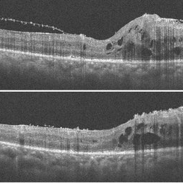
 Pre-Op-Fern-Like-Appearance-of-VMT
Feb 7 2018 by Ogugua Ndubuisi Okonkwo, MD, FRCS (Edin), FASRS
Right eye pre operative OCT of a 73-year-old woman with age related maculopathy and progressively worsening Vitreomacular Traction (VMT) , requiring surgical release of VMT. Fern like VMT is seen and undulating RPE layer showing the drusenoid deposits.
Photographer: Oreoluwa , Eye Foundation Retina Institute . Lagos
Condition/keywords: drusenoid deposit, vitreomacular traction (VMT)
-
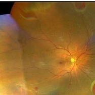
 Age-Related-Maculopathy and Vitreo Macular Adhesion in Left Eye
Feb 7 2018 by Ogugua Ndubuisi Okonkwo, MD, FRCS (Edin), FASRS
Left eye Fundus photograph of a 73-year-old woman with age related maculopathy , progressive drusenoid deposits and Vitreomacular Adhesion (VMA) monitored over a 10 year duration.
Photographer: Okonkwo Ogugua, Eye Foundation Retina Institute . Lagos
Condition/keywords: drusenoid deposit, vitreomacular traction (VMT)
-
 Age-Related-Maculopathy with Advanced Vitreo Macular Traction in Right Eye
Feb 7 2018 by Ogugua Ndubuisi Okonkwo, MD, FRCS (Edin), FASRS
Right eye Fundus photograph of a 73-year-old woman with age related maculopathy, progressive drusenoid deposits and progressively worsening Vitreomacular Traction (VMT) monitored over a 10 year duration. Requiring surgical release of VMT.
Photographer: Okonkwo Ogugua, Eye Foundation Retina Institute . Lagos
Condition/keywords: drusenoid deposit, vitreomacular adhesion, vitreomacular traction (VMT)
-
 Rhegmatogenous Retinal Detachment- Macula-On
Sep 14 2021 by Ogugua Ndubuisi Okonkwo, MD, FRCS (Edin), FASRS
Preoperative Optical Coherence Tomography (OCT) of the right eye of a 35-year-old male showing detached retina ( subretinal fluid elevates the retina), sparing the macula.
Photographer: Oreoluwa Olabode , Eye Foundation Hospital, Lagos
Imaging device: Optovue Avanti RTVue
Condition/keywords: re-attached retinal detachment (RRD)
-
 Persistence of Sub Retinal Fluid Post Retinal Reattachment Surgery
Sep 14 2021 by Ogugua Ndubuisi Okonkwo, MD, FRCS (Edin), FASRS
Postoperative Optical Coherence Tomography (OCT) of the right eye of a 35-year-old male showing persistence of subretinal fluid in the original area of the retinal detachment.
Photographer: Oreoluwa , Eye Foundation Hospital, Lagos
Imaging device: Optovue Avanti RTVue
Condition/keywords: re-attached retinal detachment (RRD), subretinal fluid
-
 Post retinal Reattachment Surgery Glaucoma with Progression
Sep 14 2021 by Ogugua Ndubuisi Okonkwo, MD, FRCS (Edin), FASRS
Change analysis of a 35-year-old male who developed glaucoma post retinal reattachment surgery with the progression of optic nerve damage despite maximum medical therapy.
Photographer: Oreoluwa , Eye Foundation Hospital, Lagos
Imaging device: Optovue Avanti RTVue
Condition/keywords: retina surgery complications
-
 Macular Hole Retinal Detachment
Sep 14 2021 by Ogugua Ndubuisi Okonkwo, MD, FRCS (Edin), FASRS
Preoperative optical coherence tomogram (OCT) of the right eye in a 65-year-old male. This shows a detached retina with significant subretinal fluid in the macular area. There is a full-thickness defect and discontinuity of the foveomacula, which represents a detached macular hole.
Photographer: Oreoluwa Olabode , Eye Foundation Hospital, Lagos.
Imaging device: Optovue Avanti RTVue.
Condition/keywords: macular hole retinal detachment, retinal detachment with retinal defect
-
 Post Retinal Reattachment Surgery Epiretinal Membrane
Sep 14 2021 by Ogugua Ndubuisi Okonkwo, MD, FRCS (Edin), FASRS
Postoperative optical coherence tomography (OCT) of the right eye in a 65-year-old male who had retinal reattachment surgery for a macular hole retinal detachment. This OCT scan shows epiretinal membrane and intraretinal cystic fluid spaces.
Photographer: Oreoluwa Olabode , Eye Foundation Hospital, Lagos.
Imaging device: Optovue Avanti RTVue.
Condition/keywords: epiretinal membrane (ERM), macular hole retinal detachment, Retinal Reattachment surgery
-
 Macular Hole-RRD
Feb 19 2024 by Ogugua Ndubuisi Okonkwo, MD, FRCS (Edin), FASRS
Fundus photograph of a 60-year-old male myope, who has multiple retina tears with rolled edges in the peripheral retina, associated with a macular hole and retinal detachment.
Photographer: Zainab Ogunsanu, Eye Foundation Hospital & Eye Foundation Retina Institute, Lagos.
Imaging device: ZEISS CLARUS 700
Condition/keywords: Retinal detachment with macular hole
-
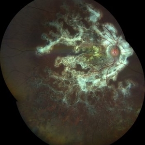 Serpiginous Choroidopathy
Mar 21 2024 by Ogugua Ndubuisi Okonkwo, MD, FRCS (Edin), FASRS
This is a right eye widefield fundus photograph of a 13-year-old male with a peripapillary ring of fibrotic scar that extends subretinally in finger-like projects along the vascular arcades and into the macula, with an extension of the scarring into the inferior retina, where it appears as a pigmented mottling.
Photographer: Zainab Ogunsanu, Eye Foundation Hospital & Eye Foundation Retina Institute, Lagos
Imaging device: ZEISS CLARUS 700
Condition/keywords: serpiginous like choroiditis
-
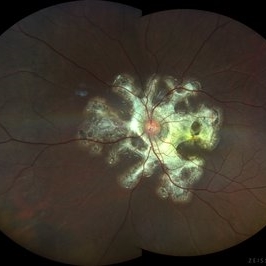 Serpiginious Choroidopathy
Mar 21 2024 by Ogugua Ndubuisi Okonkwo, MD, FRCS (Edin), FASRS
This is a left eye widefield fundus photograph of a 13-year-old male with a peripapillary ring of fibrotic scar that extends subretinally in finger-like projects along the vascular arcades and into the macula.
Photographer: Zainab Ogunsanu, Eye Foundation Hospital & Eye Foundation Retina Institute, Lagos.
Imaging device: ZEISS CLARUS 700
Condition/keywords: serpiginous like choroiditis
A project from the American Society of Retina Specialists