-
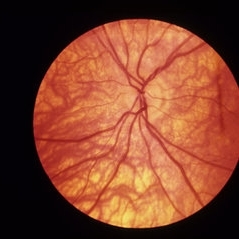
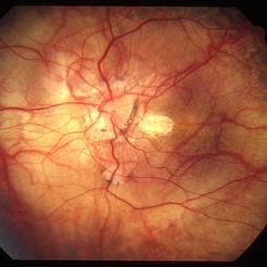
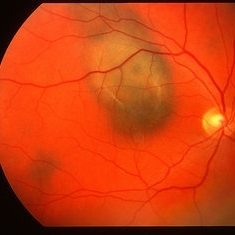
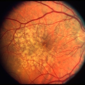
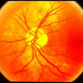
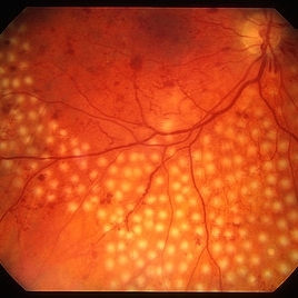
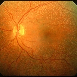
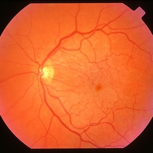
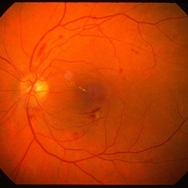
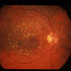 Dry AMD
Jun 4 2014 by Henry J. Kaplan, MD
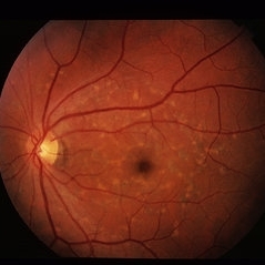
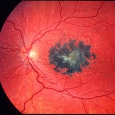
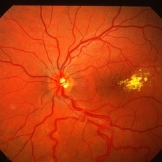
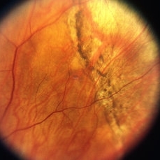
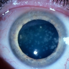
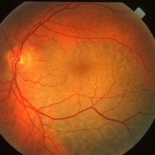
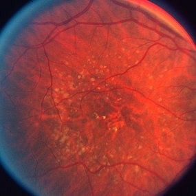
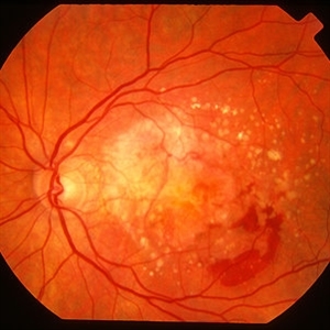
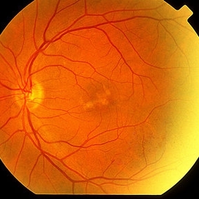
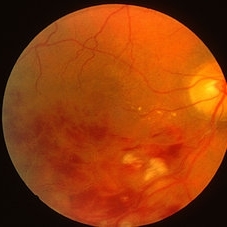
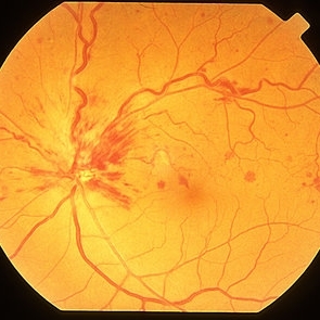
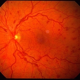
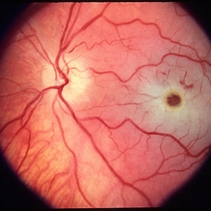
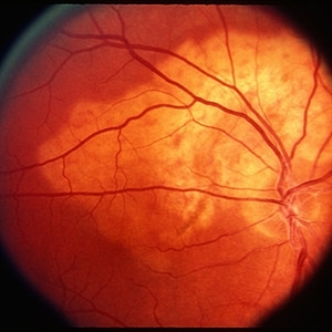
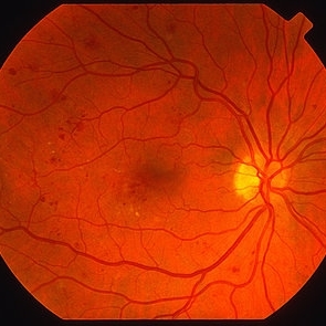
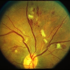
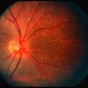
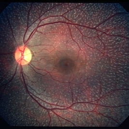
Multiple hard and calcified drusen. #1
Condition/keywords: age-related macular degeneration (AMD), calcified drusen, drusen, dry age-related macular degeneration (dry AMD)
-
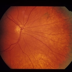
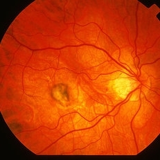
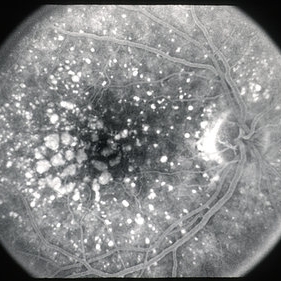
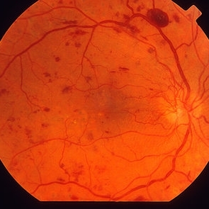
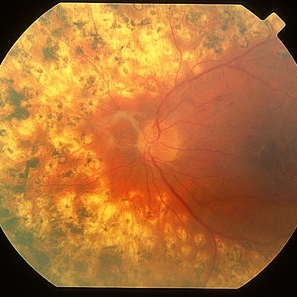
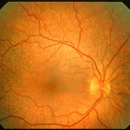
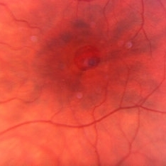
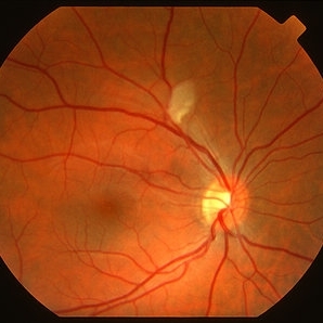
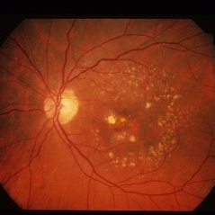 Dry AMD
Jun 4 2014 by Henry J. Kaplan, MD
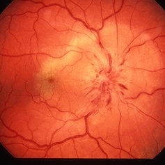
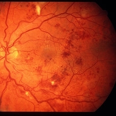
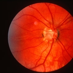
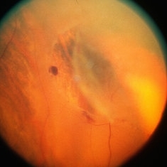
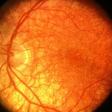
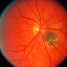
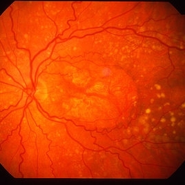
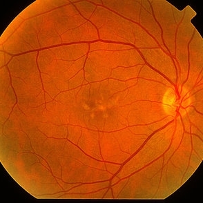
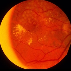
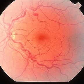
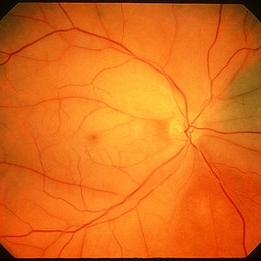
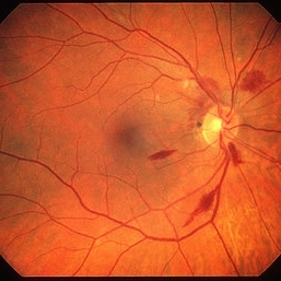
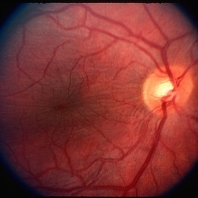
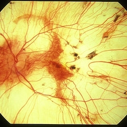
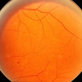
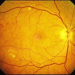
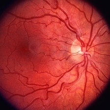
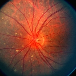
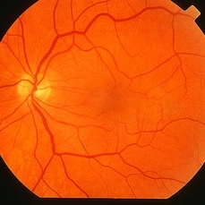
Multiple drusen with RPE changes in the macula #2.
Condition/keywords: age-related macular degeneration (AMD), dry age-related macular degeneration (dry AMD)
-
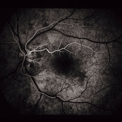
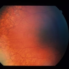
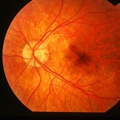
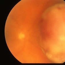
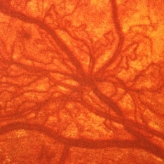
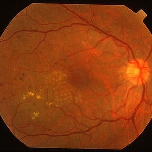
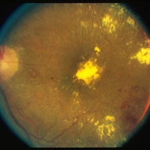
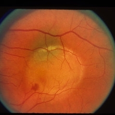 Fibrovascular PED
Jun 4 2014 by Henry J. Kaplan, MD
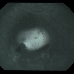
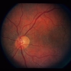
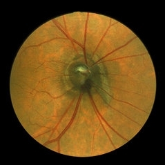
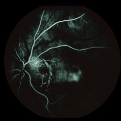
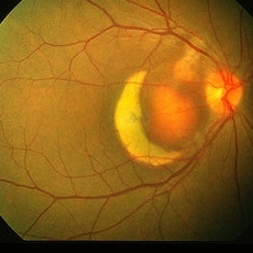
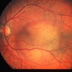
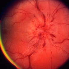
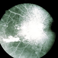
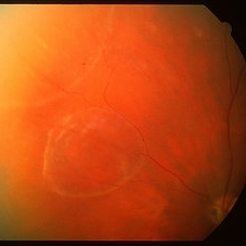
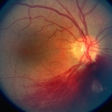
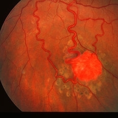
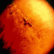
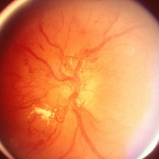
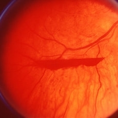
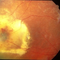
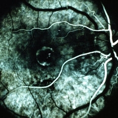
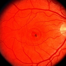
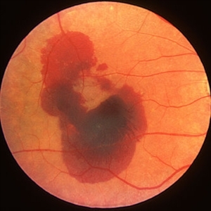
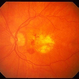
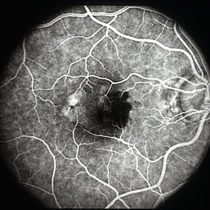
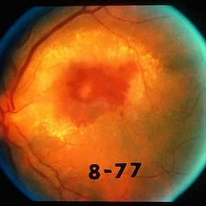
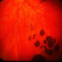
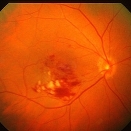
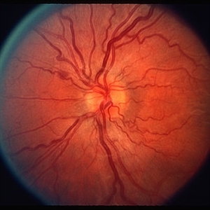
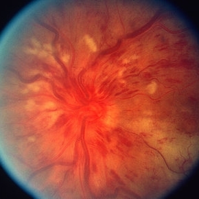
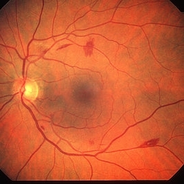
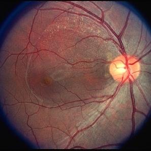
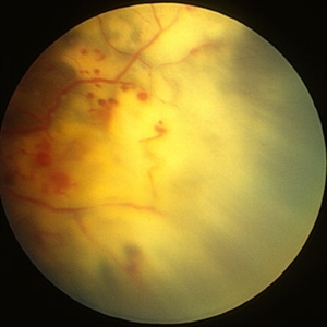
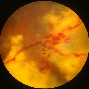
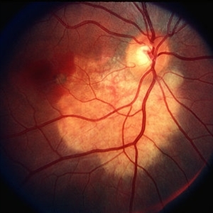
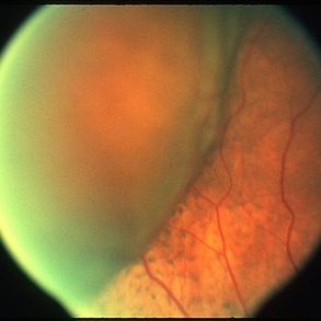
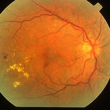
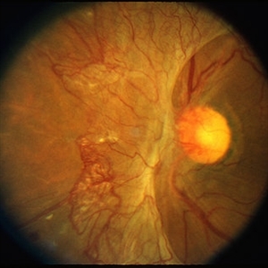
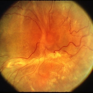
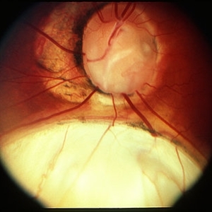
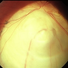
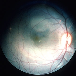
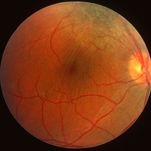
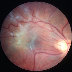
Fundus photograph of a large fibrovascular pigment epithelial detachment with inferonasal hemorrhage. #1
Condition/keywords: pigment epithelial detachment (PED), vascularized pigment epithelial detachment (PED)
-
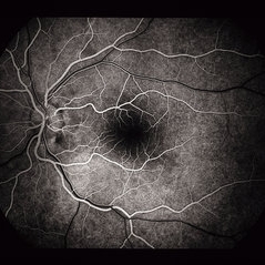
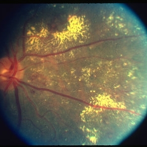
 Fibrovascular PED
Jun 4 2014 by Henry J. Kaplan, MD
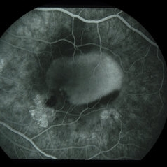
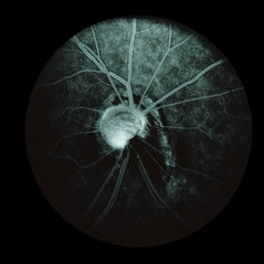
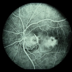
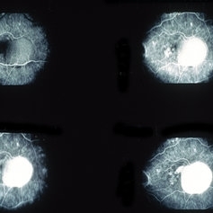
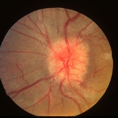
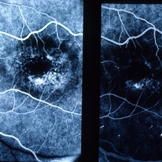
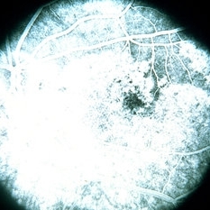
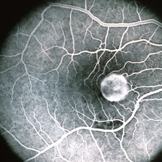
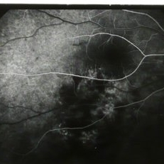
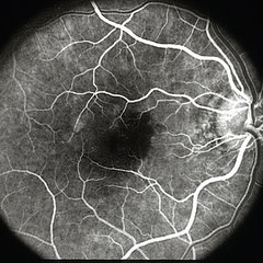
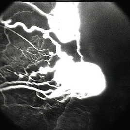
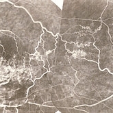
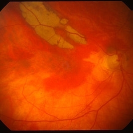
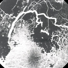
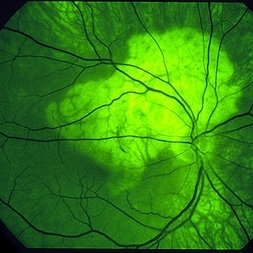
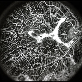
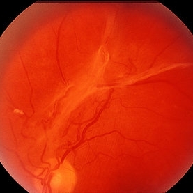
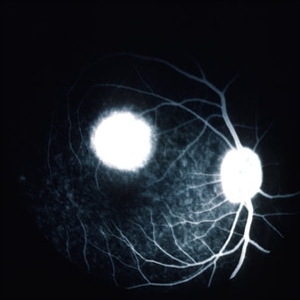
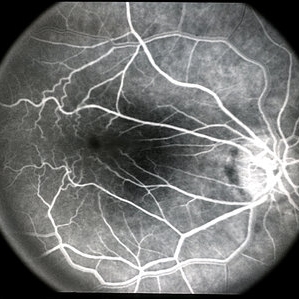
Fluorescein angiography in AV phase demonstrates filling of the PED with an inferonasal notch secondary to the fibrovascular tissue. #2
Condition/keywords: pigment epithelial detachment (PED), vascularized pigment epithelial detachment (PED)
-
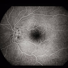
 Fibrovascular PED
Jun 4 2014 by Henry J. Kaplan, MD
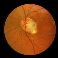
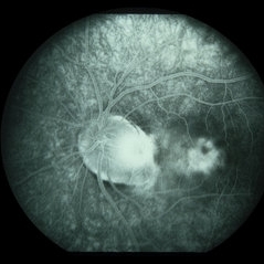
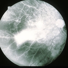
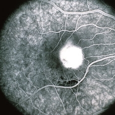
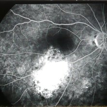
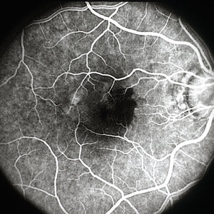
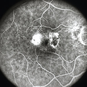
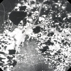
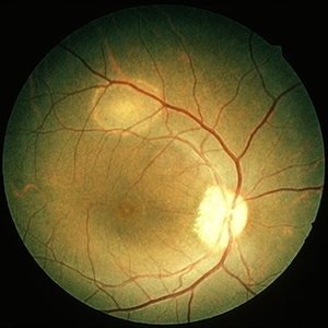
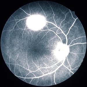
Late phase F/A in the same patient demonstrates filling of the PED with an inferonasal notch which is secondary to fibrovascular membrane and late granular hyperfluorescence of the membrane. #3
Condition/keywords: pigment epithelial detachment (PED), vascularized pigment epithelial detachment (PED)
-
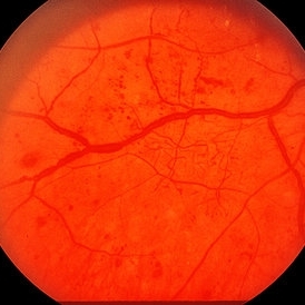
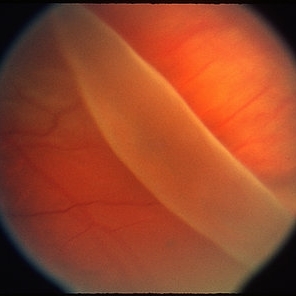
 Retinoschisis
Jun 4 2014 by Henry J. Kaplan, MD
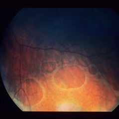
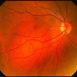
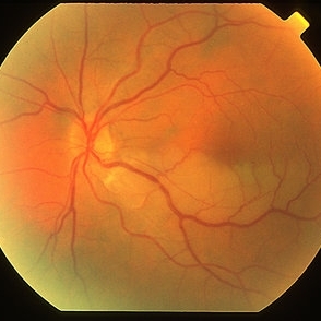
Senile degenerative peripheral retinoschisis with outer wall holes.
Condition/keywords: retinoschisis
-
 Fabry's Disease Carrier
Jun 4 2014 by Henry J. Kaplan, MD
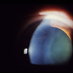
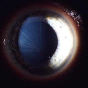
A carrier of Fabry's disease who demonstrates cornea verticillata.
Condition/keywords: cornea verticillata, Fabry disease
-
 Fabry's Disease
Jun 4 2014 by Henry J. Kaplan, MD
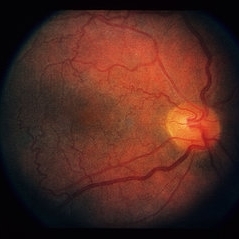
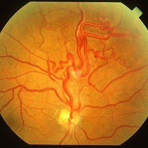
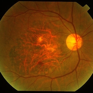
Retinal vasculature tortuosity in Fabry's disease: OD. #2
Condition/keywords: Fabry disease
-
 Fabry's Disease
Jun 4 2014 by Henry J. Kaplan, MD
Retinal vasulature tortuosity in Fabry's disease: OS . #3
Condition/keywords: Fabry disease
-
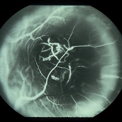
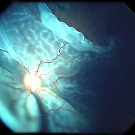
 Sickle Cell Sea Fan Retinopathy
Jun 4 2014 by Henry J. Kaplan, MD
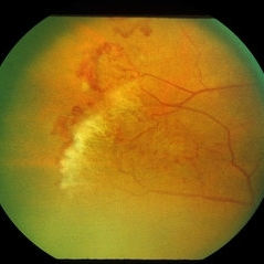
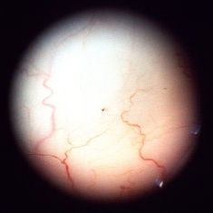
Sea fan peripheral retinal neovascularization in sickle cell anemia.
Condition/keywords: sea fan, sickle cell retinopathy
-
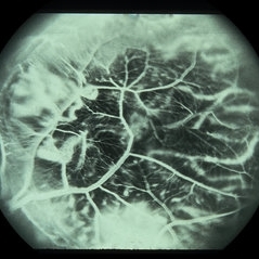
 Sickle Cell Sea Fan Retinopathy
Jun 4 2014 by Henry J. Kaplan, MD
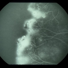
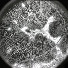
Fluorescein angiogram of the same patient shows extensive capillary non-perfusion anterior to the neovascularization area. #2
Condition/keywords: sea fan, sickle cell retinopathy
-
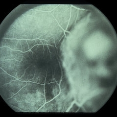
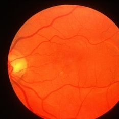
 Normal Red Free Image
Jun 4 2014 by Henry J. Kaplan, MD
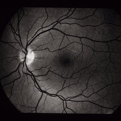
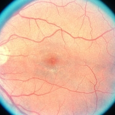
Normal Red free image OS. #1
Condition/keywords: normal eye, red-free
-
 Normal F/A
-
 Normal F/A
Jun 4 2014 by Henry J. Kaplan, MD
Laminar phase angiogram (early arteriovenous phase). #3
Condition/keywords: normal eye
-
 Normal F/A
-
 Albinism
-
 Albinism
-
 RPE Reticular Degeneration
Jun 4 2014 by Henry J. Kaplan, MD
RPE reticular degeneration.
Condition/keywords: retinal pigment epithelium, senile reticular degeneration
-
 RPE Reticular Degeneration
Jun 4 2014 by Henry J. Kaplan, MD
RPE reticular degeneration. #2
Condition/keywords: senile reticular degeneration
-
 Reticular Pattern Dystrophy
Jun 4 2014 by Henry J. Kaplan, MD
Reticular pattern dystrophy :OD. #1
Condition/keywords: pattern dystrophy, reticular pattern dystrophy
-
 Reticular Pattern Dystrophy
Jun 4 2014 by Henry J. Kaplan, MD
Reticular pattern dystrophy: OS . #2
Condition/keywords: pattern dystrophy, reticular pattern dystrophy
-
 Optic Disc Melanocytoma
Jun 4 2014 by Henry J. Kaplan, MD
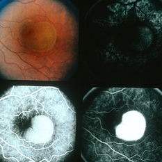
Optic disc melanocytoma with jet black pigmentation . #1
Condition/keywords: melanocytoma, optic disc melanocytoma
-
 Optic Disc Melanocytoma
Jun 4 2014 by Henry J. Kaplan, MD
Fluorescein angiogram of the same patient shows complete blockage of fluorescence around the nerve specially inferiorly secondary to melanocytoma pigmentation. #2
Condition/keywords: melanocytoma, optic disc melanocytoma
-
 Optic Disc Pit
Jun 4 2014 by Henry J. Kaplan, MD
Optic disc pit in the temporal part of optic nerve with associated CSR.
Condition/keywords: central serous retinopathy (CSR), optic disc pit
-
 Optic disc Pit and CSR
Jun 4 2014 by Henry J. Kaplan, MD
Arterial phase F/A of the same patient shows hypofluorescence of the pit area and hyperfluorescence secondary to pooling in the serous detachment part. #2
Condition/keywords: central serous retinopathy (CSR), optic disc pit
-
 Optic Disc Pit and CSR
Jun 4 2014 by Henry J. Kaplan, MD
Late AV phase F/A of the same patient with optic disc pit and CSR; intensity of hyperfluorescence in serous detachment part increased and edges of the pit are stained . #3
Condition/keywords: central serous retinopathy (CSR)
-
 Optic Disc Pit and CSR
Jun 4 2014 by Henry J. Kaplan, MD
Late phase angiogram clearly shows the abnormally large optic nerve with temporal pit which is stained. #4
Condition/keywords: central serous retinopathy (CSR), optic disc pit
-
 Choroidal Melanoma
-
 Choroidal Melanoma
May 28 2014 by Henry J. Kaplan, MD
Fluorescein angiography of a patient with choroidal melanoma clearly shows the double circulation of the retina and whithin the melanoma #2
Imaging device: Fluorescein angiography
Condition/keywords: melanoma
-
 Choroidal Melanoma
Jun 4 2014 by Henry J. Kaplan, MD
Choroidal melanoma, double circulation.later AV phase fluorescein angiography again shows the double circulation whithin the tumor and retina. #3
Condition/keywords: double circulation
-
 Choroidal Melanoma
Jun 4 2014 by Henry J. Kaplan, MD
Late phase angiogram of the same patient with choroidal melanoma, double circulation. #4
-
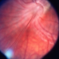 PHPV
PHPV
May 2 2013 by Henry J. Kaplan, MD
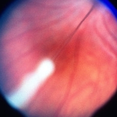
Notice the persistant fetal vasculature which traverses anteriorly. #1.
Condition/keywords: persistent fetal vasculature (PFV), persistent hyperplastic primary vitreous (PHPV)
-
 PHPV
PHPV
May 2 2013 by Henry J. Kaplan, MD
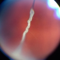
The same patient ; notice the persistant hyaloid artery which is changed to fibrotic tissue anteriorly; #2.
Condition/keywords: persistent fetal vasculature (PFV), persistent hyperplastic primary vitreous (PHPV)
-
 PHPV
PHPV
May 2 2013 by Henry J. Kaplan, MD
The same patient; hyaloid artery has changed to fibrous tissue anteriorly; #3.
Condition/keywords: hyaloid artery, persistent fetal vasculature (PFV), persistent hyperplastic primary vitreous (PHPV)
-
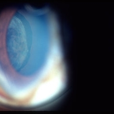
 Siderosis
May 2 2013 by Henry J. Kaplan, MD
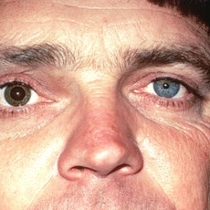
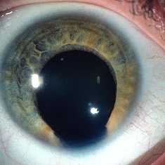
Iris heterochromia in siderosis due to retained metallic intraocular FB; notice the dilated pupil.
Condition/keywords: siderosis
-
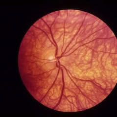
 Posterior Staphylamas
May 2 2013 by Henry J. Kaplan, MD
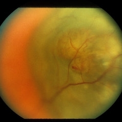
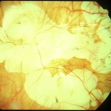
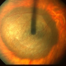
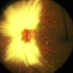
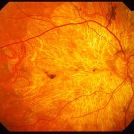
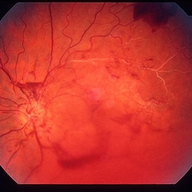
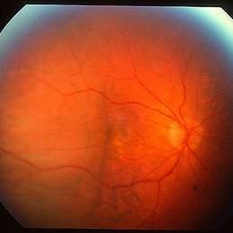
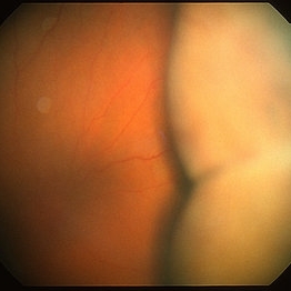
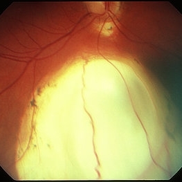
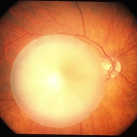
Highly myopic changes with posterior staphyloma and visible sclera.
Condition/keywords: high myopia, posterior staphyloma
-
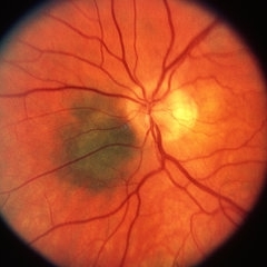
 High Myopia
May 2 2013 by Henry J. Kaplan, MD
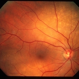
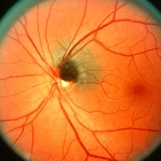
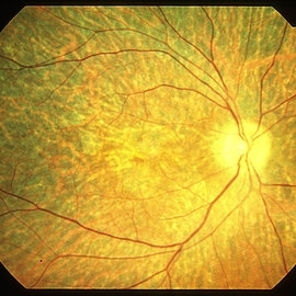
Chorioretinal atrophy in high myopia and tilted disc.
Condition/keywords: high myopia, tilted disc
-
 Myopic CNV
May 2 2013 by Henry J. Kaplan, MD
Subretinal membrane in high myopia.
Condition/keywords: myopic choroidal neovascularization (CNV)
-
 Myopic CNV
May 2 2013 by Henry J. Kaplan, MD
Choroidal neovascularization with hemorrhage in a highly myopic patient.
Condition/keywords: myopic choroidal neovascularization (CNV)
-
 Fibrovascular PED
May 2 2013 by Henry J. Kaplan, MD
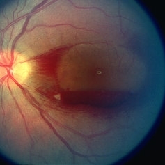
Elevated PED is visible in the temporal part of the macula; #1.
Condition/keywords: exudative age-related macular degeneration, fibrovascular pigment epithelial detachment (PED)
-
 Fibrovascular PED
May 2 2013 by Henry J. Kaplan, MD
Fluorescein angiogram of the fibrovascular PED in the same patient; early homogeneous hyperfluorescence in the PED area which is increased in fluorescence to the late phase with a granular hyperfluorescence adjacent to PED in the foveal side which starts in the mid-phase of the F/A and is increased later; #2.
Condition/keywords: exudative age-related macular degeneration, fibrovascular pigment epithelial detachment (PED)
-
 Exudative AMD
May 2 2013 by Henry J. Kaplan, MD
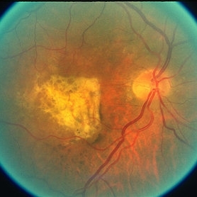
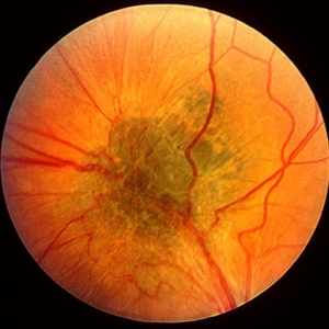
Exudative AMD as hemorrhagic PED with adjacent old subretinal hemorrhage which has turned yellow in color.
Condition/keywords: exudative age-related macular degeneration, hemorrhagic PED, pigment epithelial detachment (PED)
-
 Fibrovascular PED
May 2 2013 by Henry J. Kaplan, MD
Fundus photograph and fluorescein angiography of a fibrovascular PED with a typical notch on F/A.
Condition/keywords: exudative age-related macular degeneration, fibrovascular pigment epithelial detachment (PED), vascularized pigment epithelial detachment (PED)
-
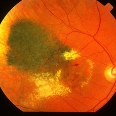
 Choroidal metastasis
May 2 2013 by Henry J. Kaplan, MD
Choroidal metastasis from breast cancer in the superior retina as a yellow lesion with Peau d'Orange appearance. Right eye.
Condition/keywords: choroidal metastasis
-
 Choroidal metastasis
May 2 2013 by Henry J. Kaplan, MD
Choroidal metastatsis in the macula as a yellow elevated mass; left eye.
Condition/keywords: choroidal metastasis
-
 Choroidal Melanoma
May 2 2013 by Henry J. Kaplan, MD
Intermediate melanotic choroidal melanoma.
-
 Choroidal melanoma; B-scan
May 2 2013 by Henry J. Kaplan, MD
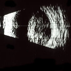
B-scan of a choroidal melanoma shows dome shaped lesion #1.
Condition/keywords: B scan ultrasound
-
 Choroidal melanoma ; A scan
May 2 2013 by Henry J. Kaplan, MD
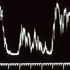
Typical decrescendo low internal reflectivity in choroidal melanoma (same patient)#2.
Condition/keywords: A-scan ultrasound
-
 Choroidal Melanoma
May 2 2013 by Henry J. Kaplan, MD
Choroidal melanoma involving the macula.
-
 Choroidal Melanoma
May 2 2013 by Henry J. Kaplan, MD
Large amelanotic choroidal melanoma extending to the pupillary area; posterior pole is out of focus because the canera is focused on the front face of the tumor.
-
 Ciliary body melanoma
May 2 2013 by Henry J. Kaplan, MD
Ciliary body melanoma visible through the pupil.
Condition/keywords: ciliary body melanoma
-
 Choroidal Melanoma
May 2 2013 by Henry J. Kaplan, MD
Peripapillary choroidal melanoma.
-
 Oxalosis
May 2 2013 by Henry J. Kaplan, MD
Crystalline retinopathy in oxalosis as a result of calcium oxalate deposits in the retina ; also deposition in RPE which causes fleck retina as pigmentary lesion in the center.
Condition/keywords: crystalline retinopathy, oxalosis
-
 AION With Ciliotretinal Artery Occlusion
May 2 2013 by Henry J. Kaplan, MD
AION accompanied by partial CRAO which is visible as retinal edema and cherry red spot.
Condition/keywords: anterior ischemic optic neuropathy, central retinal artery occlusion (CRAO)
-
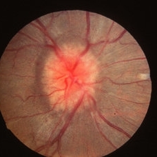
 Papillitis
May 2 2013 by Henry J. Kaplan, MD
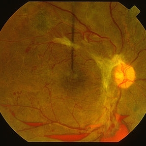
Anterior optic neuropathy or papillitis in the right eye; notice the blurred optic disc margin, engorged capillaries and flame shaped hemorrhages at the margin.
Condition/keywords: optic disc edema, optic disc swelling, papillitis
-
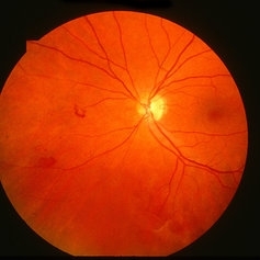
 Papilledema
May 2 2013 by Henry J. Kaplan, MD
Optic disc swelling due to RICP . Left eye; #2.
Condition/keywords: disc swelling, papilledema, raised intracranial pressure (RICP)
-
 Papilledema
May 2 2013 by Henry J. Kaplan, MD
Optic disc swelling due to RICP. Right Eye; #1.
Condition/keywords: optic disc edema, raised intracranial pressure (RICP)
-
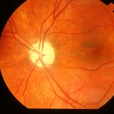 Optic Atrophy
May 2 2013 by Henry J. Kaplan, MD
Left optic atrophy as a chalky white optic nerve.
Condition/keywords: optic atrophy
-
 Radiation retinopathy
May 2 2013 by Henry J. Kaplan, MD
Radiation maculopathy after brachytherapy of a choroidal melanoma; notice the telangiectatic changes and retinal exudation.
Condition/keywords: radiation maculopathy, radiation retinopathy
-
 Radiation Retinopathy
May 2 2013 by Henry J. Kaplan, MD
Vascular changes and NVE formation in the nasal part of the retina in radiation retinopathy; #1.
Condition/keywords: radiation retinopathy
-
 Radiation Retinopathy
May 2 2013 by Henry J. Kaplan, MD
Fluorescein angiography in the same patient shows vascular occlusions and capillary non-perfusion area, telangiactatic changes and NVE as leakage of fluorescein in the same patient; #2.
Condition/keywords: radiation retinopathy
-
 Radiation Retinopathy
May 2 2013 by Henry J. Kaplan, MD
Late frame F/A clearly shows leakage from the NVE; #3.
Condition/keywords: radiation retinopathy
-
 Retinoschisis
-
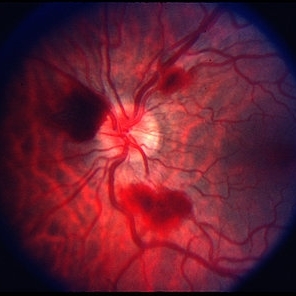
 Polycythemia Vera
May 2 2013 by Henry J. Kaplan, MD
CRVO in polycythemia vera; dilated and tortous retinal veins, hemorrhages and cotton wool spots.
Condition/keywords: central retinal vein occlusion (CRVO), polycythemia vera
-
 Thrombocytopenia
May 2 2013 by Henry J. Kaplan, MD
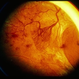
Superficial retinal hemorrhage in a patient with thrombocytopenia, right eye; #1.
Condition/keywords: thrombocytopenia
-
 Thrombocytopenia
May 2 2013 by Henry J. Kaplan, MD
Retinal and pre-retinal hemorrhage in thrombocytopenia, left eye; #2.
Condition/keywords: thrombocytopenia
-
 Myelianated Nerve Fiber Layer
May 2 2013 by Henry J. Kaplan, MD
Extensive myelinated nerve fibers.
Condition/keywords: myelinated nerve fibers
-
 Melanocytoma
May 2 2013 by Henry J. Kaplan, MD
Melanocytoma of the optic nerve head.
Condition/keywords: melanocytoma
-
 Optic pit
May 2 2013 by Henry J. Kaplan, MD
Optic pit in the inferotemporal part of the optic disc.
Condition/keywords: optic disc pit
-
 Capillary Hemongima
May 2 2013 by Henry J. Kaplan, MD
Capillary hemangioma after one session of laser treatment; #1
Condition/keywords: Von Hippel-Lindau
-
 Capillary Hemongima, Coat's Response
May 2 2013 by Henry J. Kaplan, MD
Coat's response as exudation in the macula in the same patient with retinal capillary hemangioma. Notice the dilated feeder vessles from the optic nerve infriorly; #2.
Condition/keywords: Coats' disease, Von Hippel-Lindau
-
 Sea Fan Neovascularization
May 2 2013 by Henry J. Kaplan, MD
Typical sea fan neovascularization in the peripheral retina of a patient with sickle cell disease.
Condition/keywords: sea fan, sickle cell retinopathy
-
 Lattice Degeneration
May 2 2013 by Henry J. Kaplan, MD
Pigmented lattice degeneration with lattice "wicker" caused by sclerotic blood vessels.
Condition/keywords: lattice degeneration, peripheral retinal degeneration
-
 Retinal Tear
May 2 2013 by Henry J. Kaplan, MD
Horseshoe retinal tear with small hemorrhages at the edge of the tear.
Condition/keywords: retinal tear
-
 Wagner Disease
May 2 2013 by Henry J. Kaplan, MD
Pigmented lattice degeneration in Wagner disease.
Condition/keywords: lattice degeneration, Wagner disease
-
 Bull's Eye
May 2 2013 by Henry J. Kaplan, MD
Bull's eye in chloroquine toxicity; #1.
Condition/keywords: bull's eye maculopathy, chloroquine maculopathy, chloroquine toxicity
-
 Bull's Eye
May 2 2013 by Henry J. Kaplan, MD
Fluorescein angiography demonstrates RPE window defects and hyperfluorescence in bull's eye maculopathy; #2.
Condition/keywords: bull's eye maculopathy
-
 Wyburn Mason Syndrome
May 2 2013 by Henry J. Kaplan, MD
Racemose angioma of the retina in Wyburn Mason syndrome.
Condition/keywords: racemose hemangioma
-
 Exposed Buckle
May 2 2013 by Henry J. Kaplan, MD
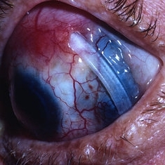
Exposed buckele with underlying scleral staphyloma.
Condition/keywords: exposed scleral buckle
-
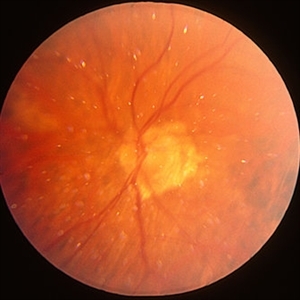
 HRC-PDR
May 2 2013 by Henry J. Kaplan, MD
Large NVD in HRC-PDR.
Condition/keywords: HRC-PDR, neovascularization of the disc (NVD)
-
 HRC-PDR
May 2 2013 by Henry J. Kaplan, MD
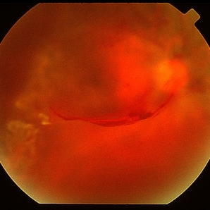
Boat shaped subyaloid hemorrhage due to underlying NVE in a patient with HRC-PDR.
Condition/keywords: HRC-PDR, subhyaloid hemorrhage
-
 Rubella cataract
May 2 2013 by Henry J. Kaplan, MD
Cataract due to rubella infection; #1.
Condition/keywords: cataract, rubella retinopathy
-
 Rubella retinopathy
May 2 2013 by Henry J. Kaplan, MD
Salt and pepper changes of RPE in rubella retinopathy in the same patient; #2.
Condition/keywords: rubella retinopathy
-
 Rubella Retinopathy
May 2 2013 by Henry J. Kaplan, MD
Fluorescein angiography of the same patient with rubella retinopathy demonstrates RPE window defects as hyperfluorescent areas; #3.
Condition/keywords: rubella retinopathy
-
 Choroidal Nevus
May 2 2013 by Henry J. Kaplan, MD
Large flat choroidal nevus involving the posterior pole with overlying drusen.
Condition/keywords: choroidal nevus
-
 Choroidal Nevus
May 2 2013 by Henry J. Kaplan, MD
Juxtapapillary choroidal nevus with overlying drusen.
Condition/keywords: choroidal nevus
-
 Leber's Miliary Aneurysm
May 2 2013 by Henry J. Kaplan, MD
Peripheral telangiectatic lesions and exudation in adult coat;s disease or Leber's Miliary Aneurysm.
-
 cavernous hemangioma
May 2 2013 by Henry J. Kaplan, MD
Typical grape like appearance of cavernous hemangioma
Condition/keywords: cavernous hemangioma of the retina
-
 Idiopathic CNV
May 2 2013 by Henry J. Kaplan, MD
Idiopathic CNV in a young patient with subretinal fluid around the lesion and hemorrhages inferiorly; #1.
Condition/keywords: idiopathic choroidal neovasculization (CNV)
-
 Idiopathic CNV
May 2 2013 by Henry J. Kaplan, MD
Fluorescein angiogram in the same patient; arterial phase: hyperfluorescence in the area of the CNV as a well delineated lesion (classic); #2.
Condition/keywords: idiopathic choroidal neovasculization (CNV)
-
 Idiopathic CNV
May 2 2013 by Henry J. Kaplan, MD
Fluorescein angiogram; arteriovenous phase shows increased hyperfluorescence in the lesion; notice the straightened vessels due to epiretinal membrane formation. Hypofluorescent area is due to masking effect of blood; #3.
Condition/keywords: epiretinal membrane (ERM), idiopathic choroidal neovasculization (CNV)
-
 Idiopathic CNV
May 2 2013 by Henry J. Kaplan, MD
Fluorescein angiogram; late phase shows leakage of fluorescein as blurred margins of the CNV; #4.
Condition/keywords: idiopathic choroidal neovasculization (CNV)
-
 Choroidal Hemangioma
Mar 29 2013 by Henry J. Kaplan, MD
Orange-red choroidal hemangioma extending from the optic nerve toward inferotemporal arcade involving the macula.#1
-
 Central areolar atrophy
May 2 2013 by Henry J. Kaplan, MD
Central areolar atrophy; GA like lesion in younger age patient.
Condition/keywords: central areolar choroidal dystrophy (CACD)
-
 Choroidal Hemangioma
Apr 19 2013 by Henry J. Kaplan, MD
Fluorescein angiography of the choroidal hemangioma in the same patient shows hyperfluorescent spots starting in the early arterial phase. #2
-
 Choroidal Hemangioma
Apr 19 2013 by Henry J. Kaplan, MD
Late arteriovenous phase of fluorescein angiogram in the same patient with chroidal hemangioma shows increased hyperfluorescence and leakage from the lesion. #3
-
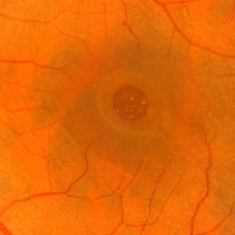
 Dry Age-Related Macular Degeneration
Mar 29 2013 by Henry J. Kaplan, MD
Multiple drusens and RPE changes in dry AMD, some of the drusens are calcified.
Condition/keywords: age-related macular degeneration (AMD), dry age-related macular degeneration (dry AMD)
-
 Dry Age-Related Macular Degeneration
Mar 29 2013 by Henry J. Kaplan, MD
Dry AMD with multiple confluent soft drusens
Condition/keywords: age-related macular degeneration (AMD), dry age-related macular degeneration (dry AMD)
-
 Dry Age-Related Macular Degeneration
Mar 29 2013 by Henry J. Kaplan, MD
Late fluorescein angiography demonstrates multiple stained soft drusens in the same patient #2.
Condition/keywords: age-related macular degeneration (AMD), dry age-related macular degeneration (dry AMD)
-
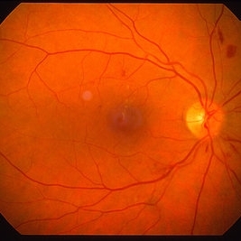
 Geographic Atrophy
Mar 29 2013 by Henry J. Kaplan, MD
Large GA involving the whole posterior pole.
Condition/keywords: dry age-related macular degeneration (dry AMD), geographic atrophy
-
 Geographic Atrophy
Mar 29 2013 by Henry J. Kaplan, MD
Dry AMD as geographic atrophy.
Condition/keywords: geographic atrophy
-
 Age Related Macular Degeneration
Mar 29 2013 by Henry J. Kaplan, MD
Geographic atrophy with small hemorrhages due to subretinal neovascular membrane development.
Condition/keywords: choroidal neovascularization (CNV), geographic atrophy
-
 Age-Related Macular Degeneration
Mar 29 2013 by Henry J. Kaplan, MD
Fundus photograph of a patient with exudative AMD which demonstrated multiple drusen some of them calcified , scar tissue and hemorrhages in the inferotemporal part due to active choroidal neovascularization.
Condition/keywords: age-related macular degeneration (AMD), choroidal neovascularization (CNV), exudative age-related macular degeneration
-
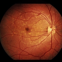
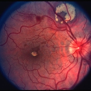
 Dry Age-Related Macular Degeneration
Mar 29 2013 by Henry J. Kaplan, MD
Fundus photograph of a patient with dry AMD demonstrates multiple drusen, RPE change and geographic atrophy; notice that the patient has also familial or dominant drusen most prominant in nasal retina.
Condition/keywords: age-related macular degeneration (AMD), dry age-related macular degeneration (dry AMD), geographic atrophy
-
 Exudative Age-Related Macular Degeneration
Mar 29 2013 by Henry J. Kaplan, MD
Exudative AMD with huge subretinal hemorrhage.
Condition/keywords: age-related macular degeneration (AMD), choroidal neovascularization (CNV), exudative age-related macular degeneration, subretinal hemorrhage
-
 Recurrence of CNV After Laser
Mar 29 2013 by Henry J. Kaplan, MD
Recurrence of CNV at the nasal edge of previously treated lesion with laser (old treatment modality long times ago).
Condition/keywords: choroidal neovascularization (CNV), exudative age-related macular degeneration
-
 End Stage Age-Related Macular Degeneration
Mar 29 2013 by Henry J. Kaplan, MD
End stage of exudative AMD as disciform scar and chorioretinal anastomosis.
Condition/keywords: age-related macular degeneration (AMD), disciform scar, exudative age-related macular degeneration
-
 CNV
CNV
Mar 29 2013 by Henry J. Kaplan, MD
Fluorescein angiogram of CNV #1.
Condition/keywords: choroidal neovascularization (CNV), exudative age-related macular degeneration
-
 CNV
CNV
Mar 29 2013 by Henry J. Kaplan, MD
Fluorescein angiogram of CNV #2.
Condition/keywords: choroidal neovascularization (CNV), exudative age-related macular degeneration
-
 CNV
CNV
Mar 29 2013 by Henry J. Kaplan, MD
Fluorescein angiogram of CNV #3.
Condition/keywords: choroidal neovascularization (CNV), exudative age-related macular degeneration
-
 CNV
CNV
Mar 29 2013 by Henry J. Kaplan, MD
Fluorescein angiogram of CNV #4.
Condition/keywords: choroidal neovascularization (CNV), exudative age-related macular degeneration
-
 Arterial Macro Aneurysm
Mar 29 2013 by Henry J. Kaplan, MD
Large arterial macroaneurysm with hemorrhage and surrounding exudates.
Condition/keywords: retinal arterial macroaneurysm
-
 Arterial Macroaneurysm
Mar 29 2013 by Henry J. Kaplan, MD
Typical arterial macroaneurysm surrounded by lipid exudates and edema.
Condition/keywords: macroaneurysm, retinal arterial macroaneurysm
-
---thumb.jpg/image-square;max$300,300.ImageHandler) Arterial Macroaneurysm
Arterial Macroaneurysm
Apr 3 2013 by Henry J. Kaplan, MD
Fluorescein angiogram of a retinal arterial macroaneurysm with both subretinal (subretinal blocked fluorescence in the superior part) and subhyaloid hemorrhage (boat shaped blocked fluorescence in the inferior part).
Condition/keywords: macroaneurysm, retinal arterial macroaneurysm
-
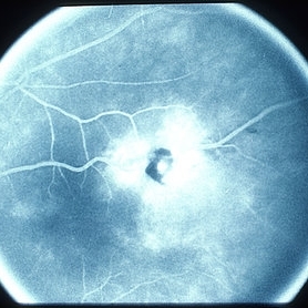 Arterial Macro Aneurysm
Mar 29 2013 by Henry J. Kaplan, MD
Fluorescein angiogram of arterial macro-aneurysm with severe leakage of fluorescein and slight blocked fluorescence due to hemorrhage.
Condition/keywords: retinal arterial macroaneurysm
-
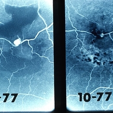 Arterial Macro Aneurysm
Mar 29 2013 by Henry J. Kaplan, MD
Fluorescein angiogram of retinal arterial macroaneurysm in 9 month follow up which shows spontaneous regression and resolving hemorrhages in 1977.
Condition/keywords: retinal arterial macroaneurysm
-
 Asteroid Hyalosis
Mar 29 2013 by Henry J. Kaplan, MD
Milder form of asteroid hyalosis.
Condition/keywords: asteroid hyalosis
-
 Asteroid Hyalosis
Mar 29 2013 by Henry J. Kaplan, MD
Multiple shiny spots of Asteroid hyalosis in the vitreous cavity.
Condition/keywords: asteroid hyalosis
-
---thumb.jpg/image-square;max$300,300.ImageHandler) Capillay Hemangioma Von Hippel
Capillay Hemangioma Von Hippel
Mar 29 2013 by Henry J. Kaplan, MD
Capillary hemangioma in the temporal macular area, notice the dilated feeder vessels which are the inferior arcade vessles.
Condition/keywords: Von Hippel-Lindau
-
 Capillary Hemangioma
Mar 29 2013 by Henry J. Kaplan, MD
Fluorescein angiography of two adjacent capillary hemangiomas.
Condition/keywords: Von Hippel-Lindau
-
---thumb.jpg/image-square;max$300,300.ImageHandler) Angioid Streaks
Angioid Streaks
Mar 29 2013 by Henry J. Kaplan, MD
Angioid streaks; Right Eye #1.
Condition/keywords: angioid streaks
-
---thumb.jpg/image-square;max$300,300.ImageHandler) Angioid Streaks
Angioid Streaks
-
---thumb.jpg/image-square;max$300,300.ImageHandler) Choroidal Neovascularization and Angiod Streaks
Choroidal Neovascularization and Angiod Streaks
Mar 29 2013 by Henry J. Kaplan, MD
End stage disciform scar due to CNV in a patient with angioid streaks.
Condition/keywords: angioid streaks, choroidal neovascularization (CNV)
-
---thumb.jpg/image-square;max$300,300.ImageHandler) Anterior Ischemic Optic Neuropathy
Anterior Ischemic Optic Neuropathy
Mar 29 2013 by Henry J. Kaplan, MD
Anterior Ischemic Optic Neuropathy; notice the typical pale optic disc swelling and faint splinter hemorrhages.
Condition/keywords: anterior ischemic optic neuropathy
-
 RPE Hypertrophy
Mar 29 2013 by Henry J. Kaplan, MD
Typical bear tracks in RPE hypertrophy.
Condition/keywords: retinal pigment epithelium (RPE) hypertrophy
-
 Pattern Dystrophy
Mar 29 2013 by Henry J. Kaplan, MD
Pattern dystrophy (butterfly type) #1.
Condition/keywords: pattern macular dystrophy
-
 Pattern Dystrophy
Mar 29 2013 by Henry J. Kaplan, MD
Pattern dystrophy #2.
Condition/keywords: pattern macular dystrophy
-
 BRVO
BRVO
Mar 29 2013 by Henry J. Kaplan, MD
Inferotemporal BRVO.
Condition/keywords: branch retinal vein occlusion (BRVO)
-
 Macular BRVO
Mar 29 2013 by Henry J. Kaplan, MD
Typical macular BRVO.
Condition/keywords: branch retinal vein occlusion (BRVO)
-
 BRVO Complications
Mar 29 2013 by Henry J. Kaplan, MD
Old superotemporal BRVO as a sclerotic vessel with NVD and NVE and vitreous hemorrhage and a preretinal hemorrhage.
Condition/keywords: branch retinal vein occlusion (BRVO), neovascularization (NV), neovascularization of the disc (NVD), vitreous hemorrhage
-
 Branch Retinal Vein Occlusion
Mar 29 2013 by Henry J. Kaplan, MD
BRVO with macular edema treated with MPC before introduction of anti-VEGF drugs ( right after treatment).
Condition/keywords: branch retinal vein occlusion (BRVO)
-
 Old BRVO
Mar 29 2013 by Henry J. Kaplan, MD
Angiogram of a patient with old BRVO shows typical collateral vessel formation in the temporal area.
Condition/keywords: branch retinal vein occlusion (BRVO), collateral retinal vessel, collaterals
-
 Impending CRVO
Mar 29 2013 by Henry J. Kaplan, MD
Dilated and tortous retinal veins and scant hemorrhage in impending CRVO.
Condition/keywords: central retinal vein occlusion (CRVO)
-
 Impending CRVO
Mar 29 2013 by Henry J. Kaplan, MD
Venous dilation and torousity and faint spot hemorrhages in a patient with impending CRVO.
Condition/keywords: central retinal vein occlusion (CRVO)
-
 Non Ischemic Hemi-CRVO
Mar 29 2013 by Henry J. Kaplan, MD
Non-ischemic CRVO: blurred disc margins, dilated and tortous veins and scattered hemorrhages in the superior half of the retina.
Condition/keywords: branch retinal vein occlusion (BRVO), central retinal vein occlusion (CRVO), hemi CRVO, non-ischemic central retinal vein occlusion (CRVO)
-
 Non Ischemic CRVO
Mar 29 2013 by Henry J. Kaplan, MD
Non ischemic CRVO.
Condition/keywords: central retinal vein occlusion (CRVO)
-
 CRVO
CRVO
Mar 29 2013 by Henry J. Kaplan, MD
Full blown ischemic CRVO with disc swelling, dilated and tortous veins, scattered hemorrhages and multiple cotton wool spots.
Condition/keywords: central retinal vein occlusion (CRVO), ischemic CRVO
-
 CRAO
CRAO
Mar 29 2013 by Henry J. Kaplan, MD
CRAO with arterial narrowing, disc pallor,retinal edema, cherry red spot and plaques in the inferonasal artery; notice the choroidal nevus in superonasal retina.
Condition/keywords: central retinal artery occlusion (CRAO), cherry red spot
-
 Branch Retinal Artery Occlusion
Mar 29 2013 by Henry J. Kaplan, MD
Inferotemporal BRAO.
Condition/keywords: branch retinal artery occlusion (BRAO)
-
 CRAO
CRAO
Mar 29 2013 by Henry J. Kaplan, MD
Central retinal artery occlusion with a cherry red spot in the fovea; notice the disc pallor.
Condition/keywords: central retinal artery occlusion (CRAO), cherry red spot
-
 Pancytopenia
Mar 29 2013 by Henry J. Kaplan, MD
Multiple blot and flame shaped hemorrhages in a patient with pancytopenia #1.
Condition/keywords: pancytopenia, retinal hemorrhage
-
 Pancytopenia
Mar 29 2013 by Henry J. Kaplan, MD
Multiple blot and flame shaped hemorrhages in a patient with pancytopenia #2.
Condition/keywords: pancytopenia, retinal hemorrhage
-
 Choroidal Hemorrhage
Mar 29 2013 by Henry J. Kaplan, MD
Traumatic choroidal hemorrhage with possible underlying choroidal ruptures.
Condition/keywords: choroidal hemorrhage
-
 CSR
CSR
Mar 29 2013 by Henry J. Kaplan, MD
Large serous detachment with multiple yellow spots of fibrin deposits.
Condition/keywords: central serous chorioretinopathy (CSCR)
-
 Retinal Cavernous Hemangioma
Mar 29 2013 by Henry J. Kaplan, MD
Fluorescein angiogram of retinal cavernous hemangioma shows slow filling with early blocked fluorescence #1.
Condition/keywords: cavernous hemangioma of the retina
-
 Cavernous Hemangioma
Mar 29 2013 by Henry J. Kaplan, MD
Fluorescein angiogram of retinal cavernous hemangioma in later phase shows typical grape like lesions with fluid levels which are hyperfluorescent in top part and hypo in bottom part due to seperation of serum from blood in these slow filling angiomas #2.
Condition/keywords: cavernous hemangioma of the retina
-
 Coat's Disease
Mar 29 2013 by Henry J. Kaplan, MD
Typical peripheral telangiectatic vessles and subretinal exudates.
Condition/keywords: Coats' disease
-
 Coat's Disease
Mar 29 2013 by Henry J. Kaplan, MD
Typical telangiectatic lesions and exudation in coat`s disease.
Condition/keywords: Coats' disease
-
 Retinal Folds
Mar 29 2013 by Henry J. Kaplan, MD
Retinal folds as fine wrinklings.
Condition/keywords: retinal fold
-
 Choroidal Osteoma
Mar 29 2013 by Henry J. Kaplan, MD
Typical choroidal osteoma as yellow subretinal lesion around optic nerve with scalloped border and mild pigmentation on the surface.
Condition/keywords: choroidal osteoma
-
 Choroidal Osteoma
Mar 29 2013 by Henry J. Kaplan, MD
Autofluorescence in choroidal osteoma.
Condition/keywords: autofluorescence imaging, choroidal osteoma
-
 Choroidal Osteoma with CNV
Mar 29 2013 by Henry J. Kaplan, MD
Typical choroidal osteoma complicated by CNV as subretinal hemorrhage in the macular edge of the lesion.
Condition/keywords: choroidal neovascularization (CNV), choroidal osteoma
-
 Choroidal Hemangioma
Mar 29 2013 by Henry J. Kaplan, MD
B-scan of the same patient showing the lesion with high internal reflectivity #2.
Condition/keywords: B scan ultrasound
-
---thumb.jpg/image-square;max$300,300.ImageHandler) Choroidal Hemangioma
Choroidal Hemangioma
Apr 3 2013 by Henry J. Kaplan, MD
Typical A-scan spikes of high internal reflectivity in choroidal hemangioma.
Condition/keywords: A-scan ultrasound
-
 Choroideremia
Mar 29 2013 by Henry J. Kaplan, MD
Choroideremia, notice the residual small central island.
Condition/keywords: choroideremia
-
 Choroideremia
Mar 29 2013 by Henry J. Kaplan, MD
Angiogram of choroideremia shows hypofluorescence due to filling defect 9choriocapillary loss) except for a small central island #1.
Condition/keywords: choroideremia
-
 Choroideremia
Mar 29 2013 by Henry J. Kaplan, MD
Angiogram of the same patient in a later phase shows a small central island and multiple peripheral and a residual area inferiorly #2.
Condition/keywords: choroideremia
-
 Choroidal Detachment
Mar 29 2013 by Henry J. Kaplan, MD
Mild choroidal detachment temporally, macula invived.
Condition/keywords: choroidal detachment
-
 Choroidal Detachment
Mar 29 2013 by Henry J. Kaplan, MD
One quadrant choroidal detachment as brownish convex lesion.
Condition/keywords: choroidal detachment
-
 Choroidal Detachment
Mar 29 2013 by Henry J. Kaplan, MD
Choroidal detachment as two adjacent brownish convex lesions.
Condition/keywords: choroidal detachment
-
 Diabetes NPDR
Mar 29 2013 by Henry J. Kaplan, MD
Moderate NPDR.
Condition/keywords: nonproliferative diabetic retinopathy
-
 NPDR
NPDR
Mar 29 2013 by Henry J. Kaplan, MD
Multiple microaneurysms visible as small round dot lesions.
Condition/keywords: nonproliferative diabetic retinopathy
-
 Severe NPDR
Mar 29 2013 by Henry J. Kaplan, MD
Severe NPDR , IRMA visible inferonasally.
Condition/keywords: nonproliferative diabetic retinopathy
-
 Diabetes NPDR
Mar 29 2013 by Henry J. Kaplan, MD
Moderated NPDR.
Condition/keywords: diabetic mellitus, nonproliferative diabetic retinopathy
-
 Diabetes NPDR
Mar 29 2013 by Henry J. Kaplan, MD
NPDR.
Condition/keywords: cotton wool spots, nonproliferative diabetic retinopathy
-
 Venous Beading
Mar 29 2013 by Henry J. Kaplan, MD
Typical venous beading in severe NPDR.
Condition/keywords: venous beading
-
 Diabetes HRC-PDR
Mar 29 2013 by Henry J. Kaplan, MD
Small NVD on the optic nerve more than 1/4 DA.
Condition/keywords: HRC-PDR, neovascularization of the disc (NVD)
-
 Diabetes HRC-PDR
Mar 29 2013 by Henry J. Kaplan, MD
NVD on the optic disc extended superiorly and temporally.
Condition/keywords: neovascularization of the disc (NVD)
-
 PDR
PDR
-
 Venous Beading & NVE
Mar 29 2013 by Henry J. Kaplan, MD
Typical venous beading and NVE in a diabetic patient with PDR.
Condition/keywords: neovascularization (NV), venous beading
-
 Preproliferative Diabetic Retinopathy
Mar 29 2013 by Henry J. Kaplan, MD
Multiple cotton wools in a patient with pre-proliferative diabetic retinopathy.
Condition/keywords: cotton wool spots, nonproliferative diabetic retinopathy, pre-proliferative diabetic retinopathy
-
 PDR
PDR
Mar 29 2013 by Henry J. Kaplan, MD
Temporal retinal NVE in early PDR.
Condition/keywords: neovascularization (NV)
-
 Advanced Active PDR
Mar 29 2013 by Henry J. Kaplan, MD
Extensive NVD-FPD and NVE-FPE in a diabetic patient.
Condition/keywords: foveal photoreceptor defect, FPE, neovascularization (NV), neovascularization of the disc (NVD)
-
 PDR
PDR
Mar 29 2013 by Henry J. Kaplan, MD
Fibrous proliferation (FPE) in a patient with PDR.
Condition/keywords: fibrous proliferation, FPE
-
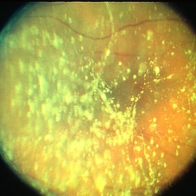
 TRD
TRD
Mar 29 2013 by Henry J. Kaplan, MD
Advanced fibrous proliferation and TRD in diabetes.
Condition/keywords: tractional retinal detachment
-
 Advanced Active PDR
Mar 29 2013 by Henry J. Kaplan, MD
Large active NVEs with fibrous proliferations in diabetes.
Condition/keywords: fibrous proliferation, neovascularization (NV)
-
 HRC--PDR
Mar 29 2013 by Henry J. Kaplan, MD
Vitreous hemorrhage in a patient with HRC-PDR.
Condition/keywords: HRC-PDR, vitreous hemorrhage
-
 Advanced PDR
Mar 29 2013 by Henry J. Kaplan, MD
Large FPD, FPE, NVE and multiple boat shaped subhyaloid hemorrhages inferiorly, ischemic retina and multiple occluded sclerotic vessels.
Condition/keywords: foveal photoreceptor defect, subhyaloid hemorrhage
-
 PRP laser
Mar 29 2013 by Henry J. Kaplan, MD
Right after PRP laser in PDR.
Condition/keywords: laser photocoagulation, pan-retinal photocoagulation (PRP)
-
 Regressed PDR
Mar 29 2013 by Henry J. Kaplan, MD
Regressed PDR after full PRP, notice the FPD on the optic nerve.
Condition/keywords: pan-retinal photocoagulation (PRP), regressed
-
 MPC for CSME
Mar 29 2013 by Henry J. Kaplan, MD
Right after MPC for CSME in diabetes (before the introduction of anti-VEGFs).
Condition/keywords: clinically significant macular edema (CSME), diabetic macular edema, multifocal chorioretinitis (MCP)
-
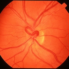 Venous Loop
Mar 29 2013 by Henry J. Kaplan, MD
Congenital anomaly: venous loop on the optic disc.
Condition/keywords: venous loop
-
 Bergmeister's Papillae
Mar 29 2013 by Henry J. Kaplan, MD
Remnants of fetal hyaloid artery as fibrous tuft called Bergmeister`s papillae on the optic disc.
Condition/keywords: Bergmeister's Papillae, hyaloid artery
-
 Vascular Tortousity
Mar 29 2013 by Henry J. Kaplan, MD
Vascular tortuosity as congenital anomaly in this patient without any underlying etiology.
Condition/keywords: vascular tortousity
-
 Iris Coloboma
-
 Coloboma
Mar 29 2013 by Henry J. Kaplan, MD
Optic disc and inferonasal choroidal coloboma in the same patient #2.
Condition/keywords: coloboma, coloboma of choroid, coloboma of optic disc
-
 Coloboma
Mar 29 2013 by Henry J. Kaplan, MD
Large choroidal coloboma.
Condition/keywords: coloboma, coloboma of choroid
-
 Coloboma
Mar 29 2013 by Henry J. Kaplan, MD
Coloboma involving optic nerve and inferior choroid.
Condition/keywords: coloboma of choroid, coloboma of optic disc
-
 Dropped nucleus
Mar 29 2013 by Henry J. Kaplan, MD
Dropped nucleus on posterior segment.
Condition/keywords: dropped nucleus
-
 Fabry's Disease
Mar 29 2013 by Henry J. Kaplan, MD
Cornea verticillata in fabry syndrome.
Condition/keywords: cornea verticillata, Fabry disease
-
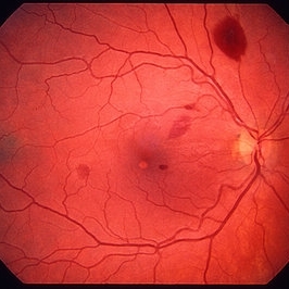
 Fabry`s disease
Mar 29 2013 by Henry J. Kaplan, MD
Vascular tortousity in Fabry`s disease
Condition/keywords: Fabry disease, vascular tortousity
-
 Fabry's Disease
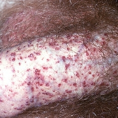
Mar 29 2013 by Henry J. Kaplan, MD
Typical angiokeratoma lesions in fabry`s disease.
Condition/keywords: Fabry disease
-
 Leukemia
Mar 29 2013 by Henry J. Kaplan, MD
Multiple blot hemorrhages in a patient with leukemia #1.
Condition/keywords: leukemia
-
 Leukemia
Mar 29 2013 by Henry J. Kaplan, MD
Retinal hemorrhages in leukemia #2.
Condition/keywords: leukemia, retinal hemorrhage
-
 Leukemia
Mar 29 2013 by Henry J. Kaplan, MD
Subretinal hemorrhages are resolving as yellowish lesions in a patient with leukemia.
Condition/keywords: leukemia
-
 Light Toxicity
Mar 29 2013 by Henry J. Kaplan, MD
Light toxicity as oval lesion superior to fovea.
Condition/keywords: light toxicity
-
 Light Toxicity
Mar 29 2013 by Henry J. Kaplan, MD
Angiogram of light toxicity demonstrates hyperfluorescence in the involved area.
Condition/keywords: light toxicity
-
 Light Toxicity
Mar 29 2013 by Henry J. Kaplan, MD
Microscope light toxicity in superior arcade.
Condition/keywords: light toxicity
-
 Light Toxicity
Mar 29 2013 by Henry J. Kaplan, MD
Angiogram of light toxicity shows hyperfluorescent lesion in the involved area.
Condition/keywords: light toxicity
-
 Hodgkin's Disease Stage IV
Mar 29 2013 by Henry J. Kaplan, MD
Multiple subretinal infiltrations in patient with disseminated Hodgkin`s disease #1.
Condition/keywords: Hodgkins lymphoma
-
 Hodgkin's Disease State IV
Mar 29 2013 by Henry J. Kaplan, MD
Stage IV Hodgkin`s lymphoma with multiple subretinal infiltrations #2.
Condition/keywords: Hodgkins lymphoma, lymphoma
-
 Combined Hamartoma of Retina and RPE
Mar 29 2013 by Henry J. Kaplan, MD
Hamartoma visible as a grreen lesion on superior arcade with ERM formation and dragging of the macula.
Condition/keywords: combined hamartoma
-
 Combined Hamartoma of Retina and RPE
Mar 29 2013 by Henry J. Kaplan, MD
Greenish lesion on the arcade with epiretinal membrane formation, vessels inside the lesion are contracted and those outside are distracted.
Condition/keywords: combined hamartoma
-
 Giant Retinal Tear
Mar 29 2013 by Henry J. Kaplan, MD
RRD with giant retinal tear.
Condition/keywords: re-attached retinal detachment (RRD), retinal tear
-
 Giant Retinal Tear
-
 Fundus Albipunctatus
Mar 29 2013 by Henry J. Kaplan, MD
Typical fundus albipunctatus a kind of stationary night blindness; notice the normal disc and vessels #1.
Condition/keywords: fundus albipunctatus
-
 Fundus Albipunctatus
Mar 29 2013 by Henry J. Kaplan, MD
Typical fundus albipunctatus a kind of stationary night blindness ; notice the normal disc and vessels #2.
Condition/keywords: fundus albipunctatus
-
 Fundus Albipunctatus
Mar 29 2013 by Henry J. Kaplan, MD
Fundus albipunctatus (one of the stationary night blindness syndromes with multiple white dots in the periphery and normal optic disc and vessels).
Condition/keywords: fundus albipunctatus
-
 Macular Pucker
Mar 29 2013 by Henry J. Kaplan, MD
Large epiretinal membrane with straightened vessels in the papillomacular bundle and distorsion of retinal vessels.
Condition/keywords: epiretinal membrane (ERM), macular pucker
-
 Macular Pucker
Mar 29 2013 by Henry J. Kaplan, MD
Angiogram of a macular pucker better demonstrates vessel straightening in papillomacular bundleand tortousity in the area of membrane.
Condition/keywords: epiretinal membrane (ERM), macular pucker
-
 Macular Pucker
Mar 29 2013 by Henry J. Kaplan, MD
Macular pucker.
Condition/keywords: epiretinal membrane (ERM), macular pucker
-
 Macular Pucker
Mar 29 2013 by Henry J. Kaplan, MD
Epiretinal membrane with pseudohole formation.
Condition/keywords: epiretinal membrane (ERM), macular pucker
-
 Macular Hole
-
 Macular Hole
Mar 29 2013 by Henry J. Kaplan, MD
Chronic macular hole with drusen like deposits and surrounding cuffing of subretinal fluid.
Condition/keywords: macular hole
-
 Siegrist Streaks
Mar 29 2013 by Henry J. Kaplan, MD
Typical Siegrist streaks in hypertensive choridopathy; hyperpigmentations in a linear fashion along choroidal vessels , a rare finding.
Condition/keywords: hypertensive choroidopathy, Siegrist Streaks
-
 Hypertensive Retinopathy
Mar 29 2013 by Henry J. Kaplan, MD
Hypertensive retinopathy grade III #1.
Condition/keywords: hypertensive retinopathy
-
 Hypertensive Retinopathy
Mar 29 2013 by Henry J. Kaplan, MD
Grade III hypertensive retinopathy #2.
Condition/keywords: hypertensive retinopathy
-
 Hypertensive Retinopathy
Mar 29 2013 by Henry J. Kaplan, MD
Cotton wool spot in grade III hypertensive retinopathy.
Condition/keywords: hypertension, hypertensive retinopathy
-
 Hypertensive Retinopathy
Mar 29 2013 by Henry J. Kaplan, MD
Severe hypertensive retinopathy with macular exudates #2.
Condition/keywords: hypertensive retinopathy
-
 Hypertensive Retinopathy
Mar 29 2013 by Henry J. Kaplan, MD
Severe hypertensive retinopathy #1.
Condition/keywords: hypertensive retinopathy
-
---thumb.jpg/image-square;max$300,300.ImageHandler) Optic Disc Drusen
Optic Disc Drusen
Mar 27 2013 by Henry J. Kaplan, MD
An 11-year-old boy presented with transient blurry vision, VA:20/20 bilaterally. He has pseudo optic disc swelling only in the right eye ; margin is blurred but the pattern of vessels are normal and there are some yellowish deposits on the superior of ON #1. AF, B-scan, CT scan, and VF are uploaded in the following slides.
Condition/keywords: drusen of optic disc, optic disc drusen, optic nerve drusen
-
---thumb.jpg/image-square;max$300,300.ImageHandler) Optic Disc Drusen
Optic Disc Drusen
Mar 27 2013 by Henry J. Kaplan, MD
Autofluorescence imaging shows heper AF on the optic nerve head specially superiorly due to drusen in the same patient #2.
Imaging device: Heidelberg spectralis
Condition/keywords: drusen of optic disc, optic disc drusen, optic nerve drusen
-
 Optic Disc Drusen
Mar 27 2013 by Henry J. Kaplan, MD
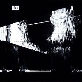
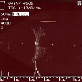
B-scan of the same patient with low gain (48db), demonstrates high spike on the optic nerve head due to drusen #3.
Condition/keywords: drusen of optic disc, optic disc drusen, optic nerve drusen
-
 Optic Disc Drusen
Mar 27 2013 by Henry J. Kaplan, MD
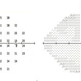
Perimetry demonstrates slightly enlarged blind spot in the same patient #5.
Condition/keywords: drusen of optic disc, optic disc drusen, optic nerve drusen
-
 Orbital CT Scan in Optic Nerve Drusen
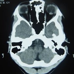
Mar 27 2013 by Henry J. Kaplan, MD
Axial CT scan of orbit demonstrates high density spot on optic nerve head on both sides #4.
Condition/keywords: drusen of optic disc, optic disc drusen, optic nerve drusen
A project from the American Society of Retina Specialists