-
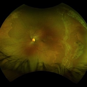
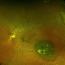 Congenital Hypertrophy of the Retinal Pigment Epithelium Wide Field Optomap
Sep 24 2019 by Sophia El Hamichi, MD
A 52-year-old female followed for 2 temporal lesions of CHRPE OD and white without pressure.
Photographer: Sophia El Hamichi,MD, Murray Ocular Oncology and Retina, Miami
Condition/keywords: congenital hypertrophy of the retinal pigment epithelium (CHRPE), Optomap, ultra-wide field imaging, white without pressure
-
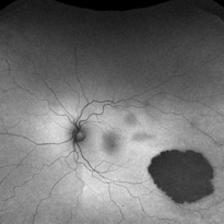 Congenital Hypertrophy of the Retinal Pigment Epithelium Autofluorescence Optomap
Sep 24 2019 by Sophia El Hamichi, MD
A 52-year-old female followed for 2 temporal lesions of CHRPE OD and white without pressure.
Photographer: Sophia El Hamichi, MD, Murray Ocular Oncology and Retina, Miami
Condition/keywords: autofluorescence imaging, congenital hypertrophy of the retinal pigment epithelium (CHRPE), Optomap, ultra-wide field imaging, white without pressure
-
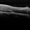
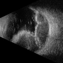 Suprachoroidal Hemorrhage
Nov 16 2019 by Sophia El Hamichi, MD
Ultrasound of the right eye of 43-year-old male presenting with suprachoroidal hemorrhage, note the multilobulated heterogenous echogenic mass aspect of the choroid
Photographer: Fiona J Ehlies, Murray Ocular Oncology and Retina, Miami
Condition/keywords: B scan ultrasound, suprachoroidal hemorrhage
-
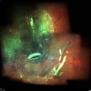
 Amelanotic Bear Tracks of the Retina
Jan 13 2020 by Sophia El Hamichi, MD
Fundus photograph of a 5-year-old patient with amelanotic bear tracks of the retina OD. No family history of colon cancer reported.
Photographer: Abby Orcutt-Hayes, Murray Ocular Oncology and Retina
Condition/keywords: amelanotic, bear tracks, congenital hypertrophy of the retinal pigment epithelium (CHRPE), pediatic retina
-
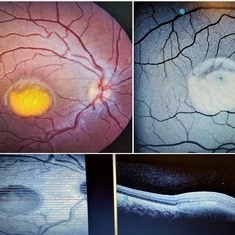 Best Vitelliform Macular Dystrophy
Mar 17 2020 by Sophia El Hamichi, MD
Classic "egg yolk" presentation in a 16-year-old female with best disease.
Condition/keywords: autofluorescence imaging, Best disease, optical coherence tomography (OCT), vitelliform macular dystrophy
-
 Myopia with Lattice Degeneration and White Without Pressure in the Setting of Marfan's Syndrome
Aug 31 2020 by Sophia El Hamichi, MD
A 1-year-old female with Marfan's syndrome, myopia OU, congenital nystagmus and exotopia OD. Ultra-wide field imaging of her left eye showed lattice degeneration with atrophic retinal holes temporally, in addition to multiple sections of white without pressure.
Imaging device: Optos
Condition/keywords: atrophic retinal hole, lattice degeneration, Marfan's syndrome, myopia, Optos, ultra-wide field imaging
-
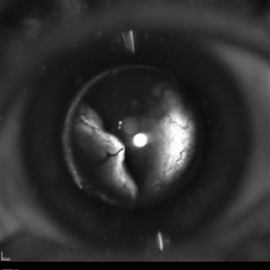
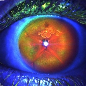 Iris Bombe
Sep 15 2020 by Sophia El Hamichi, MD
A 23-year-old female with Coats' disease OS.
Photographer: Belinda Rodriguez, Murray Ocular Oncology and Retina, Miami
Condition/keywords: Coats' disease, iris bombe, silicone oil
-
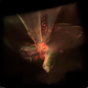
 Large Retinal Fold
Sep 22 2020 by Sophia El Hamichi, MD
A 69-year-old female, with a history of choroidal melanoma in her left eye with exudative detachment, underwent tumor laser ablation. She then developed a complex combined tractional and rhegmatogenous retinal detachment with a giant retinal tear. The patient underwent surgical repair of her retinal detachment with pars plana vitrectomy and silicone oil. In the post-op, the patient developed large retinal folds masking the optic nerve depicted in the fundus photograph.
Photographer: Belinda Rodriguez, Murray Ocular Oncology and Retina, Miami
Condition/keywords: melanoma, pars plana vitrectomy (PPV), retinal fold, silicone oil
-
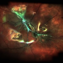
 Large Retinal Fold Masking the Optic Nerve
Sep 22 2020 by Sophia El Hamichi, MD
A 69-year-old female, with a history of choroidal melanoma in her left eye with exudative detachment, underwent tumor laser ablation. She then developed a complex combined tractional and rhegmatogenous retinal detachment with a giant retinal tear. The patient underwent surgical repair of her retinal detachment with pars plana vitrectomy and silicone oil. In the post-op, the patient developed large retinal folds masking the optic nerve depicted in the OCT photograph.
Photographer: Belinda Rodriguez, Murray Ocular Oncology and Retina, Miami
Condition/keywords: giant retinal tear, melanoma, pars plana vitrectomy (PPV), retinal fold, silicone oil
-
 Total Rhegmatogenous and Tractional Retinal Detachment Following Choroidal Melanoma Laser Ablation Treatment
Sep 22 2020 by Sophia El Hamichi, MD
A 69-year-old female, with a history of choroidal melanoma in her left eye with exudative detachment, underwent tumor laser ablation. She then developed a complex combined tractional and rhegmatogenous retinal detachment with a giant retinal tear.
Photographer: Belinda Rodriguez, Murray Ocular Oncology and Retina, Miami
Condition/keywords: anterior segment, melanoma
-
 Total Rhegmatogenous and Tractional Retinal Detachment Following Choroidal Melanoma Laser Ablation Treatment
Sep 22 2020 by Sophia El Hamichi, MD
A 69-year-old female, with a history of choroidal melanoma in her left eye with exudative detachment, underwent tumor laser ablation. She then developed a complex combined tractional and rhegmatogenous retinal detachment with a giant retinal tear.
Photographer: Belinda Rodriguez, Murray Ocular Oncology and Retina, Miami
Condition/keywords: tractional retinal detachment
-
 Fibrosis and Traction Following Traction Retinal Detachment Repair
Oct 13 2020 by Sophia El Hamichi, MD
A 29-year-old female with a history of diabetes mellitus type 1, presented with proliferative diabetic retinopathy OU and tractional retinal detachment OD. The patient underwent retinal detachment repair with pars plana vitrectomy, endolaser and silicone oil placement. After one month of her surgery, the patient presented with retinal fibrosis and tractions depicted in the image.
Photographer: Belinda Rodriguez, Murray Ocular Oncology and Retina, Miami
Condition/keywords: pars plana vitrectomy (PPV), post-op, proliferative diabetic retinopathy (PDR), proliferative vitreoretinopathy (PVR), tractional retinal detachment
-
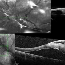 Fibrosis and Traction Following Traction Retinal Detachment Repair
Oct 13 2020 by Sophia El Hamichi, MD
A 29-year-old female with a history of diabetes mellitus type 1, presented with proliferative diabetic retinopathy OU and tractional retinal detachment OD. The patient underwent retinal detachment repair with pars plana vitrectomy, endolaser and silicone oil placement. After one month of her surgery, the patient presented with retinal fibrosis and traction. The image on the top shows the OCT of the fibrosis post op, that was not present in the pre op (OCT image on the bottom).
Photographer: Belinda Rodriguez, Murray Ocular Oncology and Retina, Miami
Condition/keywords: optical coherence tomography (OCT), pars plana vitrectomy (PPV), proliferative diabetic retinopathy (PDR), proliferative vitreoretinopathy (PVR), tractional retinal detachment
-
 Perivascular Bone Spicule Changes
Mar 1 2021 by Sophia El Hamichi, MD
A 19-year-old female African-American, who is followed for lattice degeneration and bone spicule changes OU. VA 20/20 OU. The bone spicule changes are stable throughout her follow-ups
Condition/keywords: bone spicule, lattice degeneration, Optos, perivascular, white without pressure
-
 Ultra-Wide Field Fundus Photography Showing Lattice Degeneration
Mar 22 2021 by Sophia El Hamichi, MD
Lattice degeneration, atrophic holes, white without pressure OS in a 19-year-old female.
Condition/keywords: atrophic retinal hole, Optos, peripheral lattice degeneration, ultra-wide field imaging, white without pressure
A project from the American Society of Retina Specialists