-
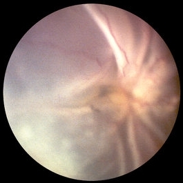 PVR Retinal Detachment with subretinal bands Slide 1
Oct 22 2012 by Ronald C. Gentile, MD
Total retinal detachment with pre-retinal and sub-retinal proliferation. The subretinal bands have a napkin ring configuration posteriorly with the macula folded and dragged above the optic nerve.
Photographer: The New York Eye & Ear Infirmary Department of Medical Imaging
Condition/keywords: proliferative vitreoretinopathy (PVR), subretinal bands
-
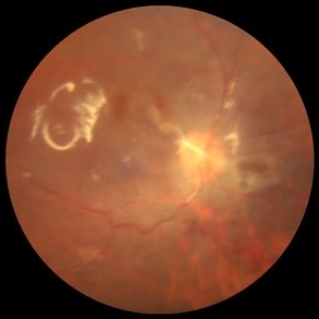 PVR Retinal Detachment with subretinal bands Slide 2
Oct 22 2012 by Ronald C. Gentile, MD
Postoperative fundus photo of the posterior pole with flat retina. As noted by the retinal surface reflex, silicone oil tamponade was used.
Photographer: The New York Eye & Ear Infirmary Department of Medical Imaging
Condition/keywords: proliferative vitreoretinopathy (PVR), subretinal bands
-
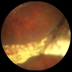 PVR Retinal Detachment with subretinal bands Slide 3
Oct 22 2012 by Ronald C. Gentile, MD
Postoperative fundus photo of the margin of the inferior nasal retinectomy site. The retinectomy was used intra-operatively to flap the retina over and remove the subretinal bands. The scleral buckle effect and acute white endolaser marks are present at the edge of the retinectomy.
Photographer: The New York Eye & Ear Infirmary Department of Medical Imaging
Condition/keywords: proliferative vitreoretinopathy (PVR), subretinal bands
-
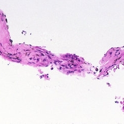 PVR Retinal Detachment with subretinal bands Slide 4
Oct 22 2012 by Ronald C. Gentile, MD
Histo-pathology of the subretinal bands revealed the presence of a fibrocellular membrane. The cells were predominately retinal pigment epithelium cells with myofibroblastic differentiation. Collagen deposition with occasional inflammatory cells and pigment were noted.
Photographer: The New York Eye & Ear Infirmary Department of Pathology and Laboratory Medicine
Condition/keywords: proliferative vitreoretinopathy (PVR), subretinal bands
A project from the American Society of Retina Specialists