-
 Multiple Myeloma with Cytomegalovirus Retinitis
Apr 5 2018 by Kim Barrett
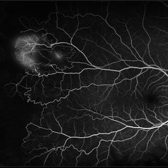
Ultra-wide field fluorescein angiogram of a 77-year-old male with multiple myeloma. Patient's angiogram presented significant peripheral retinal ischemia and cystoid macular edema. Patient tested positive for polymerase chain reaction, confirming cytomegalovirus retinitis. Patient is being treated with intravitreal ganciclovir and his current vision is 20/200.
Photographer: Kim Barrett, COA
Imaging device: Optos
Condition/keywords: cystoid macular edema (CME), intravitreal ganciclovir, myeloma, peripheral ischemia, positive polymerase chain reaction (PCR), ultra-wide field imaging
-
 Disseminated Chorioretinitis With Unknown Etiology
Apr 5 2018 by Kim Barrett
Ultra-wide field fluorescein angiogram of a 31-year-old female with intermittent pain in her left eye. Her condition has been managed in Liberia until recently when she moved to the United States. She suffers from multiple modalities including central retinal artery occlusion, posterior synechiae of the iris, interstitial keratitis, disseminated chorioretinitis, as well as HIV. An infectious cause is high on the differential in light of her HIV status. DDx: hypertensive crisis, an embolism (? IV drug use), coagulopathy, trauma, infectious. Blood work was normal. Her current vision is 20/30 right eye and 20/400 left eye.
Photographer: Kim Barrett, COA
Imaging device: Optos
Condition/keywords: central retinal artery occlusion (CRAO), chorioretinal scar, ciliary artery sparring, disseminated chorioretinitis, HIV, left eye, optic atrophy, staining
-
 Central Retinal Artery Occlusion
Apr 20 2018 by Kim Barrett
64-year-old female woke with no vision in her right eye. This image was taken at 6:11 minutes and the vessels have not filled. Patient has been treated with PRP laser and anti-VEGF therapy. Current vision is CF @ 2 ft.
Photographer: Kim Barrett C.O.A.
Imaging device: Heidelberg
Condition/keywords: central retinal artery occlusion (CRAO), diabetes, hypertension, smoker, uncontrolled
-
 Coats' Disease
Aug 24 2018 by Kim Barrett
Montage fluorescein angiography of 14-year-old male with Coats' Disease of the left eye. Multiple focal laser treatments. Current uncorrected visual acuity is 20/15-1 OU.
Photographer: Kim Barrett, C.O.A. Retina Specialist of Michigan
Imaging device: Heidelberg Spectralis
Condition/keywords: adolescent, Coats' disease, Heidelburg Spectralis, laser photocoagulation, left eye, macroaneurysm, montage
-
 Coats' Disease
Jul 16 2019 by Kim Barrett
Ultra-wide field fluorescein angiogram of a 23-year-old male with Coats' disease, presented with distorted vision affecting his left eye. He reported seeing flashes and floaters since January of 2019, but the flashes had resolved. He was treated with Intravitreal Preservative Free Triamcinolone in the office and scheduled for PRP laser in the near future.
Photographer: Kim Barrett
Imaging device: Optos
Condition/keywords: Coats' disease, inferior retina, ischemia, left eye, Optos, ultra-wide field imaging
-
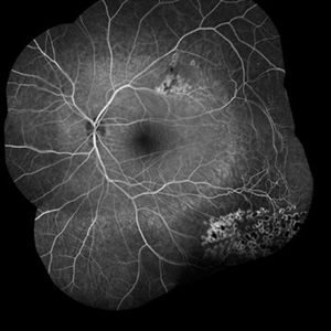
 Optic Nerve Pit Left Eye
Feb 15 2021 by Kim Barrett
A 14-year-old male presented with vision loss and VF defect. Patient was treated for presumed amblyopia with patching since age 4. He has had neurologic care for post traumatic skull fracture and brain bleed in 2012. Patient has a superior hemifield defect OS on HVF. IOP's WNL. There are vessels emanating from the optic pit OS. Patient is at risk of serous detachment. Current VA 20/20-2+2
Photographer: Kim Barrett C.O.A. Retina Specialist of Michigan, Grand Rapids, MI
Imaging device: Optos California
Condition/keywords: amblyopia, hemifield, Humphrey visual field, nerve, optic nerve pit, visual field defect
-
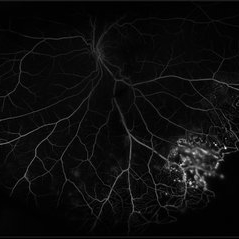
 Optic Nerve Pit Right Eye
Feb 15 2021 by Kim Barrett
A 14-year-old male presented with vision loss and VF defect. Patient was treated for presumed amblyopia with patching since age 4. He has had neurologic care for post traumatic skull fracture and brain bleed in 2012. IOP's WNL. OD is without retinoschisis or subretinal fluid. Patient is at risk of serous detachment. Current VA OD 20/200+1 PH 20/80.
Photographer: Kim Barrett C.O.A. Retina Specialist of Michigan, Grand Rapids, MI
Imaging device: Optos California
Condition/keywords: amblyopia, hemifield, Humphrey visual field, nerve, optic nerve pit, visual field defect
-
 Sickle Cell Retinopathy
Feb 15 2021 by Kim Barrett
24-year-old female with Sickle Cell Retinopathy, stage 3. She confirms she has the trait as well as her grandmother, mother and a sibling. She has seafan neovascularization superotemporal OD. Current VA is 20/20. Photo is pre-PRP laser with areas of non-profusion temporally.
Photographer: Kim Barrett C.O.A. Retina Specialist of Michigan, Grand Rapids, MI
Imaging device: Optos California
Condition/keywords: neovascularization (NV), pan-retinal photocoagulation (PRP), sickle cell retinopathy, stage 3, trait
A project from the American Society of Retina Specialists