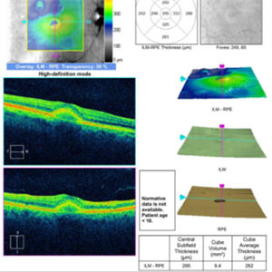
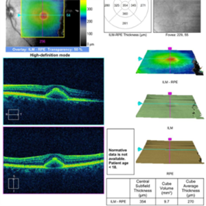
This 68 year old man was referred with a maculopathy but with normal visual acuity. He is thought to have multifocal Best Disease; there is no evidence of inflammation or uveitis and no history of cancer. Macular exam reveals yellow subretinal pigment clumping in each macula, including the foveal area. OCT shows moderate hyper-reflective PED, with no evidence of subretinal fluid or macular edema. Angiography reveals blocking defects due to pigment clumping with no evidence of CNVM.
-
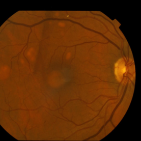 Multifocal Best Disease
Jan 31 2015 by Thomas A. Ciulla, MD, MBA, FASRS
Macular exam reveals yellow subretinal pigment clumping in each macula, including the foveal area.
Photographer: Charlotte Harris
Condition/keywords: adult vitelliform dystrophy, Best disease
-
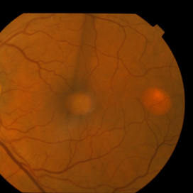 Multifocal Best Disease
Jan 31 2015 by Thomas A. Ciulla, MD, MBA, FASRS
Macular exam reveals yellow subretinal pigment clumping in each macula, including the foveal area.
Photographer: Charlotte Harris
Condition/keywords: adult vitelliform dystrophy, Best disease
-
 Multifocal Best Disease
Jan 31 2015 by Thomas A. Ciulla, MD, MBA, FASRS
Macular exam reveals yellow subretinal pigment clumping in each macula, including the foveal area.
Photographer: Charlotte Harris
Condition/keywords: adult vitelliform dystrophy, Best disease
-
 Multifocal Best Disease
Jan 31 2015 by Thomas A. Ciulla, MD, MBA, FASRS
Macular exam reveals yellow subretinal pigment clumping in each macula, including the foveal area.
Photographer: Charlotte Harris
Condition/keywords: adult vitelliform dystrophy, Best disease
-
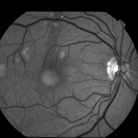
 Multifocal Best Disease
Jan 31 2015 by Thomas A. Ciulla, MD, MBA, FASRS
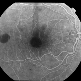
Angiography reveals blocking defects due to pigment clumping with no evidence of CNVM.
Photographer: Charlotte Harris
Condition/keywords: adult vitelliform dystrophy, Best disease
-
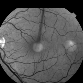
 Multifocal Best Disease
Jan 31 2015 by Thomas A. Ciulla, MD, MBA, FASRS
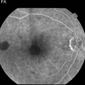
Angiography reveals blocking defects due to pigment clumping with no evidence of CNVM.
Photographer: Charlotte Harris
Condition/keywords: adult vitelliform dystrophy, Best disease
-
 Multifocal Best Disease
Jan 31 2015 by Thomas A. Ciulla, MD, MBA, FASRS
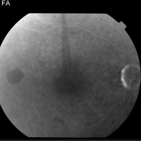
Angiography reveals blocking defects due to pigment clumping with no evidence of CNVM.
Photographer: Charlotte Harris
Condition/keywords: adult vitelliform dystrophy, Best disease
-
 Multifocal Best Disease
Jan 31 2015 by Thomas A. Ciulla, MD, MBA, FASRS
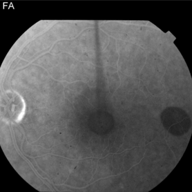
Angiography reveals blocking defects due to pigment clumping with no evidence of CNVM.
Photographer: Charlotte Harris
Condition/keywords: adult vitelliform dystrophy, Best disease
-
 Multifocal Best Disease
Jan 31 2015 by Thomas A. Ciulla, MD, MBA, FASRS
OCT shows moderate hyper-reflective PED, with no evidence of subretinal fluid or macular edema.
Condition/keywords: adult vitelliform dystrophy, Best disease
-
 Multifocal Best Disease
Jan 31 2015 by Thomas A. Ciulla, MD, MBA, FASRS
OCT shows moderate hyper-reflective PED, with no evidence of subretinal fluid or macular edema.
Condition/keywords: adult vitelliform dystrophy, Best disease