 Initializing download.
-
 By Olivia Rainey
Retina Specialists of Michigan
Co-author(s): Scott Westhouse, DO - Uploaded on Jul 5, 2022.
- Last modified by Joshua Friedman on Jul 7, 2022.
- Rating
- Appears in
- Miscellaneous
- Condition/keywords
- vascular disorders of iris and ciliary body, vascular tuft, vascular anomaly, infrared image, near infrared autofluorescence (NIRAF), fluorescein angiogram (FA), anterior segment, slit lamp photo, heidelberg spectralis
- Photographer
- Olivia Rainey, OCT-C, COA
- Imaging device
- Heidelberg Spectralis, Slit Lamp with Samsung Galaxy 7
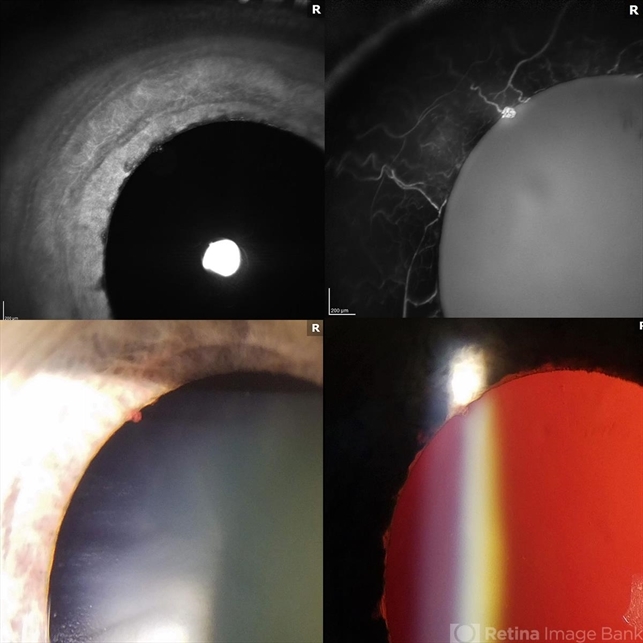
- Description
- Anterior segment imaging of a 66-year-old male with Vascular Disorders of Iris and Ciliary Body affecting his right eye. The physician stated that the findings are most consistent with a benign vascular tuft at the pupillary margin. The patient presented at the office with 20/20 vision in both eyes and had no ocular complaints at the time of his appointment.

---thumb.jpg/image-square;max$79,0.ImageHandler "Polypoidal Choroidal Vasculopathy: Case 1 - Image 3 of 7")


---thumb.jpg/image-square;max$79,0.ImageHandler "Choroideremia - Scleral Stain")
---thumb.jpg/image-square;max$79,0.ImageHandler "Choroideremia")
---thumb.JPG/image-square;max$79,0.ImageHandler "Carotid Cavernous Fistula")