File number: 94589
Comments
-
 Manish Nagpal, MD, FRCS (UK), FASRS (October 6 2022)
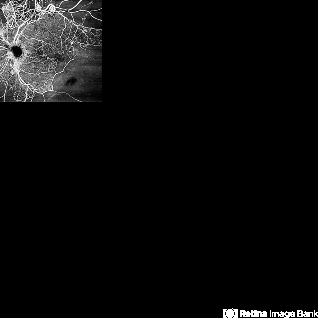
fantastic capture clearly demarcating the capillary non perfusion areas and the intertwined networks of capillaries and new vessels
Sign in to comment.
 Initializing download.
-
 By JORGE SOBERANES
Asociacion para Evitar la Ceguera en México
Co-author(s): Julian García MD - Uploaded on Jun 13, 2022.
- Last modified by Joshua Friedman on Sep 30, 2022.
- Image of the week
-
Oct 2, 2022
View all images of the week - Rating
- Appears in
- Central retinal vein occlusion
- Condition/keywords
- OCT angiography, Central vein oclussion, Shunts, neovascularization, retina
- Photographer
- Jorge I. Soberanes MD
- Imaging device
-
Optical coherence tomography system
PLEX Elite 9000, Zeiss - Description
- A 63 year old man with a central retinal vein oclussion. In the OCT angiogram we could observe retinal isquemia, neovascularization and arteriovenous shunts.


---thumb.jpg/image-square;max$79,0.ImageHandler "Retinoblastoma To Chemothermotherapy")
---thumb.jpg/image-square;max$79,0.ImageHandler "Retinoblastoma To Chemothermotherapy")


