 Initializing download.
-
 By ruth pav
Co-author(s): Ruth Pav, Rambam medical center,Hifa Israel. - Uploaded on May 31, 2014.
- Last modified by Caroline Bozell on Jun 2, 2014.
- Rating
- Appears in
- Miscellaneous
- Condition/keywords
- retina
- Photographer
- Ruth Pav, Rambam medical center,Hifa Israel.
- Imaging device
-
Fundus camera
Zeiss FF4 - Description
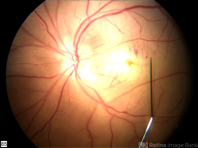
- A 32-year-old woman with a history of drug abuse was admitted due to acute manifestation of multiple infarcts, including acute stroke, splenic and renal infarcts, and multiple cutaneous hematomas. Due to decreased vision in her left eye the patient was referred for ophthalmic evaluation. On exam, visual acuity was 6/10 in the right eye and no light perception in her left eye. Ophthalmoscopic examination was normal in the right eye but showed pallor of the optic nerve head with attenuated retinal vessels in the left eye. Fluorescein angiography showed an oval area of hyperfluorescence from from non-perfusion involving the macular center with staining of overlying retinal capillaries.


---thumb.jpg/image-square;max$79,0.ImageHandler "Retinoblastoma To Chemothermotherapy")
---thumb.jpg/image-square;max$79,0.ImageHandler "Retinoblastoma To Chemothermotherapy")


