 Initializing download.
-
 By Purva Patwari
Patwari Retina Clinic - Uploaded on Apr 23, 2019.
- Last modified by Caroline Bozell on Apr 23, 2019.
- Rating
- Appears in
- Patwari Retina Clinic
- Condition/keywords
- choroiditis, choroidal granuloma, tubercular choroidal granuloma, tuberculosis, granulomatous choroiditis
- Photographer
- Dr Purva Patwari, Patwari Retina Center
- Imaging device
-
Fundus camera
Zeiss Visu 500 - Description
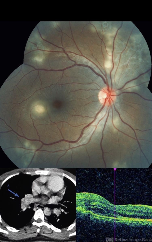
- 22-year-old male patient presented with blurring of vision in the right eye noticed since last one week. He was asymptomatic a week ago when he noticed the blurring in his right eye. On examination his vision was 6/6 in both eyes. Anterior segment was normal. Posterior segment was normal for the left eye. Right eye examination revealed a clear vitreous cavity with choroidal granulomas scattered throughout the fundus. The present picture shows choroidal granulomas with OCT segment passing through the parafoveal lesion showing subretinal fluid accumulation and corresponding thickening of the retinal layers. CT scan reveals heterogeneously enhancing lymph nodes showing conglomerationin the hilar region-possibility of tubercular etiology.


---thumb.JPG/image-square;max$79,0.ImageHandler "Tubercular choroidal granuloma")


---thumb.JPG/image-square;max$79,0.ImageHandler "Blastomycosis")
---thumb.JPG/image-square;max$79,0.ImageHandler "Tubercular choroidal granuloma")
---thumb.JPG/image-square;max$79,0.ImageHandler "Tubercular choroidal granuloma")
---thumb.JPG/image-square;max$79,0.ImageHandler "Sarcoidosis")


