-
 Branch Retinal Vein Occlusion
Sep 28 2017 by Purva Patwari
Patient came for routine work.
Photographer: Dr Purva Patwari,Patwari Retina Clinic,Ahmedabad
Imaging device: Zeiss
Condition/keywords: neovascularization (NV)
-
 Neovascularisation of Disc
Oct 3 2017 by Purva Patwari
52-year-old with uncontrolled DM, unilateral NVD, other eye moderate NPDR.
Photographer: Dr Purva Patwari, Patwari Retina Clinic, Ahmedabad INDIA
Condition/keywords: neovascularization of the disc (NVD)
-
 Neovascularisation of Disc
Oct 3 2017 by Purva Patwari
52-year-old with uncontrolled DM, unilateral NVD, other eye moderate NPDR.
Photographer: Dr Purva Patwari, Patwari Retina Clinic, Ahmedabad INDIA
Condition/keywords: neovascularization (NV), neovascularization of the disc (NVD)
-
 Laser in a Patient With Branch Vein Occlusion
Dec 24 2017 by Purva Patwari
Post laser photocoagulation in a patient with branch vein occlusion.
Photographer: Dr Purva Patwari, Patwari Retina Center, Ahmedabad, Gujarat , India
Condition/keywords: laser photocoagulation
-
 Infero Nasal Vein Occlusion
Dec 24 2017 by Purva Patwari
Female patient on routine eye exam
Photographer: Dr Purva Patwari, Patwari Retina Center, Ahmedabad, Gujarat , India
-
 Nevus
Nevus
Dec 24 2017 by Purva Patwari
Female patient for routine Ophthalmic check-up
Photographer: Dr Purva Patwari, Patwari Retina Center, Ahmedabad, Gujarat , India
Condition/keywords: nevus
-
 Hypertensive Retinopathy
Dec 24 2017 by Purva Patwari
52-year-old female diagnosed of hypertension by retina evaluation.
Photographer: Dr Purva Patwari, Patwari Retina Center, Ahmedabad, Gujarat , India
Imaging device: ZEISS VISU500
Condition/keywords: hypertensive retinopathy, neovascularization elsewhere (NVE), Roth spots
-
 Supero Temporal Branch Vein Occlusion
Dec 24 2017 by Purva Patwari
Fundus photo of a 56-year-old male patient
Photographer: Dr Purva Patwari, Patwari Retina Center, Ahmedabad, Gujarat , India
-
 Proliferative Diabetic Retinopathy
Jan 2 2018 by Purva Patwari
63-year-old male patient on presentation. Vision was 6/6, known diabetic since 10 years.
Photographer: Dr Purva Patwari, Patwari Retina Center, Ahmedabad, Gujarat , India
Imaging device: ZEISS VISU 500
Condition/keywords: proliferative diabetic retinopathy (PDR)
-
 Human Shoe Tear
Jan 2 2018 by Purva Patwari
Asymptomatic horse shoe tear found on preoperative cataract assessment of a 54-year-old male patient. Laser barrage was done and he underwent phacoemulsification surgery a month later.
Photographer: Dr Purva Patwari, Patwari Retina Center, Ahmedabad, Gujarat , India
Imaging device: ZEISS VISU 500
-
 Choroidal Neovacular Membrane
Feb 4 2018 by Purva Patwari
78-year-old female with gradual progressive loss of vision since last 5 years.
Photographer: Dr Purva Patwari
Condition/keywords: choroidal neovascular membrane (CNVM)
-
 Subretinal Hemorrhage
Feb 4 2018 by Purva Patwari
65-year-old female with sudden loss of vision.
Photographer: Dr Purva Patwari
Condition/keywords: subretinal hemorrhage
-
 Stargardts Disease
Mar 26 2018 by Purva Patwari
16-year-old girl with defective vision, gradually progressive since childhood.
Photographer: Dr Purva Patwari, Patwari Retina Center, Ahmedabad, Gujarat , India
Condition/keywords: macular dystrophy, Stargardt disease
-
 Stargardts Disease
Mar 26 2018 by Purva Patwari
16-year-old girl with defective vision, gradually progressive since child hood.
Photographer: Dr Purva Patwari, Patwari Retina Center, Ahmedabad, Gujarat , India
Condition/keywords: macular dystrophy, Stargardt disease
-
 Stargardts Disease
Mar 26 2018 by Purva Patwari
16-year-old girl with defective vision, gradually progressive since childhood.
Photographer: Dr Purva Patwari, Patwari Retina Center, Ahmedabad, Gujarat , India
Condition/keywords: Stargardt disease
-
 NVD
NVD
Mar 26 2018 by Purva Patwari
65-year-old male patient diabetic since 10 years, presented with mild dip in vision corresponding to the cataractous changes. On thorough examination he had florid NVD with sclerosed sup temporal vessel. HbA1c of 11.3 and nephropathy all diagnosed by us during the routine investigations for his retinopathy.
Photographer: Dr Purva Patwari, Patwari Retina Center, Ahmedabad, Gujarat , India
Condition/keywords: neovascularization (NV), neovascularization of the disc (NVD)
-
 IJFT
IJFT
Mar 26 2018 by Purva Patwari
Defective vision.
Photographer: Dr Purva Patwari, Patwari Retina Center, Ahmedabad, Gujarat , India
Condition/keywords: abnormal retinal vessel, IJFT, IJT
-
 IJFT
IJFT
Mar 26 2018 by Purva Patwari
48-year-old bank employee presented with gradual decrease of vision in both eyes VA OU 6/18 Other eye was operated for cataract surgery but no visual gain. She is a known hypertension patient with an axial length of arount 20mm.
Photographer: Dr Purva Patwari, Patwari Retina Center, Ahmedabad, Gujarat , India
Condition/keywords: idiopathic juxta foveal telenjectasia, IJFT, IJT
-
 Optic Disc Coloboma`
Mar 26 2018 by Purva Patwari
16-year-old female patient with vision of 6/60 presented with diminished vison. Other eye was normal.She had a normal birth history and developmental milestone. Look at the optic disc coloboma extending upto the macula. Intercalary membrane looks normal.
Photographer: Dr Purva Patwari, Patwari Retina Center, Ahmedabad, Gujarat , India
Imaging device: ZEISS VISU 500
Condition/keywords: coloboma, coloboma of optic disc, optic disc
-
 Moderate Non proliferative Diabetic Retinopathy
Mar 26 2018 by Purva Patwari
Nonproliferative diabetic retinopathy
Photographer: Dr Purva Patwari, Patwari Retina Center, Ahmedabad, Gujarat , India
Imaging device: ZEISS VISU 500
Condition/keywords: nonproliferative diabetic retinopathy
-
 Post Pars Plana Vitrectomy
Mar 31 2018 by Purva Patwari
40-year-old high myope, axial lenght of 31mm had retinal detachment. She underwent PPV, PFCL, endolaser +silicon oil implantation . The pic depicts 3 months post operative.
Photographer: Dr Purva Patwari,Patwari Retina Center,Ahmedabad,India
Condition/keywords: re-attached retinal detachment (RRD)
-
 Subhyaloid Hemorrhage
Apr 3 2018 by Purva Patwari
43-year-old male, known case of hypertension presented with sudden loss of vision for last 2 days.
Photographer: Dr Purva Patwari,Patwari Retina Center,Ahmedabad,India
Condition/keywords: subhyaloid hemorrhage
-
 Self Settled RD
Apr 6 2018 by Purva Patwari
24-year-old with defective vision in LE since childhood. On examination his BCVA was RE 6/6, LE CF. LE >45 degree exo., extra ocular movements were full in both eyes. Fundus photo revealed the rest.
Photographer: Dr Purva Patwari,Patwari Retina Center,Ahmedabad,India
Imaging device: ZEISS VISU 500
Condition/keywords: spontaneous retinal reattachment, subretinal bands
-
 Post Retinal Detachment
Apr 12 2018 by Purva Patwari
67-year-old patient one eye 2 months post RD surgery, present vision is 6/18,there is ERM but patient is comfortable.
Photographer: Dr Purva Patwari,Patwari Retina Center,Ahmedabad,India
Imaging device: Zeiss Visucam 500
Condition/keywords: silicone oil
-
 Choroiditis
Jun 13 2018 by Purva Patwari
A 25-year-old male patient presented with defective vision noticed since last 2 weeks. No significant past history. Anterior segment was normal. Vision RE 6/6, LE 6/60. Other eye was normal on examination.
Photographer: Dr Purva Patwari, Patwari Retina Center, Ahmedabad, Gujarat , India
Imaging device: Zeiss Visucam 500
Condition/keywords: choroiditis, disseminated choroiditis
-
 Choroidal Granuloma
Apr 23 2019 by Purva Patwari
22-year-old male patient presented with blurring of vision in the right eye noticed since last one week. He was asymptomatic a week ago when he noticed the blurring in his right eye. On examination his vision was 6/6 in both eyes. Anterior segment was normal. Posterior segment was normal for the left eye. Right eye examination revealed a clear vitreous cavity with choroidal granulomas scattered throughout the fundus. The present picture shows choroidal granulomas with OCT segment passing through the parafoveal lesion showing subretinal fluid accumulation and corresponding thickening of the retinal layers. CT scan reveals heterogeneously enhancing lymph nodes showing conglomerationin the hilar region-possibility of tubercular etiology.
Photographer: Dr Purva Patwari, Patwari Retina Center
Imaging device: Zeiss Visu 500
Condition/keywords: choroidal granuloma, choroiditis, granulomatous choroiditis, tubercular choroidal granuloma, tuberculosis
-
 Horse Shoe Tear
Sep 16 2017 by Purva Patwari
Asymptomatic horse shoe tear found on preoperative cataract assessment of a 54-year-old male patient. Laser barrage was done and he underwent Phacoemulsification surgery a month later.
Photographer: Dr Purva Patwari,Patwari Retina Center,Ahmedabad,India
Imaging device: Zeiss Visucam 500
-
 Toxoplasma
Sep 16 2017 by Purva Patwari
Head light in the fog appearance.
Photographer: Dr Purva Patwari,Patwari Retina Center,Ahmedabad,India
Condition/keywords: toxoplasmosis retinitis
-
 Subhyaloid Hemorrhage With Flat Neovascular Vessels
Sep 26 2017 by Purva Patwari
60-year-old patient with uncontrolled diabetes.
Photographer: Dr Purva Patwari, Patwari Retina Clinic,Ahmedabad, India
Imaging device: Zeiss
Condition/keywords: flat neovascularization
-
 Post Treatment
Sep 26 2017 by Purva Patwari
60-year-old patient with uncontrolled diabetes.
Photographer: Dr Purva Patwari, Patwari Retina Clinic,Ahmedabad , India
Condition/keywords: flat neovascularization
-
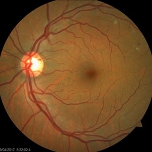
 AV Anastomosis
Sep 19 2017 by Purva Patwari
54-year-old female post cataract surgery.
Photographer: Dr Purva Patwari, Patwari Retina Center,Ahmedabad
Imaging device: Zeiss visu 500
Condition/keywords: arteriovenous anastomosis
A project from the American Society of Retina Specialists