 Initializing download.
-
 By Gustavo Uriel Fonseca Aguirre
Hospital de la Luz - Uploaded on Apr 9, 2025.
- Last modified by Joshua Friedman on Apr 10, 2025.
- Rating
- Appears in
- Miscellaneous
- Condition/keywords
- diabetic retinopathy, tractional retinal detachment, Vitreous hemorrhage
- Photographer
- Gustavo U. Fonseca Aguirre, Hospital Conde de Valenciana, Ciudad de México
- Imaging device
- Ultrasonography device
- Description
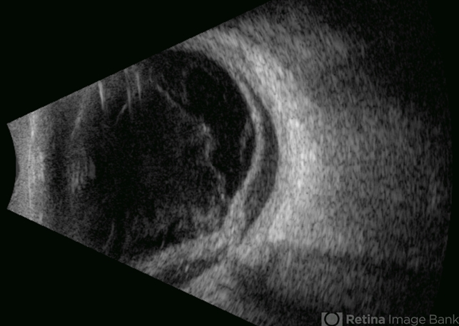
- B-mode ultrasound of a patient with long-standing poorly controlled diabetes demonstrates characteristic findings of advanced proliferative diabetic retinopathy. The examination reveals moderate vitreous hemorrhage appearing as diffuse hyperechoic opacities throughout the vitreous cavity, along with a posterior hyaloid membrane densely infiltrated by hemorrhagic material, showing irregular thickening and increased reflectivity. A mild subhyaloid hemorrhage is visible as a subtle hyphema-like space anterior to the retinal surface. The study documents a total tractional retinal detachment, evidenced by rigid retinal folds with clear insertion points of vitreous strands, accompanied by a significant subretinal hemorrhage seen as a prominent hyperechoic collection beneath the elevated retina. These findings collectively illustrate the severe vitreoretinal interface pathology characteristic of end-stage diabetic eye disease, with predominant tractional components and distinct echographic stratification of hemorrhagic layers - from anterior vitreous involvement to deeper subretinal blood accumulation.


---thumb.jpg/image-square;max$79,0.ImageHandler "Complications of ARN, TRD")

