-
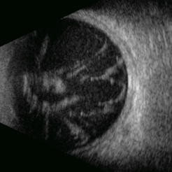
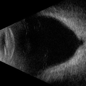
 Submacular Hemorrhage
Sep 24 2024 by Gustavo Uriel Fonseca Aguirre
24-year-old patient, submacular hemorrhage is observed in the left eye secondary to blunt ocular trauma.
Photographer: Gustavo U. Fonseca Aguirre, Fundación Hospital Nuestra Señora de la Luz, Ciudad de México
Condition/keywords: submacular hemorrhage
-
 Ocular Toxoplasmosis
Sep 24 2024 by Gustavo Uriel Fonseca Aguirre
24-year-old patient with a history of retinochoroiditis due to toxoplasmosis in the right eye, a focus of retinochoroiditis reactivation of toxoplasmosis is observed.
Photographer: Gustavo U. Fonseca Aguirre, Fundación Hospital Nuestra Señora de la Luz, Ciudad de México
Condition/keywords: Kyrieleis arteritis, toxoplasmosis reactivation
-
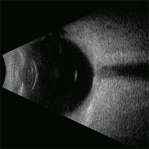
 Coats Disease
Sep 24 2024 by Gustavo Uriel Fonseca Aguirre
A 5-year-old male patient with no ophthalmological history, diagnosed with Coats disease in the right eye.
Photographer: Gustavo U. Fonseca Aguirre, Fundación Hospital Nuestra Señora de la Luz, Ciudad de México
Condition/keywords: Coats' disease
-
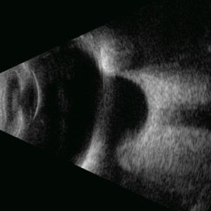
 Branch Retinal Artery Occlusion
Sep 24 2024 by Gustavo Uriel Fonseca Aguirre
60-year-old male patient diagnosed with diabetes with superior temporal retinal artery branch occlusion.
Photographer: Gustavo U. Fonseca Aguirre, Fundación Hospital Nuestra Señora de la Luz, Ciudad de México
Condition/keywords: branch retinal vein occlusion (BRVO)
-
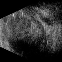
 Optic Nerve Head Avulsion
Sep 24 2024 by Gustavo Uriel Fonseca Aguirre
A 14-year-old male with a history of blunt ocular trauma in the right eye presented partial avulsion of the optic nerve head and submacular hemorrhage that was managed with neumatic displacement.
Photographer: Gustavo U. Fonseca Aguirre, Fundación Hospital Nuestra Señora de la Luz, Ciudad de México
Condition/keywords: optic nerve head avulsion
-
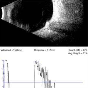
 Lacteocrumenasia
Mar 11 2025 by Gustavo Uriel Fonseca Aguirre
A 75-year-old female with a history of cataract surgery with intraocular lens implantation 20 years ago presented with progressive visual loss. On slit lamp examination, opaque material was found in the capsular bag behind the intraocular lens. Ultrasound biomicroscopy revealed hyperechoic material contained in the temporal-posterior sector of the capsular bag corresponding to lacteocrumenasia.
Photographer: Gustavo U. Fonseca Aguirre, Hospital Conde de Valenciana, Ciudad de México
Condition/keywords: Lacteocrumenasia, ultrasound biomicroscopy
-
 Hyaloid Butterfly
Mar 13 2025 by Gustavo Uriel Fonseca Aguirre
Axial ultrasound showing a phakic eye with vitreous hemorrhage, hyaloids impregnated with blood, hyalochisis (butterfly-shaped), subhyaloid hemorrhage, and retinal tractions involving the macular area.
Photographer: Gustavo U. Fonseca Aguirre, Hospital Conde de Valenciana, Ciudad de México
Condition/keywords: Hyaloschisis, Vitreous hemorrhage
-
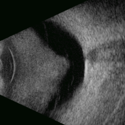
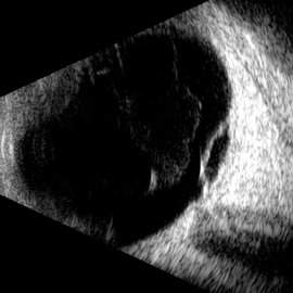
 Three Kisses
Mar 18 2025 by Gustavo Uriel Fonseca Aguirre
Cross-section of a B-mode ultrasound showing a kiss-shaped choroidal detachment; three lobes, giving the appearance of three kisses.
Photographer: Gustavo U. Fonseca Aguirre, Hospital Conde de Valenciana, Ciudad de México
Condition/keywords: Kissing Choroidal Detachment
-
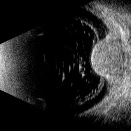
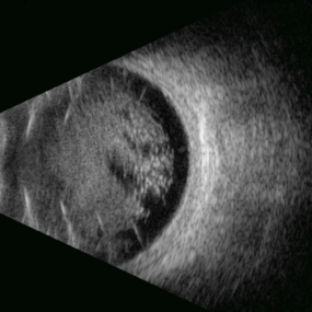 Disco Ball
Mar 19 2025 by Gustavo Uriel Fonseca Aguirre
Cross-section of B-mode ultrasound showing intense asteroid hyalosis.
Photographer: Gustavo U. Fonseca Aguirre, Hospital Conde de Valenciana, Ciudad de México
Condition/keywords: Asteroid hyalosis
-
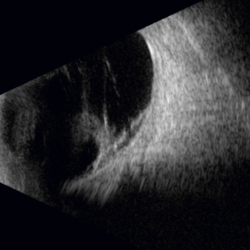
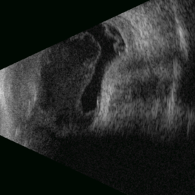
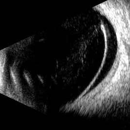
 Ectopia Lentis
Mar 21 2025 by Gustavo Uriel Fonseca Aguirre
Cross-section of B-mode ultrasound showing the lens in the lower part of the vitreous cavity.
Photographer: Gustavo U. Fonseca Aguirre, Hospital Conde de Valenciana, Ciudad de México
Condition/keywords: ectopia lentis
-
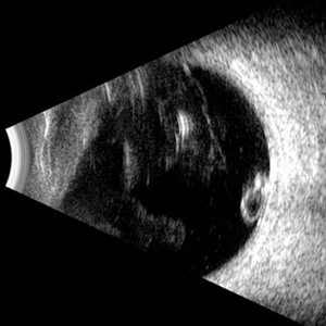
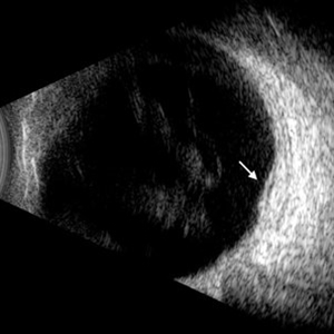
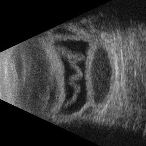
 Vasoproliferative Tumor
Mar 25 2025 by Gustavo Uriel Fonseca Aguirre
Patient diagnosed with pars planitis and a history of phacovitrectomy. Longitudinal B-scan section showing a very pronounced, homogeneous tumor lesion in the periphery. The A-scan revealed high average reflectivity with an irregular internal structure.
Photographer: Gustavo U. Fonseca Aguirre, Hospital Oftalmológico de la Luz, Ciudad de México
Condition/keywords: pars planitis, Vasoproliferative Tumor
-
 Melanocytoma
Mar 25 2025 by Gustavo Uriel Fonseca Aguirre
Longitudinal B-scan echogram shows mildly elevated lesion overlying surface of optic nerve. A-scan shows regular internal structure and high reflectivity of lesion.
Photographer: Gustavo U. Fonseca Aguirre, Hospital Conde de Valenciana, Ciudad de México
Condition/keywords: Melanocytoma
-
 IOL in Vitreous Cavity
Mar 25 2025 by Gustavo Uriel Fonseca Aguirre
Peripheral cross-section of B-mode ultrasound showing IOL dislocated into the vitreous cavity.
Photographer: Gustavo U. Fonseca Aguirre, Hospital Conde de Valenciana, Ciudad de México
Condition/keywords: IOL, vitreous cavity
-
 Macular Hole in Tractional Retinal Detachment
Apr 1 2025 by Gustavo Uriel Fonseca Aguirre
B-scan findings in a diabetic patient reveal vitreous hemorrhage, blood-soaked hyaloids, and tractional retinal detachment with an associated macular hole in the posterior pole.
Photographer: Gustavo U. Fonseca Aguirre, Hospital Conde de Valenciana, Ciudad de México
Condition/keywords: macular hole, tractional retinal detachment
-
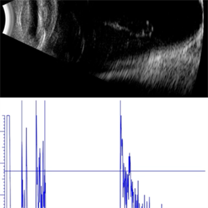
 IOL Reverberation
Apr 3 2025 by Gustavo Uriel Fonseca Aguirre
Top: Axial B-scan ultrasonography demonstrating an IOL with associated vitreous reverberation artifacts. Bottom: A-mode tracing revealing characteristic spike patterns consistent with IOL-induced reverberation.
Photographer: Gustavo U. Fonseca Aguirre, Hospital Conde de Valenciana, Ciudad de México
Condition/keywords: IOL, ultrasound
-
 Intraocular Foreign Body
Apr 3 2025 by Gustavo Uriel Fonseca Aguirre
B-mode ultrasonography of an eye with a 1-year history of suspected blunt trauma revealed an incidental intraocular foreign body within the vitreous cavity.
Photographer: Gustavo U. Fonseca Aguirre, Hospital Conde de Valenciana, Ciudad de México
Condition/keywords: intraocular foreign body
-
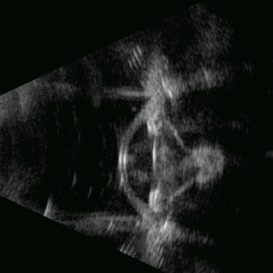
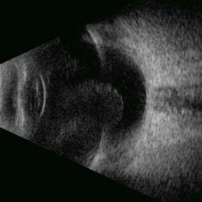
 Cyclodialysis
Apr 3 2025 by Gustavo Uriel Fonseca Aguirre
Ultrasound biomicroscopy (UBM) of a blunt-traumatized eye revealing cyclodialysis, zonular disruption with lenticular ectopia, and anterior chamber vitreous prolapse.
Photographer: Gustavo U. Fonseca Aguirre, Hospital Conde de Valenciana, Ciudad de México
Condition/keywords: cyclodialysis
-
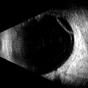
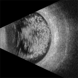 Asteroidal Hyalosis
Apr 7 2025 by Gustavo Uriel Fonseca Aguirre
B-mode ultrasound reveals an eye with significant asteroid hyalosis, with no relevant pathological history.
Photographer: Gustavo U. Fonseca Aguirre, Hospital Conde de Valenciana, Ciudad de México
Condition/keywords: asteroid hyalosis
-
 Calcification of the Retina
Apr 7 2025 by Gustavo Uriel Fonseca Aguirre
B-mode ultrasound of a vitrectomized eye reveals emulsified silicone oil in the vitreous cavity, retinal detachment, and calcification of the retina and optic nerve head.
Photographer: Gustavo U. Fonseca Aguirre, Hospital Conde de Valenciana, Ciudad de México
Condition/keywords: calcification, Retina detachment, vitrectomy
-
 Stag Horn
Apr 8 2025 by Gustavo Uriel Fonseca Aguirre
B-mode ultrasound of a young male patient with bilateral panuveitis (currently under investigation) reveals intense vitritis with islands of preserved vitreous and partial posterior hyaloid detachment, creating a characteristic "stag horn" appearance.
Photographer: Gustavo U. Fonseca Aguirre, Hospital Conde de Valenciana, Ciudad de México
Condition/keywords: Panuveitis
-
 Retinal Detachment Associated With a Posterior Staphyloma
Apr 9 2025 by Gustavo Uriel Fonseca Aguirre
B-mode axial ultrasound scan of a highly myopic eye shows a posterior staphyloma with an associated macular hole-induced retinal detachment.
Photographer: Gustavo U. Fonseca Aguirre, Hospital Conde de Valenciana, Ciudad de México
Condition/keywords: high myopia, posterior staphyloma, rhegmatogenous retinal detachment
-
 Advanced Proliferative Diabetic Retinopathy
Apr 9 2025 by Gustavo Uriel Fonseca Aguirre
B-mode ultrasound of a patient with long-standing poorly controlled diabetes demonstrates characteristic findings of advanced proliferative diabetic retinopathy. The examination reveals moderate vitreous hemorrhage appearing as diffuse hyperechoic opacities throughout the vitreous cavity, along with a posterior hyaloid membrane densely infiltrated by hemorrhagic material, showing irregular thickening and increased reflectivity. A mild subhyaloid hemorrhage is visible as a subtle hyphema-like space anterior to the retinal surface. The study documents a total tractional retinal detachment, evidenced by rigid retinal folds with clear insertion points of vitreous strands, accompanied by a significant subretinal hemorrhage seen as a prominent hyperechoic collection beneath the elevated retina. These findings collectively illustrate the severe vitreoretinal interface pathology characteristic of end-stage diabetic eye disease, with predominant tractional components and distinct echographic stratification of hemorrhagic layers - from anterior vitreous involvement to deeper subretinal blood accumulation.
Photographer: Gustavo U. Fonseca Aguirre, Hospital Conde de Valenciana, Ciudad de México
Condition/keywords: diabetic retinopathy, tractional retinal detachment, Vitreous hemorrhage
-
 Traumatic Posterior Capsular Rupture
Apr 9 2025 by Gustavo Uriel Fonseca Aguirre
Immersion B-mode ultrasound in a patient with blunt ocular trauma demonstrates an isolated posterior lens capsule rupture accompanied by phacodonesis.
Photographer: Gustavo U. Fonseca Aguirre, Hospital Conde de Valenciana, Ciudad de México
Condition/keywords: blunt trauma, Posterior Capsular Rupture
-
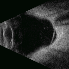
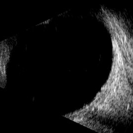
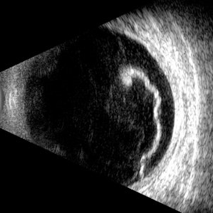
Hemorrhagic Choroidal Detachment
Apr 14 2025 by Gustavo Uriel Fonseca Aguirre
This B-mode transverse ultrasound scan demonstrates a hemorrhagic choroidal detachment with a characteristic wreath-like configuration, accompanied by concurrent retinal detachment. The choroidal lesion shows dome-shaped elevation with heterogeneous internal reflectivity, while the detached retina appears as a hyperechoic, undulating membrane.
Photographer: Gustavo U. Fonseca Aguirre, Hospital Conde de Valenciana, Ciudad de México
Condition/keywords: hemorrhagic choroidal detachment
-
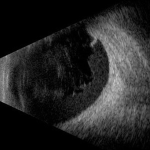
 PCR
PCR
Apr 17 2025 by Gustavo Uriel Fonseca Aguirre
B-mode transverse ultrasound scan of an eye with recent posterior capsule rupture during phacoemulsification shows hyperechoic punctate echoes in the vitreous (consistent with residual viscoelastic material) along with lens fragments in the subhyaloid space.
Photographer: Gustavo U. Fonseca Aguirre, Hospital Conde de Valenciana, Ciudad de México
Condition/keywords: Posterior capsule rupture (PCR)
-
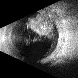
 Proliferative Vitreoretinopathy
Apr 17 2025 by Gustavo Uriel Fonseca Aguirre
This B-mode transverse ultrasound scan depicts a post-vitrectomy eye with recurrent retinal detachment in a patient with diabetic retinopathy history. The image reveals fresh vitreous cavity hemorrhage and subretinal bleeding, along with subretinal proliferative bands (PVR strands). These findings indicate complicated tractional re-detachment with active hemorrhagic components.
Photographer: Gustavo U. Fonseca Aguirre, Hospital Conde de Valenciana, Ciudad de México
Condition/keywords: proliferative vitreoretinopathy (PVR)
-
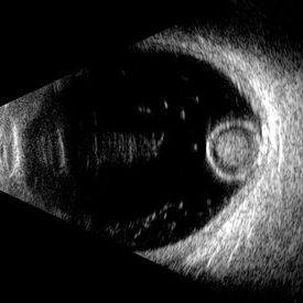
Vitreous Waltz vs Retinal Rigidity
Apr 18 2025 by Gustavo Uriel Fonseca Aguirre
B-mode dynamic ultrasound of an eye with vitreous hemorrhage shows hyaloid traction inducing retinal detachment in diabetic retinopathy. The video clearly delineates all anatomical compartments: vitreous, subhyaloid, and subretinal spaces. Characteristic movement patterns are observed - the vitreous demonstrates smooth, wide excursions while the detached retina shows shorter, stiffer motions -confirming tractional pathology.
Condition/keywords: diabetic retinopathy, retinal detachment
-
 Retinoschisis
Apr 21 2025 by Gustavo Uriel Fonseca Aguirre
This B-mode longitudinal ultrasound scan reveals a peripheral temporal retinoschisis, demonstrating a characteristic thin, dome-shaped separation of the retinal layers without associated subretinal fluid or vitreous traction. The lesion shows smooth, convex contours with maintained structural integrity of both retinal layers.
Photographer: Gustavo U. Fonseca Aguirre, Hospital Conde de Valenciana, Ciudad de México
Condition/keywords: retinoschisis
-
 Scleral Buckling
Apr 21 2025 by Gustavo Uriel Fonseca Aguirre
This B-mode axial ultrasound scan shows an eye with a scleral buckle in place for previous rhegmatogenous retinal detachment. The image demonstrates the characteristic indentation of the ocular wall at the buckle site, with proper retinal reattachment.
Photographer: Gustavo U. Fonseca Aguirre, Hospital Conde de Valenciana, Ciudad de México
Condition/keywords: scleral buckling
-
 Synchysis Scintillans
Apr 21 2025 by Gustavo Uriel Fonseca Aguirre
This B-mode axial ultrasound scan demonstrates synchysis scintillans, characterized by multiple hyperechoic mobile opacities within the vitreous cavity. The particles exhibit a distinctive 'snowglobe' motion pattern during dynamic assessment, with gravitational settling in dependent areas. The vitreous framework appears liquefied without associated tractional changes.
Photographer: Gustavo U. Fonseca Aguirre, Hospital Conde de Valenciana, Ciudad de México
Condition/keywords: synchysis scintillans
-
 Posterior Nodular Scleritis
Apr 23 2025 by Gustavo Uriel Fonseca Aguirre
This B-mode ultrasound scan demonstrates a posterior scleral nodule accompanied by vitritis, serous retinal detachment, and annular choroidal detachment. The nodule appears as a localized hypoechoic scleral thickening, while the serous retinal detachment shows a smooth convex configuration. The choroidal detachment presents with the characteristic ring-shaped elevation, suggesting significant intraocular inflammation or hypotony.
Photographer: Gustavo U. Fonseca Aguirre, Hospital Conde de Valenciana, Ciudad de México
Condition/keywords: posterior nodular scleritis, posterior scleritis
-
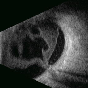
 Cyclic Membrane
Apr 23 2025 by Gustavo Uriel Fonseca Aguirre
This UBM scan reveals pars planitis with characteristic findings: an inflammatory pupillary membrane, a cataractous lens, and cyclitic membrane causing ciliary body detachment and traction. The lens demonstrates spherical deformation due to zonular laxity from ciliary body traction.
Photographer: Gustavo U. Fonseca Aguirre, Hospital Conde de Valenciana, Ciudad de México
Condition/keywords: cyclic membrane, pars planitis
-
 Diabetic Macular Edema
Apr 28 2025 by Gustavo Uriel Fonseca Aguirre
This B-mode longitudinal ultrasound scan demonstrates irregular macular thickening with homogeneous medium-to-high internal reflectivity, consistent with diabetic macular edema. The lesion shows poorly defined borders and absence of cystic spaces or subretinal fluid on dynamic evaluation.
Photographer: Gustavo U. Fonseca Aguirre, Hospital Conde de Valenciana, Ciudad de México
Condition/keywords: diabetic macular edema
-
 Vitreous Bands
Apr 28 2025 by Gustavo Uriel Fonseca Aguirre
This B-mode transversal ultrasound scan shows condensed vitreous bands with anterior traction toward the cornea, accompanied by vitreous cellularity, in a patient with corneal perforation secondary to bacterial keratitis. The findings indicate severe intraocular inflammation with potential vitreous involvement.
Photographer: Gustavo U. Fonseca Aguirre, Hospital Conde de Valenciana, Ciudad de México
Condition/keywords: keratitis, vitreous bands
-
Posterior Hyphema
Apr 29 2025 by Gustavo Uriel Fonseca Aguirre
This kinetic B-mode ultrasound scan (inferior transverse view) reveals combined vitreous and subhyaloid hemorrhage, accompanied by a mobile posterior hyphema level. The dynamic evaluation shows dependent blood shifting with positional changes, confirming fresh hemorrhage without organization.
Condition/keywords: diabetic retinopathy
-
 Weiss Ring
Apr 29 2025 by Gustavo Uriel Fonseca Aguirre
This B-mode axial ultrasound scan demonstrates the Weiss ring, visualized as a circular hyperechoic structure in the vitreous cavity, representing the detached posterior vitreous face with the optic disc insertion site. The ring shows mild mobility on dynamic assessment without retinal traction.
Photographer: Gustavo U. Fonseca Aguirre, Hospital Conde de Valenciana, Ciudad de México
Condition/keywords: Weiss ring
-
 Deep Scleral Buckle
May 5 2025 by Gustavo Uriel Fonseca Aguirre
This B-mode axial ultrasound scan shows an eye with a scleral buckle in place for previous rhegmatogenous retinal detachment. The image demonstrates the characteristic indentation of the ocular wall at the buckle site, with proper retinal reattachment.
Photographer: Gustavo U. Fonseca Aguirre, Hospital Conde de Valenciana, Ciudad de México
Condition/keywords: scleral buckle
-
 Scleral Rupture
May 9 2025 by Gustavo Uriel Fonseca Aguirre
This B-mode longitudinal ultrasound scan reveals dense vitreous hemorrhage, subretinal fluid, annular choroidal detachment, and scleral wall discontinuity with adjacent scleral folds. These findings indicate severe ocular trauma with probable scleral rupture and multi-compartment involvement.
Photographer: Gustavo U. Fonseca Aguirre, Hospital Conde de Valenciana, Ciudad de México
Condition/keywords: ocular trauma, scleral rupture
-
 Macular Hole
May 9 2025 by Gustavo Uriel Fonseca Aguirre
This B-mode longitudinal ultrasound scan demonstrates a full-thickness macular hole, appearing as a well-defined hypoechoic defect in the retinal surface with elevated edges.
Photographer: Gustavo U. Fonseca Aguirre, Hospital Conde de Valenciana, Ciudad de México
Condition/keywords: full thickness macular hole, macular hole
-
 Hemorrhagic Vitreous Detachment
May 21 2025 by Gustavo Uriel Fonseca Aguirre
This B-mode longitudinal ultrasound scan shows a hemorrhagic vitreous detachment with the peripheral hyaloid strongly adherent to a retinal break. Associated vitreous and subhyaloid hemorrhage are present, indicating acute vitreoretinal traction.
Photographer: Gustavo U. Fonseca Aguirre, Hospital Conde de Valenciana, Ciudad de México
Condition/keywords: Hemorrhagic Vitreous Detachment
-
 Glass
Glass
May 21 2025 by Gustavo Uriel Fonseca Aguirre
This B-mode transverse ultrasound scan reveals an intraocular glass foreign body secondary to penetrating trauma, with associated vitreous and subhyaloid hemorrhage. The glass fragments appear as hyperechoic linear structures in both the vitreous cavity and the retinachoroidal complex.
Photographer: Gustavo U. Fonseca Aguirre, Hospital Conde de Valenciana, Ciudad de México
Condition/keywords: glass, intraocular foreign body
-
 RRD in Posterior Staphyloma
May 21 2025 by Gustavo Uriel Fonseca Aguirre
This B-mode axial ultrasound scan of a highly myopic eye demonstrates a prominent posterior staphyloma with an associated inferior retinal detachment sparing the macular region.
Photographer: Gustavo U. Fonseca Aguirre, Hospital Conde de Valenciana, Ciudad de México
Condition/keywords: high myopia, posterior staphyloma, Retina detachment
-
 Morgagnian Ghost in the Deep
Jul 3 2025 by Gustavo Uriel Fonseca Aguirre
This B-mode para-axial ultrasound scan shows a posteriorly dislocated lens with cortical liquefaction, a dense nucleus, and an intact capsular bag. Vitreous bands are visible extending from the anterior to posterior segments. These findings were bilateral and not associated with trauma or prior surgery.
Photographer: Gustavo U. Fonseca Aguirre, Hospital Conde de Valenciana, Ciudad de México
Condition/keywords: ectopia lentis, morgagnian cataract
-
 Macular Retinoschisis
Jul 3 2025 by Gustavo Uriel Fonseca Aguirre
This B-mode longitudinal ultrasound scan reveals macular retinoschisis, demonstrating a characteristic splitting of retinal layers with a smooth, dome-shaped elevation. The lesion shows maintained structural integrity of both inner and outer retinal walls without associated subretinal fluid or vitreous traction.
Photographer: Gustavo U. Fonseca Aguirre, Hospital Conde de Valenciana, Ciudad de México
Condition/keywords: macular retinoschisis
-
 Diabetic Macular Edema
Jul 3 2025 by Gustavo Uriel Fonseca Aguirre
This B-mode longitudinal ultrasound scan demonstrates diabetic macular edema with mild subretinal fluid accumulation, appearing as a subtle hypoechoic space beneath the neurosensory retina. The macular region shows retinal thickening and heterogeneous medium reflectivity, consistent with active exudative changes (arrow). No vitreomacular traction is observed.
Photographer: Gustavo U. Fonseca Aguirre, Hospital Conde de Valenciana, Ciudad de México
Condition/keywords: diabetic macular edema
-
 Choroidal Melanoma
Jul 3 2025 by Gustavo Uriel Fonseca Aguirre
This B-mode transverse ultrasound scan shows asteroid hyalosis with partial posterior vitreous detachment. A dome-shaped choroidal melanoma is observed in the inferior quadrant (preequatorial to equatorial region), appearing as a solid, regularly bordered lesion with heterogeneous internal structure and mild acoustic attenuation. Standardized A-mode reveals medium-to-low internal reflectivity. The tumor measures 11.62 mm in base diameter and 6.60 mm in height. The retina and choroid remain attached, with minimal suprachoroidal fluid in the inferior quadrant.
Photographer: Gustavo U. Fonseca Aguirre, Hospital Conde de Valenciana, Ciudad de México
Condition/keywords: choroidal melanoma
-
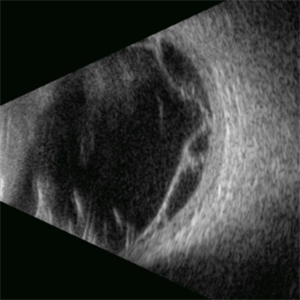
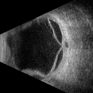
 Retinal Dialysis
Jul 5 2025 by Gustavo Uriel Fonseca Aguirre
This B-mode longitudinal ultrasound scan demonstrates a retinal dialysis, appearing as a linear discontinuity at the ora serrata with associated vitreous base avulsion. The scan reveals mild subretinal fluid extending from the dialysis site with macular involvement.
Photographer: Gustavo U. Fonseca Aguirre, Hospital Conde de Valenciana, Ciudad de México
Condition/keywords: retinal dialysis
-
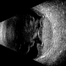
 Giant Retinal Tear
Jul 5 2025 by Gustavo Uriel Fonseca Aguirre
This B-mode longitudinal ultrasound scan reveals a giant retinal tear, demonstrating a circumferential retinal flap with rolled edges extending over M-X to M-I. The vitreous shows diffuse hemorrhage and anterior-posterior traction strands inserting at the tear margins. The remaining retina appears attached without subretinal fluid.
Photographer: Gustavo U. Fonseca Aguirre, Hospital Conde de Valenciana, Ciudad de México
Condition/keywords: giant retinal tear
-
Color Central Retinal Artery
Aug 13 2025 by Gustavo Uriel Fonseca Aguirre
Color Doppler ultrasound of the central retinal artery.
Condition/keywords: Central retinal artery, color doppler
-
Color Rhegmatogenous Retinal Detachment
Aug 13 2025 by Gustavo Uriel Fonseca Aguirre
Color Doppler ultrasound of Rhegmatogenous Retinal Detachment.
Condition/keywords: color doppler, Rhegmatogenous retinal detachment
-
 Hot-Dog Like Choroidal Detachment
Aug 19 2025 by Gustavo Uriel Fonseca Aguirre
This B-mode transverse ultrasound scan reveals a bullous hemorrhagic choroidal detachment with an associated closed-funnel retinal detachment featuring central retinal folds. The choroidal detachment demonstrates convex, lobular elevation with heterogeneous internal reflectivity due to blood accumulation.
Photographer: Gustavo U. Fonseca Aguirre, Hospital Conde de Valenciana, Ciudad de México
Condition/keywords: hemorrhagic choroidal detachment
-
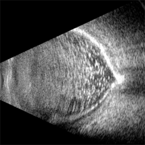
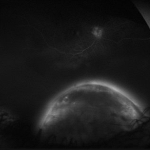 Full Moon
Aug 20 2025 by Gustavo Uriel Fonseca Aguirre
This ultra-widefield fluorescein angiography reveals a hyperfluorescent peripheral inferior choroidal melanoma. The lesion demonstrates early heterogeneous hyperfluorescence with progressive late staining and diffuse leakage.
Photographer: Gustavo U. Fonseca Aguirre, Hospital Conde de Valenciana, Ciudad de México
Condition/keywords: choroidal melanoma
-
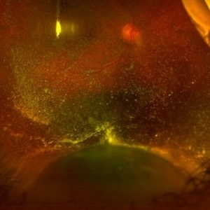 Jupiter and the Sun
Aug 20 2025 by Gustavo Uriel Fonseca Aguirre
This ultra-widefield fundus photograph demonstrates a peripheral inferior choroidal melanoma with overlying asteroid hyalosis. The lesion shows characteristic pigmentation and irregular borders, while the asteroid bodies appear as numerous refractile opacities distributed throughout the vitreous cavity.
Photographer: Gustavo U. Fonseca Aguirre, Hospital Conde de Valenciana, Ciudad de México
Condition/keywords: asteroid hyalosis, choroidal melanoma
-
 Posterior Staphyloma + ON-Coloboma
Aug 20 2025 by Gustavo Uriel Fonseca Aguirre
This axial B-scan reveals a highly myopic eye with a posterior staphyloma and an associated optic nerve coloboma. The staphyloma appears as a deep scleral outpouching adjacent to the optic disc, while the coloboma demonstrates a focal posterior excavation with retrobulbar extension.
Photographer: Gustavo U. Fonseca Aguirre, Hospital Conde de Valenciana, Ciudad de México
Condition/keywords: optic nerve coloboma, posterior staphyloma
-
 Localized Retinal Adhesion Amidst Recurrent Detachment
Aug 25 2025 by Gustavo Uriel Fonseca Aguirre
This longitudinal B-scan reveals a recurrent rhegmatogenous retinal detachment in a patient previously treated with phacovitrectomy, scleral buckle, and 360° endolaser at the equator. The retina has redetached except in the region where the scleral buckle and endolaser were applied, which remains adhered.
Photographer: Gustavo U. Fonseca Aguirre, Hospital Conde de Valenciana, Ciudad de México
Condition/keywords: Recurrent retinal detachment, scleral buckle
-
 Small Retinoschisis
Aug 30 2025 by Gustavo Uriel Fonseca Aguirre
This longitudinal B-scan reveals a small inferotemporal peripheral retinoschisis, appearing as a smooth, thin-walled bipartite retinal separation without associated subretinal fluid or vitreous traction. The lesion demonstrates characteristic acoustic homogeneity and minimal mobility on dynamic evaluation.
Photographer: Gustavo U. Fonseca Aguirre, Hospital Conde de Valenciana, Ciudad de México
Condition/keywords: retinoschisis
-
 AL 39.16 mm
Sep 10 2025 by Gustavo Uriel Fonseca Aguirre
This axial B-scan reveals an elongated globe with an axial length of 39.16 mm, consistent with high axial myopia. Posterior staphyloma and scleral thinning are observed, though the retina remains attached.
Photographer: Gustavo U. Fonseca Aguirre, Hospital Conde de Valenciana, Ciudad de México
Condition/keywords: high myopia
-
 Type 1 Aneurysmal Neovascularization
Sep 22 2025 by Gustavo Uriel Fonseca Aguirre
This transverse B-scan demonstrates vitreous hemorrhage, a bullous retinal detachment involving the macula, and dense subretinal hemorrhage, consistent with type 1 aneurysmal neovascularization. The scan reveals significant exudative activity with multi-level bleeding.
Photographer: Gustavo U. Fonseca Aguirre, Hospital Conde de Valenciana, Ciudad de México
Condition/keywords: polypoidal choroidal vasculopathy (PCV), Type 1 Aneurysmal Neovascularization
-
 Persistent Fetal Vasculature
Sep 27 2025 by Gustavo Uriel Fonseca Aguirre
This longitudinal B-scan reveals Persistent Fetal Vasculature, demonstrating a hyperechoic band extending from the optic nerve head to the posterior lens capsule in a 2-year-old child.
Photographer: Gustavo U. Fonseca Aguirre, Hospital Conde de Valenciana, Ciudad de México
Condition/keywords: persistent fetal vasculature (PFV)
-
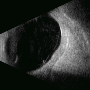
 Tennis Ball
Oct 4 2025 by Gustavo Uriel Fonseca Aguirre
This longitudinal B-scan reveals a serous choroidal detachment exhibiting a smooth, convex, and dome-shaped elevation, creating a tennis ball-like appearance due to its symmetrical and rounded contour.
Photographer: Gustavo U. Fonseca Aguirre, Hospital Conde de Valenciana, Ciudad de México
Condition/keywords: serous choroidal detachment
-
 RD in a Silicone Oil-filled Eye
Oct 4 2025 by Gustavo Uriel Fonseca Aguirre
This transverse B-scan reveals a retinal detachment in a silicone oil-filled eye.
Photographer: Gustavo U. Fonseca Aguirre, Hospital Conde de Valenciana, Ciudad de México
Condition/keywords: Retinal detachment under Silicon Oil
-
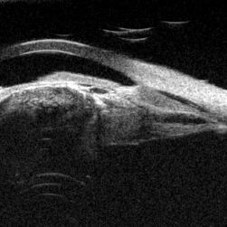
 Optic Nerve Drusen
Oct 8 2025 by Gustavo Uriel Fonseca Aguirre
This longitudinal B-scan demonstrates an optic nerve head drusen, appearing as a hyperechoic, well-defined focus at the optic disc with mild acoustic shadowing. The drusen exhibits a rounded contour and is superficial to the lamina cribrosa.
Photographer: Gustavo U. Fonseca Aguirre, Hospital Conde de Valenciana, Ciudad de México
Condition/keywords: optic nerve drusen
A project from the American Society of Retina Specialists