 Initializing download.
-
 By KANWALJEET HARJOT MADAN, M.S. (Ophthalmology); FAICO (Vitreous - Retina)
Thind Eye Hospital, Jalandhar City (Punjab). India. - Uploaded on Apr 8, 2025.
- Last modified by Joshua Friedman on Apr 9, 2025.
- Rating
- Appears in
- Retina Images
- Condition/keywords
- Retinopathy of Prematurity
- Photographer
- Dr. Kanwaljeet Harjot Madan, Thind Eye Hospital, Jalandhar City (Punjab) INDIA.
- Imaging device
-
Fundus camera
Zeiss Fundus Camera - Description
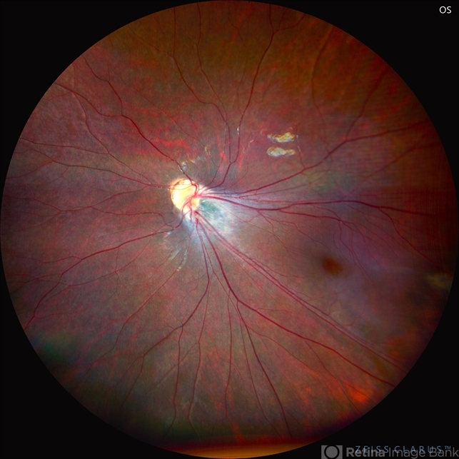
- This is the fundus picture of right eye (RE) of a 4 years female child presented with outward deviation of right eye. Her parents also complained of diminution of vision in both eyes. On examination, her best corrected vision in RE was hand movements close to face and was 20/80 in LE. Posterior segment exam revealed presence of macular scar in RE and presence of dry retinal fold with dragging of retinal vessels. LE fundus revealed presence of nasal drag of optic disc. Parents gave history of untreated ROP as an infant. Retinopathy of Prematurity (ROP) is a Vaso proliferative disorder of Retina occurring in premature infants. Advances in neonatal care and ROP treatment has led these babies to live longer with this disease.
")


